Normal Prostate MRI vs Cancer: Differences Explained

A normal prostate MRI usually shows no suspicious focal lesion, although it cannot rule out every prostate cancer. Prostate cancer may appear as a darker, restricted area on MRI, especially in the peripheral zone of the gland.
Key Takeaways
- A normal prostate MRI usually shows no suspicious focal lesion, although it cannot rule out every prostate cancer.
- Prostate cancer may appear as a darker, restricted area on MRI, especially in the peripheral zone of the gland.
- PI-RADS scores communicate how likely a visible MRI finding is to represent clinically significant cancer.
- Benign prostate enlargement and inflammation can sometimes resemble cancer on MRI.
- A targeted biopsy is commonly recommended when MRI, PSA and clinical findings suggest a meaningful risk of cancer.
A prostate MRI can show whether the prostate has a typical appearance or contains areas that may need closer assessment for cancer. It is a powerful diagnostic tool, but MRI findings alone do not always confirm or exclude prostate cancer; results are interpreted alongside PSA, examination findings and, when needed, biopsy.
Normal prostate MRI vs cancer: the main difference
In normal prostate MRI vs cancer comparisons, the central difference is how prostate tissue looks and behaves on several MRI image sequences. A normal scan generally shows prostate zones with expected shape, borders and signal patterns, without a distinct area that appears suspicious. Cancer may create a focal area with abnormal signal, reduced water movement within tissue and early enhancement after contrast, depending on the MRI protocol used.
However, MRI does not diagnose cancer with complete certainty. Small cancers, low-grade cancers and some cancers in challenging locations may not be clearly visible. Conversely, non-cancerous conditions such as benign prostate enlargement, inflammation or previous bleeding can produce changes that resemble cancer. A radiologist interprets MRI together with prostate-specific antigen (PSA) results, medical history, rectal examination and prior biopsy findings.
Multiparametric MRI, often called mpMRI, combines detailed anatomical images with functional sequences. This approach helps clinicians identify areas that may warrant a targeted biopsy and may also help some people avoid an immediate biopsy when the MRI and overall risk assessment are reassuring.
Can you tell prostate cancer from an MRI?
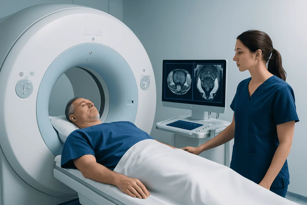
An MRI can identify areas that are suspicious for clinically significant prostate cancer, meaning cancer more likely to grow, spread or require treatment. It can also show whether a suspicious lesion appears confined to the prostate or may extend beyond the gland. For these reasons, prostate MRI is an important part of modern prostate cancer assessment.
Nevertheless, MRI cannot by itself prove that a suspicious area is cancer. The only way to confirm the diagnosis is to examine prostate tissue under a microscope, usually through a biopsy. When a lesion is seen, doctors may use MRI-targeted biopsy techniques to sample that specific area, often alongside standard systematic samples.
A reassuring MRI can lower the likelihood of significant cancer, but it should not be considered a stand-alone all-clear result. A person with a persistently rising PSA, strong family history, concerning examination or inherited cancer-risk mutation may still need follow-up or biopsy despite a low-suspicion MRI.
What is the difference between a normal prostate MRI and cancer?

A normal prostate MRI commonly shows a smooth outer contour and expected internal anatomy. The peripheral zone, where many prostate cancers begin, often appears relatively bright on T2-weighted images. The transition zone, around the urethra, may look more mixed in older adults because benign prostate enlargement is common.
Prostate cancer often appears as a focal darker area on T2-weighted images, particularly in the peripheral zone. It may also show restricted diffusion, meaning water molecules move less freely within tightly packed cells, and it can enhance differently after contrast. Radiologists assess the lesion’s size, location, edges and possible involvement of the capsule, seminal vesicles or nearby structures.
Reports often use the Prostate Imaging Reporting and Data System, known as PI-RADS, to standardize findings. PI-RADS 1 and 2 indicate a very low or low likelihood of clinically significant cancer, PI-RADS 3 is indeterminate, and PI-RADS 4 or 5 indicates increasing suspicion. The score estimates risk; it is not a cancer stage or final diagnosis.
- Normal or low-suspicion MRI: no clear suspicious lesion or only changes likely to be benign.
- Suspicious MRI: a defined lesion with features associated with clinically significant cancer.
- Confirmed cancer: cancer cells identified on biopsy and graded by a pathologist.
Can an MRI tell the difference between BPH and prostate cancer?
MRI can often help distinguish benign prostatic hyperplasia (BPH) from prostate cancer, but the distinction is not always straightforward. BPH is a non-cancerous enlargement of the prostate that usually develops in the transition zone. It can cause urinary symptoms such as a weaker stream, hesitancy, frequent urination or waking at night to pass urine.
BPH commonly produces multiple rounded nodules and an enlarged transition zone, whereas cancer may form a more irregular focal lesion. Yet some BPH nodules can have restricted diffusion or other features that overlap with cancer. Inflammation, known as prostatitis, can also cause abnormal MRI signals and raise the PSA level.
Radiologists use the location and full pattern of MRI findings, not one image alone, to assess whether a change is likely benign or suspicious. If uncertainty remains, a urologist may recommend PSA monitoring, repeat imaging, a targeted biopsy or other assessment. Information about benign prostatic hyperplasia can help explain the common non-cancerous causes of prostate enlargement.
How prostate MRI works and what to expect
A prostate MRI uses a strong magnetic field and radio waves to produce detailed images; it does not use ionizing radiation. Multiparametric MRI typically includes T2-weighted imaging, diffusion-weighted imaging and, in many cases, dynamic contrast-enhanced imaging. These sequences provide complementary information about prostate anatomy and tissue behavior.
It may be considered for people with an elevated or rising PSA, an abnormal digital rectal examination, a previous negative biopsy with ongoing concern, or newly diagnosed cancer that needs local staging. It can also assist with planning a targeted biopsy and, in selected cases, monitoring during active surveillance. Suitability depends on the individual’s symptoms, PSA pattern, prior results and ability to undergo MRI safely.
Before the scan, the imaging team reviews implanted devices, kidney health, allergies and any concerns about confined spaces. Some metal implants are MRI-compatible, while others require special assessment. If contrast is planned, a gadolinium-based agent may be injected through a vein; this is generally well tolerated, but the team will check for relevant kidney disease or previous contrast reactions.
During the examination, the person lies still on a padded table that moves into the scanner. The machine makes loud tapping sounds, so ear protection is provided. The scan commonly takes around 30 to 60 minutes, depending on the protocol. Staff can communicate throughout the examination, and most people return to usual activities immediately afterward.
When to worry about prostate MRI results?
A prostate MRI result deserves timely discussion with a urologist when it describes a PI-RADS 4 or 5 lesion, possible extension beyond the prostate, enlarged pelvic lymph nodes or a lesion that corresponds with a rising PSA. A PI-RADS 3 result is not a diagnosis of cancer, but it often requires individualized risk assessment because it falls into an uncertain middle category.
Other important information includes PSA density, which relates PSA to prostate size, changes in PSA over time, age, family history and genetic risk. A large prostate from BPH can raise PSA without cancer, while a relatively high PSA for the gland size may increase concern. The clinician will explain how these findings fit together rather than relying on one test result.
If biopsy is advised, MRI findings can guide sampling of the most concerning area. MRI-targeted prostate biopsy may improve the chance of finding significant disease compared with non-targeted sampling alone. If cancer is confirmed, the results from pathology, PSA and imaging guide discussions about surveillance, surgery, radiation or other treatments.
Recovery, benefits and limitations of prostate MRI
Prostate MRI itself is noninvasive, so recovery is usually immediate. There is no incision, no radiation exposure and typically no need for sedation. People can usually eat, drink, drive and resume normal routine after the scan unless their care team gives different instructions for a specific reason.
The main benefit is better risk assessment before biopsy or treatment planning. MRI can help locate suspicious tissue, provide information about the extent of known cancer and support more informed decisions about whether biopsy is appropriate. It may also help reduce unnecessary biopsies in some people with a low-suspicion scan and otherwise low clinical risk.
Limitations are equally important to understand. MRI can miss some cancers and can identify abnormalities that prove non-cancerous after biopsy. Contrast reactions are uncommon but possible, and MRI may be unsuitable or require adaptation for some implants or severe claustrophobia. The scan should therefore be viewed as one component of a careful urological evaluation, rather than a replacement for clinical follow-up.
For international patients, Acibadem International’s multidisciplinary specialists and JCI-accredited hospitals can coordinate prostate imaging, urology assessment and further evaluation when indicated.
When to seek medical care
Medical advice is appropriate for a persistently elevated or rising PSA, an abnormal prostate examination, blood in urine or semen, unexplained bone pain, unintentional weight loss, or urinary symptoms that are new, worsening or troublesome. Many urinary symptoms are caused by BPH or other non-cancerous conditions, but they should be assessed rather than self-diagnosed.
Urgent care is important if a person cannot pass urine, develops fever or chills with urinary symptoms, has severe lower abdominal pain, or experiences heavy bleeding in urine. These symptoms can have several causes and need prompt medical assessment.
A urologist can review MRI findings in context and discuss the next appropriate step. People should bring their MRI report, images if available, PSA history, biopsy reports and a list of medicines to the appointment. Questions about prostate cancer risk and further testing can then be addressed in a personalized way.
Frequently asked questions
Can a normal prostate MRI rule out cancer?
A normal or low-suspicion MRI lowers the chance of clinically significant prostate cancer, but it cannot rule out all cancer. Small, low-grade or difficult-to-see tumors may not be detected. PSA trends, examination findings and individual risk factors still guide follow-up.
What does a PI-RADS 3 prostate MRI result mean?
PI-RADS 3 means the MRI finding is indeterminate, with an intermediate likelihood of clinically significant prostate cancer. A urologist considers PSA density, family history, prior biopsy findings and other factors to decide whether biopsy, monitoring or repeat MRI is appropriate.
Does a PI-RADS 4 or 5 score mean cancer is certain?
No. PI-RADS 4 and 5 scores indicate that cancer is increasingly likely, but they do not confirm it. A biopsy is generally needed to determine whether cancer is present and, if so, its grade.
Can prostatitis look like prostate cancer on MRI?
Yes. Inflammation of the prostate can create MRI changes that overlap with cancer and may also increase PSA. Symptoms, laboratory tests, imaging features and follow-up help clinicians distinguish these possibilities, although biopsy may sometimes be needed.
How long does it take to get prostate MRI results?
Timing varies by imaging center and the complexity of the examination. A radiologist reviews the images and provides a report to the referring clinician, who explains the findings and next steps. The care team can give the most accurate expected timeframe.
Is a prostate biopsy always needed after an abnormal MRI?
Not always, but it is often recommended when MRI findings are suspicious and the overall clinical risk is significant. Decisions should consider the PI-RADS score, PSA level and density, medical history and patient preferences. A urologist can explain the likely value and possible downsides of biopsy in the individual situation.
References
- European Association of Urology
- American Urological Association
- National Cancer Institute
- Radiological Society of North America
- Prostate Imaging Reporting and Data System (PI-RADS)
This article is for general information only and is not a substitute for professional medical advice. Please consult a qualified doctor about your individual situation.
Oncology care in Turkey — second opinion and treatment plan
JCI-accredited · board-certified surgeons · reply within 24h
Add us as a Preferred Source to see more of our trusted health content across Google Search, AI Overviews and Discover.

More from the Health Library



Related Specialists

Dr. Nurcan Öztürk Yumuk
Anesthesiology
Dyt. Senem Azizzade
Nutrition & Diet
Prof. Dr. Gökhan Demir
Medical Oncology
Assoc. Prof. Dr. Tonguç Utku Yılmaz
Kidney Transplant Center