Tracheal Deviation — Explained by Medical Evidence, Not Myths

Tracheal deviation is a finding that may be noticed during a physical examination or on imaging, not a disease by itself. A shifted trachea can result from structures pushing it away or from loss of lung volume pulling it toward one side.
Key Takeaways
- Tracheal deviation is a finding that may be noticed during a physical examination or on imaging, not a disease by itself.
- A shifted trachea can result from structures pushing it away or from loss of lung volume pulling it toward one side.
- Sudden shortness of breath, chest pain, blue lips, severe distress, or deviation after an injury needs emergency medical care.
- Chest imaging, especially a chest X-ray or CT scan, helps identify the cause and guide treatment.
- Treatment focuses on the underlying condition; a tracheal shift does not always require treatment itself.
Tracheal deviation means that the trachea, or windpipe, appears shifted to one side rather than centered in the neck and upper chest. It is a clinical sign rather than a diagnosis: some causes are longstanding and non-urgent, while sudden deviation with breathing symptoms requires immediate evaluation.
What Tracheal Deviation Means
Tracheal deviation describes a position in which the trachea, commonly called the windpipe, is not in its usual central location. The trachea normally runs from the lower throat into the chest, where it divides into the two main airways leading to the lungs. A small degree of apparent asymmetry can occur because of normal anatomy, body position, or the way an image was taken. However, a clear or new shift may provide an important clue about changes in the neck, lungs, pleural space, or chest.
It is important to separate the finding from the cause. Tracheal deviation does not automatically mean a person has a severe lung problem, cancer, or an emergency. It may develop gradually from thyroid enlargement, scarring, or prior surgery, for example. In other circumstances, particularly when it appears suddenly alongside breathing difficulty or chest symptoms, it can indicate pressure within the chest that needs urgent treatment.
Clinicians assess tracheal position together with symptoms, oxygen level, breathing effort, medical history, and imaging. A person should not try to diagnose a deviated trachea by looking in a mirror or pressing deeply on the neck. The windpipe can be difficult to assess accurately outside a clinical examination, and a professional evaluation is safer and more reliable.
How a Tracheal Shift Happens
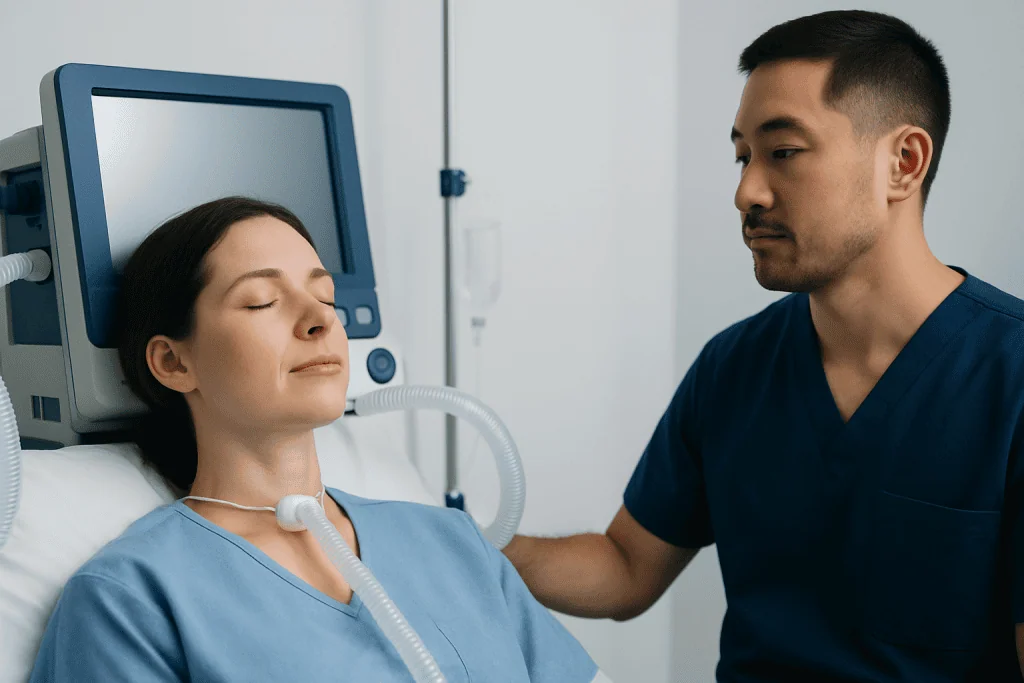
A useful way to understand tracheal deviation is to consider whether something is pushing the trachea away or whether loss of volume is pulling it toward one side. Increased pressure or volume on one side of the chest can move the trachea away from that side. Examples include a large collection of air or fluid around a lung, or a sizable mass within the chest.
By contrast, when part of a lung loses volume, the trachea may move toward the affected side. This can occur with lung collapse, substantial scarring, or after removal of part or all of a lung. These patterns are clinical principles, not a substitute for imaging: the direction and degree of shift must be interpreted in the context of the whole examination.
Changes in the neck can also alter tracheal position. An enlarged thyroid gland, a thyroid nodule, other neck masses, or previous neck surgery may gradually displace the windpipe. In many people, these changes develop slowly and are first detected during a routine examination or imaging performed for another reason.
Possible Causes and Related Symptoms

Causes of tracheal deviation range from benign structural changes to acute chest conditions. A large goiter, which is an enlarged thyroid gland, may push the trachea to one side. Other possible causes include masses in the neck or chest, enlarged lymph nodes, congenital anatomical variation, and scarring after infection, radiation therapy, or surgery. The underlying cause determines whether the finding is stable, treatable, or urgent.
Within the chest, a pleural effusion (fluid around the lung), a large pneumothorax (air around the lung), bleeding into the pleural space after injury, or a large mass may push the trachea away from the affected side. Severe lung collapse, chronic scarring, or surgical removal of lung tissue may pull it toward the affected side. A tension pneumothorax is a life-threatening form of collapsed lung in which pressure can build rapidly and affect circulation; it is treated as an emergency.
Tracheal deviation itself may cause no symptoms, especially when it develops slowly. Symptoms come more often from the underlying condition and can include cough, wheezing, noisy breathing, hoarseness, a neck lump, trouble swallowing, chest discomfort, reduced exercise tolerance, fever, or shortness of breath. Rapidly worsening breathing difficulty, particularly after chest trauma or a medical procedure, should never be attributed to a tracheal shift alone and needs immediate medical assessment.
- Gradual neck fullness or a visible lump may suggest a thyroid or other neck-related cause.
- Sudden one-sided chest pain and breathlessness may occur with a pneumothorax.
- Persistent cough, fever, or unexplained weight loss should be evaluated for their underlying cause.
- A history of lung surgery, severe infection, or chronic lung disease may help explain a stable shift on imaging.
How Doctors Confirm the Finding
A clinician may first notice possible tracheal deviation during a neck and chest examination. They may gently inspect and feel the area above the breastbone, observe breathing, listen to the lungs, and assess for neck swelling. Because body position and anatomy can make bedside assessment uncertain, imaging is generally needed to confirm whether a meaningful shift is present and to identify why it has occurred.
A chest X-ray is often the first imaging test. It can show tracheal position as well as signs of lung collapse, fluid around the lungs, changes in lung volume, or other chest abnormalities. The image must be taken and interpreted carefully, because rotation of the person during the X-ray can make the trachea appear displaced when it is not.
CT imaging of the chest or neck provides more detailed information when needed. Ultrasound may be useful for evaluating the thyroid and other neck structures. Depending on the clinical situation, doctors may also request blood tests, lung function testing, a bronchoscopy to look inside the airways, or sampling of fluid or tissue. These tests are selected to clarify the cause, not simply because deviation is visible.
Treatment Depends on the Underlying Cause
There is no single treatment for tracheal deviation. Care is directed at the condition causing the shift and at any effect it has on breathing or circulation. A stable, mild deviation associated with longstanding anatomy or prior surgery may only need observation and follow-up. The care team will consider imaging findings, symptoms, changes over time, and the person’s overall health.
When fluid around a lung is large or causing symptoms, drainage and treatment of its cause may be considered. A pneumothorax may require observation, oxygen support, aspiration, or placement of a chest tube, depending on its size, symptoms, and type. Tension pneumothorax requires immediate emergency decompression before further evaluation. Lung collapse may be managed by addressing mucus blockage, infection, airway obstruction, or another contributing condition.
For thyroid enlargement, nodules, or other neck masses, management may include monitoring, medication for a related thyroid condition, biopsy, surgery, or other specialist-directed treatment. If a mass is suspected, clinicians use appropriate imaging and tissue testing before recommending care. Treatment decisions should be individualized; not every displaced trachea requires an operation.
Supportive care may include oxygen, inhaled medicines, pain control, or treatment for infection when clinically appropriate. People should follow the plan given by their treating clinician and attend recommended follow-up imaging, especially if a shift was found incidentally and its cause is still being clarified.
What People Can Do and What to Avoid
There is no home method to reposition the trachea, and forceful neck massage, manipulation, or attempts to “straighten” the windpipe should be avoided. These approaches cannot treat an underlying chest or neck condition and could delay appropriate care. A person who has been told of a tracheal shift should ask what caused it, whether it is new or longstanding, and what follow-up is recommended.
General respiratory health measures can support overall lung health but do not replace medical treatment. Avoiding tobacco smoke and vaping, keeping recommended vaccinations up to date, managing chronic lung conditions as prescribed, and seeking care for persistent respiratory symptoms can all be helpful. People with known thyroid enlargement or chronic lung disease should report new swallowing problems, voice changes, worsening breathlessness, or a rapidly enlarging neck swelling.
Keeping a record of prior chest imaging, surgery, major lung infections, and thyroid assessments can help clinicians compare findings over time. This is particularly useful when a tracheal deviation is discovered unexpectedly. A comparison with older imaging may show that the position has been unchanged for years, which can significantly inform the evaluation.
When to Seek Medical Care
Emergency medical care is needed for sudden or severe shortness of breath, rapidly worsening chest pain, blue or gray lips, fainting, confusion, severe breathing effort, or symptoms that begin after chest injury. Emergency assessment is also important if a person develops these symptoms after a central line procedure, lung procedure, or surgery involving the chest or neck. These symptoms may reflect a serious cause that needs rapid treatment.
A prompt, non-emergency medical appointment is appropriate for persistent cough, unexplained breathlessness, a new neck lump, neck pressure, hoarseness, difficulty swallowing, repeated chest infections, or an abnormal chest X-ray report mentioning tracheal deviation. The clinician can determine whether further imaging or referral to a respiratory, ear, nose and throat, thyroid, or thoracic specialist is appropriate.
For international patients, Acibadem International’s multidisciplinary specialists and JCI-accredited hospitals can assess tracheal deviation and treat the underlying condition when care is needed. The right pathway depends on the person’s symptoms, examination, and imaging results, so an individualized medical evaluation remains essential.
Frequently asked questions
Is tracheal deviation always serious?
No. Tracheal deviation is a physical or imaging finding, not a diagnosis, and it can be longstanding or related to non-urgent structural changes. Its importance depends on the cause, how quickly it developed, and whether symptoms such as breathlessness or chest pain are present.
Can tracheal deviation be normal?
The trachea is usually near the midline, but small apparent differences may result from normal anatomy, posture, or rotation during an X-ray. A clinician can determine whether the shift is real and clinically meaningful by examining the person and reviewing properly obtained imaging.
Which direction does the trachea move in a collapsed lung?
The answer depends on the type of lung problem. Loss of lung volume, such as marked collapse of a lung segment, can pull the trachea toward the affected side, while pressure from a large pneumothorax can push it away. Imaging and the overall clinical picture are needed to distinguish these situations.
Can thyroid problems cause a deviated trachea?
Yes. A significantly enlarged thyroid gland or a large thyroid nodule can gradually displace the trachea, sometimes without causing symptoms initially. New neck swelling, trouble swallowing, voice changes, or breathing difficulty should be assessed by a healthcare professional.
How is tracheal deviation diagnosed?
A doctor may suspect it during a neck and chest examination, but chest X-ray or CT imaging is commonly used to confirm it. Additional tests, such as thyroid ultrasound, blood tests, bronchoscopy, or fluid sampling, may be used to investigate the cause.
Can a deviated trachea be corrected?
It may improve when the underlying cause is successfully treated, such as drainage of fluid around a lung or management of a compressing neck mass. Some stable deviations do not need correction if they are not causing symptoms or functional problems; the treatment plan is based on the cause and clinical findings.
References
- American College of Radiology
- American Thoracic Society
- Merck Manual Professional Edition
- National Heart, Lung, and Blood Institute
- Radiological Society of North America
This article is for general information only and is not a substitute for professional medical advice. Please consult a qualified doctor about your individual situation.
Explore treatments in Turkey — costs, top hospitals & a free quote
JCI-accredited · board-certified surgeons · reply within 24h
Add us as a Preferred Source to see more of our trusted health content across Google Search, AI Overviews and Discover.