Oligodendroglioma MRI: Preparation, Procedure and Results

MRI is the main imaging test used to investigate and monitor suspected oligodendroglioma. The scan is painless and does not use ionizing radiation, although contrast dye may be recommended.
Key Takeaways
- MRI is the main imaging test used to investigate and monitor suspected oligodendroglioma.
- The scan is painless and does not use ionizing radiation, although contrast dye may be recommended.
- MRI can show tumor size, location, swelling and change over time, but it cannot confirm the diagnosis alone.
- Oligodendroglioma is classified using tumor tissue and molecular markers, including IDH mutation and 1p/19q codeletion.
- Treatment and outlook depend on tumor grade, symptoms, location, molecular findings and whether the tumor can be safely removed.
An oligodendroglioma MRI is a brain scan that helps doctors identify a possible tumor, understand its location and features, and monitor changes over time. MRI findings can strongly suggest oligodendroglioma, but a tissue sample and molecular testing are usually needed to confirm the diagnosis and tumor grade.
Overview: what an oligodendroglioma MRI can show
An oligodendroglioma MRI is a magnetic resonance imaging scan of the brain performed when symptoms or an earlier test raise concern about a brain tumor. It creates detailed images of brain structures, allowing specialists to assess whether there is an abnormal area, where it is located, how large it is and whether it is affecting nearby tissue.
MRI is particularly useful because it provides more soft-tissue detail than a CT scan and can be repeated during follow-up without radiation exposure. It may be used before treatment, after surgery, during chemotherapy or radiotherapy, and for longer-term surveillance. However, imaging alone cannot reliably establish the exact tumor type; confirmation generally requires tumor tissue examined by a neuropathologist.
For an integrated diagnosis of oligodendroglioma, current classification relies on both microscopic examination and molecular testing. In adults, an oligodendroglioma is typically defined by an IDH mutation and whole-arm 1p/19q codeletion. These results help the care team discuss prognosis and choose appropriate treatment.
Can oligodendroglioma be seen on an MRI?
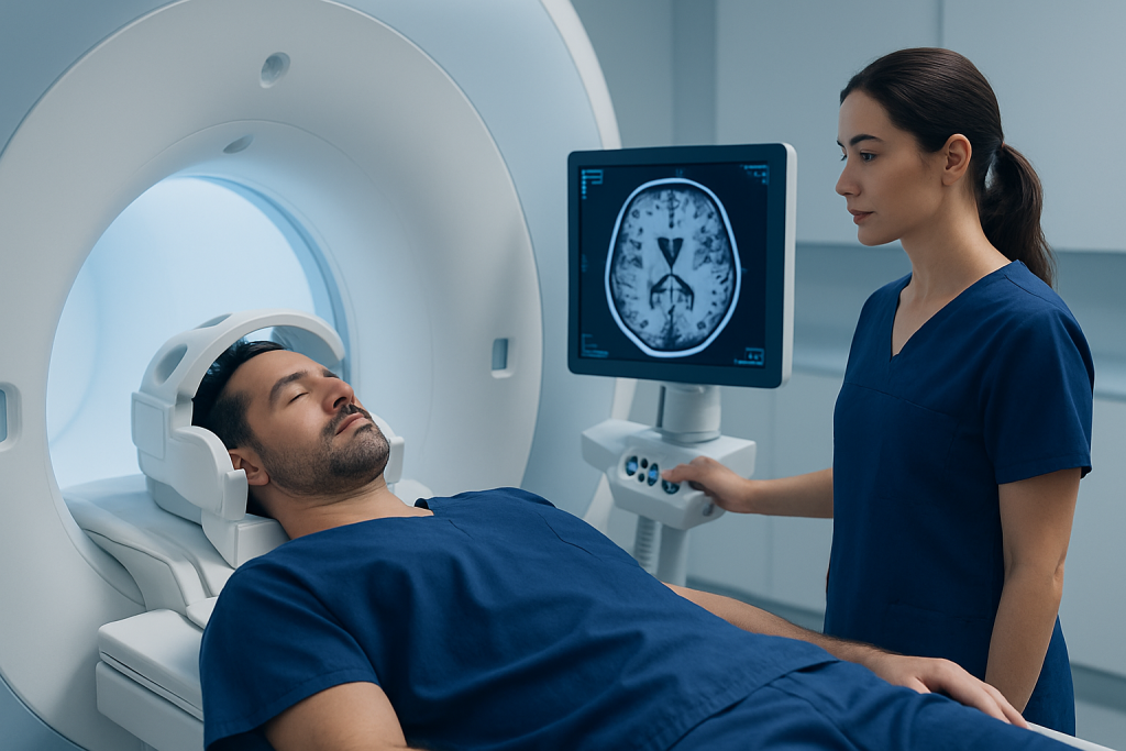
Yes. Oligodendroglioma can often be seen on an MRI as an area that looks different from normal brain tissue. Many tumors arise in the cerebral hemispheres, especially the frontal lobe, though they may occur elsewhere. The MRI helps show the lesion’s boundaries, its relationship to important brain areas and whether it is associated with swelling, pressure effects or changes in surrounding tissue.
The oligodendroglioma MRI appearance varies among individuals. On common MRI sequences, a tumor may appear darker on T1-weighted images and brighter on T2-weighted or FLAIR images. Some tumors have areas of calcification, which may be easier to recognize on CT, and some show contrast enhancement after an intravenous contrast agent is given. Enhancement can occur in lower- or higher-grade tumors, so it does not by itself determine grade.
People looking for oligodendroglioma MRI images may encounter educational image collections, including resources sometimes searched as “oligodendroglioma MRI Radiopaedia.” Such images can illustrate typical patterns, but individual scans must be interpreted in clinical context by a neuroradiologist. Other brain tumors, vascular changes, inflammation and prior treatment effects can sometimes produce overlapping appearances.
How the MRI works, who may need it and how to prepare

MRI uses a strong magnetic field and radiofrequency waves to produce detailed cross-sectional pictures of the brain. It does not use X-rays. The scanner may take images in several planes and with several sequences, each designed to highlight different features such as fluid, tissue structure, blood products or contrast uptake.
A doctor may request an oligodendroglioma brain MRI for persistent or unexplained seizures, new neurological symptoms, a suspected lesion on CT, or monitoring of a known tumor. Symptoms can include headaches with other concerning features, weakness, speech changes, changes in sensation, personality or memory changes, or visual disturbance. Symptoms alone do not mean a person has a brain tumor, but they deserve appropriate assessment.
Before the appointment, patients should tell the imaging team about any implanted device, metal fragments, prior surgery, kidney disease, pregnancy, allergies or previous reaction to MRI contrast. Many implants are MRI-compatible, but this must be checked. Patients may usually eat and drink normally unless the center gives different instructions, and they should remove metal items such as jewelry, watches, hearing aids and removable dental appliances before scanning.
- Bring previous brain scans and medical reports if available.
- Tell staff about claustrophobia or difficulty lying flat; support, relaxation measures or sedation may be available when appropriate.
- Ask whether contrast is planned and whether kidney-function testing is needed.
Step by step: what happens during an oligodendroglioma MRI
At the imaging center, the patient completes a safety screening form and changes into clothing without metal fasteners if necessary. A radiographer positions the patient on a moving table, usually lying on their back. A lightweight head coil is placed around the head to improve image quality, and cushions or straps may help the head remain still and comfortable.
The table then moves into the scanner. During image acquisition, the machine produces loud tapping or knocking sounds, so earplugs or headphones are provided. The patient can communicate with the radiographer through an intercom and may be given an alert device. Staying still is important because movement can blur the images and may require sequences to be repeated.
Some examinations include gadolinium-based contrast given through a small intravenous line, often after initial images are taken. Contrast can make certain tumor features more visible and can help assess blood-brain barrier changes. The scan commonly lasts about 30 to 60 minutes, depending on the protocol and whether contrast or advanced sequences are required.
Advanced MRI techniques may sometimes be added. These can include perfusion imaging to evaluate blood flow, diffusion imaging to assess water movement in tissue, spectroscopy to examine chemical signals, or functional MRI when surgery near language or movement areas is being considered. These methods provide useful additional information but do not replace pathology.
Results, benefits, limitations and possible risks
A neuroradiologist reviews the images and sends a report to the referring clinician. The report commonly describes the lesion’s location, dimensions, signal characteristics, calcification or bleeding features, degree of contrast enhancement, surrounding swelling and any pressure on nearby brain structures. The treating team will interpret these findings alongside symptoms, examination results and previous scans.
The major benefit of MRI is its detailed, noninvasive view of the brain. It helps specialists plan the safest path for biopsy or surgery, establish a baseline before treatment and detect changes over time. Serial MRI scans are especially important because tumor behavior and treatment effects may develop gradually.
MRI has limitations. It cannot conclusively distinguish oligodendroglioma from every other brain lesion, and it cannot assign a definitive grade without tissue analysis. After radiotherapy or chemotherapy, new or changing MRI appearances may reflect tumor progression, inflammation or treatment-related changes, sometimes requiring repeat imaging or further assessment.
MRI itself is generally safe for eligible patients. Potential concerns include anxiety in enclosed spaces, discomfort from lying still and noise exposure without proper hearing protection. Gadolinium contrast reactions are uncommon and usually mild, but serious reactions can occur rarely. The imaging team carefully reviews kidney health and other factors before contrast is used.
What is a low-grade oligodendroglioma?
A low-grade oligodendroglioma generally refers to an oligodendroglioma classified as World Health Organization grade 2. Grade 2 tumors are usually slower growing than grade 3 tumors, but they are still malignant brain tumors and require long-term specialist follow-up. The term “low grade” should not be understood to mean harmless.
Grade is determined from tumor tissue, not from MRI alone. Pathologists consider how the cells look under the microscope, whether there are features such as increased cell division or abnormal blood vessel growth, and the molecular profile. A tumor that appears relatively nonaggressive on imaging can still need biopsy or surgery to establish an accurate diagnosis.
Management may include observation with scheduled MRI for selected people, especially after extensive surgical removal and when symptoms are controlled. Others may benefit from surgery, radiotherapy, chemotherapy or a combination of approaches. Decisions are individualized through discussion among neurosurgery, neuro-oncology, radiation oncology, neuroradiology and pathology specialists.
Is oligodendroglioma grade 3 terminal?
No. Oligodendroglioma grade 3 is a serious, higher-grade brain tumor, but describing it as terminal is not medically accurate for every person. Outcomes vary substantially depending on age, overall health, tumor location, extent of surgery, response to treatment and, importantly, molecular features such as IDH mutation and 1p/19q codeletion.
Grade 3 oligodendroglioma usually requires active treatment and close follow-up. Care may involve maximal safe surgical removal followed by radiotherapy and chemotherapy, based on the individual clinical situation. The aim is to control the tumor, preserve neurological function and support quality of life.
It is understandable for patients and families to want a clear prediction. Their neuro-oncology team is best placed to explain the specific pathology results, MRI findings and treatment options in a compassionate, individualized way. Supportive care for seizures, fatigue, cognitive symptoms and emotional wellbeing is an important part of care at every stage.
Can oligodendroglioma go into remission?
Oligodendroglioma can respond well to treatment, and scans may show no visible evidence of active tumor or a prolonged period without growth. Doctors may describe this as remission, stable disease, response or no evidence of progression, depending on the clinical setting. Because these tumors can recur or change over time, continued MRI follow-up remains important even after a strong treatment response.
Complete removal may not always be possible when a tumor is close to areas controlling speech, movement, sensation or vision. In these situations, treatment focuses on achieving the greatest safe tumor reduction and using additional therapies when indicated. MRI after surgery establishes a new baseline and helps guide further decisions.
Care is usually planned by a multidisciplinary brain tumor team. Acibadem International’s multidisciplinary specialists and JCI-accredited hospitals support international patients with assessment and treatment planning for brain tumors, including coordinated imaging, pathology and specialist care.
When to seek medical care
Prompt medical assessment is advisable for a first seizure, a seizure lasting longer than five minutes, repeated seizures without full recovery, sudden weakness or numbness, new difficulty speaking, loss of consciousness, severe sudden headache, or a major change in vision or balance. Emergency services should be contacted for severe or rapidly developing neurological symptoms.
A non-urgent medical appointment is appropriate for persistent or progressively worsening headaches, recurrent episodes of confusion, subtle personality or memory changes, ongoing weakness, or new seizures that have already stopped. These symptoms have many possible causes, and evaluation helps identify the right next step.
For a known oligodendroglioma, patients should contact their treating team about new or worsening seizures, headaches, neurological changes or treatment side effects. Regularly scheduled MRI is an essential part of monitoring, and appointments should not be skipped without discussing this with the care team.
Frequently asked questions
How accurate is MRI for oligodendroglioma?
MRI is highly valuable for finding and characterizing a suspected brain tumor, defining its location and tracking it over time. However, it cannot confirm oligodendroglioma with complete certainty because other lesions can look similar. A definitive diagnosis usually requires biopsy or surgical tissue analysis with molecular testing.
Does an oligodendroglioma always enhance with MRI contrast?
No. Some oligodendrogliomas show little or no contrast enhancement, while others enhance to varying degrees. Enhancement is one feature radiologists assess, but it does not independently confirm the diagnosis or determine tumor grade.
Do I need contrast for an oligodendroglioma MRI?
Contrast is often helpful because it can reveal features that are less visible on non-contrast sequences and can support treatment planning or follow-up. Whether it is needed depends on the clinical question, prior imaging and individual safety considerations. The imaging team will review kidney function and previous contrast reactions when relevant.
How often are follow-up MRIs needed for oligodendroglioma?
The schedule varies according to tumor grade, treatment stage, symptoms and MRI stability. Scans may be more frequent after diagnosis or treatment and less frequent when results remain stable over time. A neuro-oncology team creates an individualized surveillance plan.
Can MRI tell whether an oligodendroglioma is grade 2 or grade 3?
MRI may show features that raise or lower concern for a more aggressive tumor, but it cannot reliably establish the grade alone. Grade 2 or grade 3 classification requires examination of tumor tissue by a pathologist. Molecular testing is also central to the modern diagnosis of oligodendroglioma.
What should a patient do if they feel claustrophobic during MRI?
Patients should tell the imaging provider before the appointment. The team may explain the process in detail, offer communication and comfort measures, or discuss medication or sedation if clinically appropriate. It is important not to take sedating medicine without medical guidance, especially if transport home may be needed.
References
- World Health Organization Classification of Tumours Editorial Board
- National Cancer Institute
- European Association of Neuro-Oncology
- American Cancer Society
- Radiological Society of North America
This article is for general information only and is not a substitute for professional medical advice. Please consult a qualified doctor about your individual situation.
Oncology care in Turkey — second opinion and treatment plan
JCI-accredited · board-certified surgeons · reply within 24h
Add us as a Preferred Source to see more of our trusted health content across Google Search, AI Overviews and Discover.