Ookp Surgery: Procedure, Recovery and Results

OOKP surgery is reserved for severe, bilateral corneal blindness with major ocular surface damage. The procedure is completed in stages and involves both ophthalmic and dental or maxillofacial specialists.
Key Takeaways
- OOKP surgery is reserved for severe, bilateral corneal blindness with major ocular surface damage.
- The procedure is completed in stages and involves both ophthalmic and dental or maxillofacial specialists.
- Recovery is long-term and requires frequent follow-up to monitor vision, pressure, the graft and surrounding tissues.
- Potential benefits can be substantial for carefully selected patients, but complications and further procedures are possible.
- Not all eye surgeries have the same recovery time; superficial keratectomy and keratoconus procedures generally heal much faster than OOKP surgery.
OOKP surgery, also called osteo-odonto-keratoprosthesis, is a highly specialized, staged procedure for selected people with severe corneal blindness and a damaged eye surface. It uses a small tooth-and-bone graft to support an optical cylinder, offering the possibility of useful vision when standard corneal transplantation is unlikely to succeed.
Overview: what is OOKP surgery?
OOKP surgery is a reconstructive eye procedure designed for a small group of people with severe corneal blindness in both eyes. Its full name is osteo-odonto-keratoprosthesis. The operation may be considered when the front surface of the eye is severely scarred or dry and conventional corneal transplantation is unlikely to remain clear or heal properly.
The procedure uses part of a patient’s own tooth root and surrounding jawbone to hold a tiny clear optical cylinder. This tooth-bone support is implanted into the eye after the ocular surface has been prepared, creating a stable pathway for light to enter the eye. OOKP is sometimes described as a type of biological keratoprosthesis, meaning an artificial corneal device supported by living tissue.
It is not a routine treatment for blurred vision, cataracts, or uncomplicated corneal disease. OOKP is performed only in experienced specialist centers after detailed assessment, because the surgery is complex and lifelong monitoring is essential.
How OOKP works and who may be a candidate
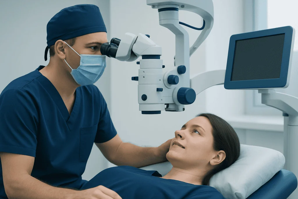
For sight to be possible, light must pass through the front of the eye and reach a functioning retina and optic nerve. In severe ocular surface disease, the cornea and protective tissues may be too damaged for standard grafting. OOKP creates a small, protected optical channel through the front of the eye while using the patient’s own tooth and bone as a durable support.
Potential candidates usually have profound vision loss in both eyes caused by conditions such as severe chemical or thermal injury, advanced cicatrizing eye disease, or severe dry-eye-related ocular surface failure. They generally need to have enough retinal and optic nerve function for visual recovery to be possible. Evaluation may also include dental assessment, imaging, eye pressure testing, ultrasound, and tests of light perception.
Suitability depends on the underlying cause of vision loss, the health of the eyelids and eye tissues, oral and dental health, general medical fitness, and the person’s ability to attend regular reviews. In some people, other ocular-surface reconstruction or corneal treatment is more appropriate. Related corneal conditions, including keratoconus, are commonly managed with less extensive approaches when the eye surface is otherwise healthy.
OOKP surgery step by step

OOKP is commonly performed in two major stages, separated by a healing period. In the first stage, the surgical team prepares the eye surface. This may involve removing scarred tissue and covering the eye with a mucous membrane graft, often taken from inside the mouth, to create a more stable and moist surface.
During the same stage or a coordinated procedure, a dentist or maxillofacial surgeon removes a suitable tooth, often a canine tooth, with a small piece of surrounding bone. The tooth-bone graft is shaped to hold the optical cylinder. It is then placed beneath the skin or soft tissue, typically in the cheek or beneath the eye muscles, so that it can become integrated with the patient’s own tissue.
After several months of healing, the graft is retrieved and implanted into the prepared eye. The optical cylinder is positioned so that light can pass through it. Additional procedures, such as lens removal, glaucoma management, or retinal assessment, may be needed depending on the individual eye. The exact sequence varies, and the treating team explains the planned stages before surgery.
Because OOKP combines ophthalmology with dental and reconstructive expertise, planning is multidisciplinary. Acibadem International’s multidisciplinary specialists and JCI-accredited hospitals assess and treat complex eye conditions for international patients, including cases that require coordinated ophthalmic and surgical care.
Recovery timeline and expected results
Recovery after OOKP surgery is gradual and involves close observation rather than a single, fixed healing date. The interval between stages is often several months, allowing the eye surface and tooth-bone graft to stabilize. After the final implantation, vision may improve as the eye heals and the optical pathway is assessed, but visual rehabilitation continues over time.
Follow-up appointments are essential. The team monitors the clarity and position of the optical cylinder, the health of the mucous membrane covering, eye pressure, inflammation, and the retina and optic nerve. Some patients need specialized imaging because routine examination can be more difficult after a keratoprosthesis procedure.
When successful in a suitable candidate, OOKP can restore useful functional vision, such as recognizing faces, navigating surroundings, or reading with visual aids. The degree of improvement is individual and depends especially on the condition of the retina and optic nerve before surgery. Glasses, magnification, low-vision rehabilitation, and adaptations for daily activities may still be helpful.
Compared with OOKP, many procedures used for corneal shape disorders have much shorter recovery periods. For example, corneal cross-linking may be used to slow progression of keratoconus, while other interventions are selected according to corneal thickness, shape, and visual needs.
Benefits, limitations and possible risks
The main potential benefit of OOKP surgery is restoration of a visual pathway in eyes where standard corneal grafts are very unlikely to work. The use of the patient’s own tooth and bone can offer a biologically compatible support for the optical cylinder in a severely compromised eye surface.
However, OOKP is major surgery and does not guarantee a specific level of sight. Possible complications include infection, inflammation, glaucoma or raised eye pressure, retinal detachment, damage to the optical cylinder, loosening or resorption of the tooth-bone graft, tissue breakdown around the implant, and vision loss from problems affecting the retina or optic nerve. Further surgery may be required.
There can also be mouth-related effects from tooth removal or mucosal graft harvesting, such as temporary discomfort, swelling, or altered sensation. The medical team considers oral health, medications, immune-related disease, and other health conditions when discussing risks. Patients should ask what warning symptoms to report, how follow-up will be arranged, and what support is available during long-term recovery.
Can I watch TV after superficial keratectomy?
After superficial keratectomy, a person may be able to watch television for short periods when comfort allows, but it is usually best to rest the eyes during the first days of healing. Light sensitivity, tearing, blurred vision, and a foreign-body sensation are common while the corneal surface regenerates. The surgeon may recommend limiting screen use if it worsens discomfort or dryness.
A protective contact lens is often placed after superficial keratectomy, and prescribed drops must be used exactly as directed. Television and other screens should not replace scheduled rest, medication use, or follow-up visits. If vision suddenly worsens, pain increases, or there is marked redness or discharge, the surgical team should be contacted promptly.
Superficial keratectomy is a surface procedure and is very different from OOKP surgery. It may be used for selected corneal surface problems, whereas OOKP is reserved for severe ocular surface failure and profound corneal blindness.
How painful is a keratectomy?
A keratectomy can cause discomfort because the corneal surface has many sensitive nerve endings. During surgery, anesthetic drops are used so the procedure itself is usually not painful. Afterward, people may experience burning, stinging, tearing, light sensitivity, or a gritty feeling for several days, particularly after surface-based procedures such as superficial keratectomy or photorefractive keratectomy.
The intensity of discomfort differs between individuals and procedures. A bandage contact lens, lubricating drops, and pain-relief medicines recommended by the clinician can help. It is important not to rub the eyes and not to use non-prescribed eye drops, as some products may slow healing or mask a problem.
Severe or worsening pain should not be assumed to be normal healing. It may signal infection, a healing problem, elevated eye pressure, or another complication and requires prompt clinical advice.
How long does it take to fully recover from eye surgery?
There is no single recovery time for eye surgery. Some procedures allow functional recovery within days or weeks, while others require months of healing and ongoing monitoring. Recovery depends on the operation, the reason for surgery, the condition of the eye beforehand, the presence of inflammation or dry eye, and whether additional treatment is needed.
For example, surface corneal procedures may have several days of discomfort and weeks of fluctuating vision, while vision after certain lens procedures can stabilize more quickly. Corneal graft surgery often takes many months for healing and visual rehabilitation. OOKP surgery has one of the longest recovery pathways because it is staged and requires lifelong surveillance after implantation.
The safest guide is the personalized plan from the operating ophthalmologist. Attending follow-up visits, using prescribed medicines, protecting the eye from injury, and asking before resuming driving, contact sports, swimming, or strenuous activity can support safe recovery.
How long does keratoconus surgery take to heal?
Healing after keratoconus surgery depends on the treatment used. Corneal cross-linking commonly causes several days of discomfort and blurred vision while the corneal surface heals, with vision and corneal stability continuing to change over weeks to months. The purpose of cross-linking is usually to strengthen the cornea and reduce the chance of further progression rather than to provide an immediate improvement in sight.
Other treatments for keratoconus have different timelines. Intracorneal ring segment procedures, specialty contact lenses, and corneal transplantation may each be considered in selected cases. Recovery after a corneal transplant is much longer and may take many months, with ongoing adjustment of vision and monitoring for graft-related complications.
A corneal specialist can explain which approach is appropriate and what healing pattern to expect. People with increasing blur, changing glasses prescriptions, ghosting, or reduced night vision should arrange an eye assessment, especially if keratoconus is known or suspected.
When to seek medical care
Anyone considering OOKP surgery should seek assessment from a cornea and ocular-surface specialist, particularly when both eyes have severe vision loss from scarring, burns, or chronic surface disease. A careful assessment can clarify whether OOKP, conventional corneal transplantation, ocular-surface reconstruction, low-vision support, or another option is most suitable.
After any eye procedure, urgent medical advice is needed for worsening pain, a sudden decrease in vision, increasing redness or swelling, pus-like discharge, flashes of light, a new curtain or shadow in vision, severe headache with nausea, or injury to the operated eye. These symptoms do not always indicate a serious complication, but they should be checked without delay.
People should keep all planned reviews even if the eye feels comfortable. Long-term follow-up is particularly important after OOKP because some complications can develop gradually and may be more manageable when identified early.
Frequently asked questions
What does OOKP stand for?
OOKP stands for osteo-odonto-keratoprosthesis. It is a complex eye reconstruction procedure that uses a tooth-and-bone graft to support a small optical cylinder in carefully selected people with severe corneal blindness.
Is OOKP surgery the same as a corneal transplant?
No. A corneal transplant replaces damaged corneal tissue with donor cornea, while OOKP creates an optical pathway using an implanted cylinder supported by the patient’s own tooth and bone. OOKP is generally considered only when standard corneal transplantation is unlikely to be successful.
Who is not likely to be suitable for OOKP surgery?
People may not be suitable if the retina or optic nerve cannot support vision, if the eye has uncontrolled disease, or if general health or oral health makes surgery unsafe. Suitability is determined through detailed specialist assessment rather than one test alone.
How long does the OOKP process take?
OOKP is usually completed in stages with a healing interval of several months between them. Follow-up continues long term because the implant, eye pressure, retinal health, and surrounding tissues need regular monitoring.
Can vision return completely after OOKP surgery?
The procedure may restore useful vision in appropriately selected patients, but it cannot promise complete or normal vision. The result depends on factors including the health of the retina and optic nerve and whether complications develop.
What should a person avoid during recovery from eye surgery?
The operating team may advise avoiding eye rubbing, swimming, dusty environments, contact sports, and strenuous activity for a defined period. Patients should use only prescribed eye medicines and ask their clinician before driving or returning to activities that could risk eye injury.
References
- American Academy of Ophthalmology
- Royal College of Ophthalmologists
- National Eye Institute
- Orphanet
- Moorfields Eye Hospital
This article is for general information only and is not a substitute for professional medical advice. Please consult a qualified doctor about your individual situation.
Explore treatments in Turkey — costs, top hospitals & a free quote
JCI-accredited · board-certified surgeons · reply within 24h
Add us as a Preferred Source to see more of our trusted health content across Google Search, AI Overviews and Discover.