Optical Coherence Tomography: An Evidence-Based Guide for Patients
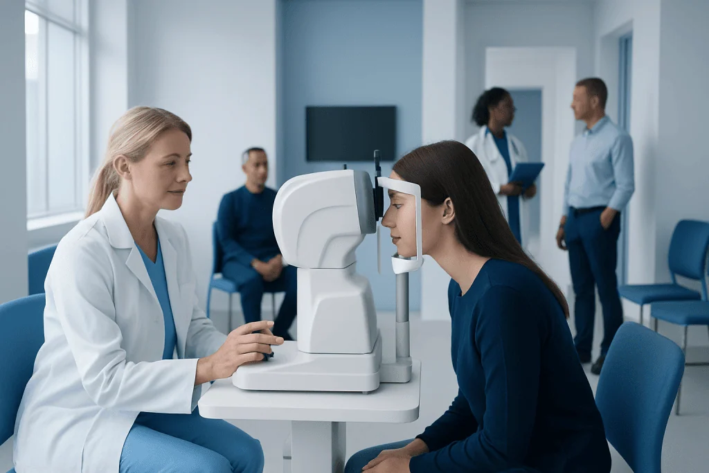
Optical coherence tomography, often called OCT, is quick, painless, and does not involve radiation. In eye care, OCT helps doctors examine the retina, macula, and optic nerve in great detail.
Key Takeaways
- Optical coherence tomography, often called OCT, is quick, painless, and does not involve radiation.
- In eye care, OCT helps doctors examine the retina, macula, and optic nerve in great detail.
- OCT can support diagnosis and follow-up for conditions such as macular degeneration, diabetic retinopathy, and glaucoma.
- The test itself usually takes only a few minutes, but results should always be interpreted by a qualified specialist.
- Pupil dilation is sometimes needed, but many OCT scans can be done without it.
Optical coherence tomography is a noninvasive imaging test that uses light waves to create detailed cross-sectional pictures of tissue, especially the retina and optic nerve. It is commonly used to help detect, monitor, and guide treatment for eye conditions, and in some settings it can also image other structures such as the coronary arteries.
What optical coherence tomography is
Optical coherence tomography is a fast, noninvasive imaging test that uses light waves to produce highly detailed cross-sectional images of body tissues. In everyday patient care, it is used most often in ophthalmology to examine the back of the eye, especially the retina, macula, and optic nerve. These images help doctors see tissue layers that cannot be assessed fully during a routine visual examination alone.
Many patients hear the term “OCT” when they are being evaluated for blurred vision, diabetes-related eye changes, glaucoma risk, or age-related macular problems. The scan itself does not touch the eye, does not use X-rays, and is usually completed within minutes. Because it provides layer-by-layer detail, it is especially useful for finding subtle changes early and for comparing scans over time.
Although eye care is the most familiar use, optical coherence tomography can also be used in some other specialties. For example, a related form of OCT may be used during certain heart procedures to look inside blood vessels. Still, for most patients, the phrase optical coherence tomography refers to an eye scan that supports diagnosis and treatment planning.
How the test works and what it can show
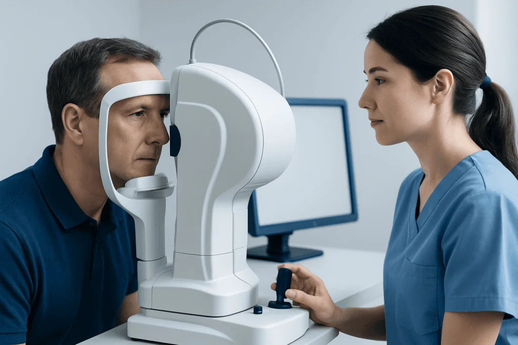
OCT works by directing light toward tissue and measuring how that light reflects back. A computer then converts this information into high-resolution images. In the eye, this allows specialists to view the thickness and structure of the retinal layers and to identify swelling, fluid, thinning, scarring, or damage to the optic nerve.
This detailed view can be valuable because some eye diseases affect very specific layers before symptoms become obvious. OCT may show fluid under or within the retina, changes related to diabetes, or loss of nerve tissue associated with glaucoma. It can also help doctors determine whether changes are stable, improving, or progressing.
In practice, OCT is often one piece of a larger evaluation rather than a stand-alone answer. Doctors combine it with symptoms, visual acuity testing, eye pressure measurement, a dilated eye exam, and sometimes additional imaging. The goal is not only to make a diagnosis, but also to understand how active the condition is and whether treatment is helping.
- Produces detailed cross-sectional images
- Helps detect subtle tissue changes
- Useful for both diagnosis and follow-up
- Most often used for retinal and optic nerve assessment
Why a doctor may recommend OCT

A doctor may recommend optical coherence tomography when symptoms or examination findings suggest a problem affecting the retina or optic nerve. Common reasons include blurred or distorted vision, changes in central vision, floaters that need further assessment, unexplained visual loss, or concern about eye disease in a person with diabetes or high eye pressure.
OCT is especially useful in conditions that can change gradually over time. It helps establish a baseline and makes future comparisons more accurate. This is important in patients being monitored for glaucoma, macular degeneration, or retinal changes related to diabetes.
Doctors may also use OCT to guide treatment decisions. For example, if retinal fluid or swelling is present, OCT can help determine whether treatment such as retina treatment may be needed and whether it is working. In patients with optic nerve concerns, OCT may add useful information alongside visual field testing and pressure measurements.
What to expect before, during, and after the scan
For most eye OCT scans, preparation is minimal. Patients are usually asked to sit in front of the machine and place the chin on a support while looking at a target. The device scans the eye in a matter of seconds, although the full visit may take longer if other tests are being performed at the same appointment.
Some patients need dilating eye drops before OCT, especially if the doctor wants a clearer view of the retina. Dilation can cause temporary light sensitivity and blurred near vision for a few hours, so arranging help with driving may be sensible depending on the individual and the clinic’s advice. If no dilation is needed, normal activities can often be resumed right away.
The scan itself is painless and non-contact in standard eye OCT. Patients may be asked to keep still and blink only at certain moments to improve image quality. After the test, a specialist reviews the images in context. Because OCT findings can be complex, patients should not try to interpret printouts or color maps on their own without professional explanation.
Conditions OCT can help evaluate
Optical coherence tomography is widely used to evaluate retinal and optic nerve conditions. One common use is assessing the macula, the central part of the retina responsible for sharp, detailed vision. OCT may show swelling, thinning, traction, or abnormal fluid in this area, all of which can affect reading and face recognition.
It is often part of the workup for diabetic eye disease, retinal vein problems, age-related changes, and inherited retinal disorders. It is also an important tool in the assessment of the optic nerve, where it can help identify structural changes linked with glaucoma and other causes of nerve damage. In some cases, OCT supports decisions about glaucoma treatment or referral for more specialized testing.
OCT does not replace a full eye examination, but it can add objective measurements and images that are difficult to obtain in other ways. Depending on the clinical question, a doctor may combine OCT with angiography, ultrasound, or a wider retinal assessment. For patients with cataracts or other media opacities, image quality can sometimes be reduced, which is one reason interpretation must be individualized.
When the main concern relates to cloudy natural lenses affecting vision, the specialist may discuss whether cataract surgery could improve overall examination quality and visual function, although this is a separate decision from the OCT test itself.
Benefits, limits, and safety
One of the main benefits of optical coherence tomography is that it is noninvasive and does not expose the patient to ionizing radiation. It is generally comfortable, repeatable, and well suited for long-term follow-up. This makes it valuable for chronic conditions where small changes matter and where treatment decisions depend on whether swelling, thinning, or fluid is increasing or decreasing.
At the same time, OCT has limits. It shows structure in great detail, but structure alone does not always explain symptoms. A scan may look stable while a patient still notices visual changes, or a scan may reveal abnormalities that need correlation with the rest of the examination. Poor fixation, dry eye, blinking, cataracts, or movement can affect image quality and lead to artifacts.
Safety is excellent in standard ophthalmic OCT because the test uses light rather than radiation and does not usually require contact with the eye. If dilation drops are used, temporary side effects such as blurred near vision and light sensitivity may occur. Patients should tell the clinic about past reactions to eye drops, current medications, and any difficulty staying still during testing.
How results are used in treatment planning
OCT results help clinicians decide whether a patient needs observation, medication, laser treatment, injections, surgery, or simply repeat imaging at a later date. In retinal disease, doctors often compare retinal thickness and fluid patterns from one visit to the next. This can be more informative than symptoms alone, especially when changes are gradual.
For glaucoma care, OCT measurements of the retinal nerve fiber layer and optic nerve can contribute to risk assessment and follow-up, but they are interpreted together with eye pressure, corneal measurements, and visual field tests. In this way, OCT supports a more complete understanding rather than serving as a single yes-or-no test for disease.
Because treatment planning depends on the whole clinical picture, it is common for a specialist to explain both what the scan shows and what it does not show. Patients benefit from asking how the findings compare with previous scans, whether changes are mild or significant, and when repeat imaging should be done. Near the end of the care pathway, centers such as Acibadem International may coordinate evaluation by multidisciplinary specialists in JCI-accredited hospitals for international patients who need diagnosis or treatment for complex eye conditions.
When to seek medical care
Patients should seek prompt medical advice if they notice sudden vision loss, a rapid increase in floaters, flashes of light, a curtain-like shadow over part of the vision, new distorted central vision, or eye pain with visual change. These symptoms can have several causes, some of which require urgent assessment and cannot be diagnosed by symptoms alone.
Medical review is also important for people with diabetes, known glaucoma, a strong family history of serious eye disease, or ongoing changes in reading vision. Even when symptoms seem mild, regular follow-up can help identify problems earlier. A doctor can advise whether an OCT scan or another test is the most appropriate next step.
For non-urgent concerns, patients may benefit from booking an eye examination if they are experiencing gradual blurring, reduced contrast, or difficulty with detailed central tasks such as reading. Early evaluation is usually the best way to understand what is happening and to discuss treatment options if needed.
Frequently asked questions
Is optical coherence tomography painful?
No. Standard eye OCT is usually painless and non-contact, meaning the device does not touch the eye. Most patients simply look at a target for a few seconds while the image is captured.
How long does an OCT scan take?
The scan itself is usually very quick, often taking only a few minutes. The full appointment may be longer if dilation, vision testing, or a full eye examination is also needed.
Does OCT use radiation?
No. Optical coherence tomography uses light waves, not X-rays or other ionizing radiation. This is one reason it can be repeated safely when doctors need to monitor changes over time.
Do all patients need dilating drops before OCT?
Not always. Some OCT scans can be performed without dilation, but drops may be recommended if the doctor needs a clearer view of the retina. If dilation is used, temporary blurred near vision and light sensitivity are common.
Can OCT diagnose glaucoma or macular degeneration by itself?
OCT provides very helpful structural information, but it is usually not the only test used to make a diagnosis. Doctors interpret OCT together with symptoms, examination findings, and other tests to reach the most accurate conclusion.
Why might an OCT scan need to be repeated?
Repeat scans help doctors compare changes over time. This is especially useful for chronic conditions where small increases in fluid, swelling, or nerve thinning can influence follow-up plans and treatment decisions.
References
- American Academy of Ophthalmology
- National Eye Institute
- Mayo Clinic
- Royal College of Ophthalmologists
- National Institute for Health and Care Excellence
This article is for general information only and is not a substitute for professional medical advice. Please consult a qualified doctor about your individual situation.
Explore treatments in Turkey — costs, top hospitals & a free quote
JCI-accredited · board-certified surgeons · reply within 24h
Add us as a Preferred Source to see more of our trusted health content across Google Search, AI Overviews and Discover.