Optical Hypertension Treatment: How It Works, Results and What to Expect
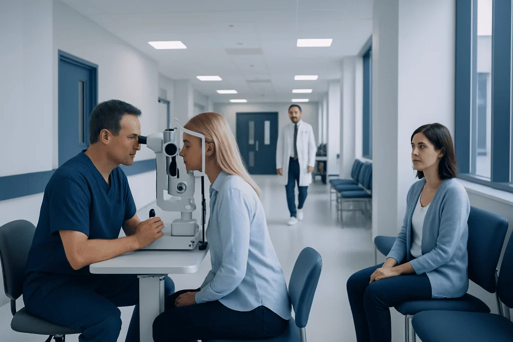
Ocular hypertension means raised intraocular pressure without confirmed glaucoma-related optic nerve damage. Most people have no symptoms, so routine eye examinations are important for detecting high eye pressure.
Key Takeaways
- Ocular hypertension means raised intraocular pressure without confirmed glaucoma-related optic nerve damage.
- Most people have no symptoms, so routine eye examinations are important for detecting high eye pressure.
- Not everyone with ocular hypertension needs immediate treatment; risk assessment guides the decision.
- Eye drops and laser trabeculoplasty are common ways to lower eye pressure, while surgery is usually reserved for selected situations.
- Regular follow-up is essential because pressure readings, optic nerve health and visual field results can change over time.
Optical hypertension treatment—more accurately called ocular hypertension treatment—aims to lower pressure inside the eye and reduce the chance of optic nerve damage and glaucoma. Care may involve regular monitoring, prescription eye drops, laser treatment, or surgery in selected cases, based on an individual’s measured risk.
Overview: What Is Optical Hypertension Treatment?
Optical hypertension treatment usually refers to ocular hypertension treatment. Ocular hypertension means the pressure inside one or both eyes, called intraocular pressure, is higher than the usual range, but examinations have not found clear glaucoma-related damage to the optic nerve or loss of vision. Treatment is designed to lower this pressure and reduce the long-term risk of developing glaucoma.
Raised eye pressure is not an emergency in most people, and it does not automatically mean glaucoma will occur. However, persistently elevated pressure can increase strain on the optic nerve. An ophthalmologist considers the pressure level alongside corneal thickness, optic nerve appearance, visual field testing, age, family history and other factors before recommending observation or active treatment.
Management is individualized. Some people benefit most from regular monitoring, while others are advised to use pressure-lowering eye drops or have laser treatment. The goal is not simply to reach one number, but to maintain eye pressure at a level that is considered safer for that person’s eyes over time.
How Ocular Hypertension Treatment Works
The eye continuously produces a clear fluid called aqueous humor. This fluid normally drains through a structure near the front of the eye called the trabecular meshwork. Eye pressure rises when fluid does not drain efficiently enough, when production is relatively high, or when both processes contribute.
Prescription eye drops lower pressure in different ways. Some reduce the amount of fluid the eye produces, while others improve fluid drainage through existing pathways or alternative drainage routes. A clinician selects treatment according to eye findings, overall health, possible side effects, other medicines and the person’s ability to use drops consistently.
Laser trabeculoplasty uses carefully controlled laser energy to improve drainage through the trabecular meshwork. It is often considered for open-angle ocular hypertension or glaucoma, either as an initial approach for suitable patients or when drops are not sufficient, not tolerated or difficult to use. In selected cases, surgical procedures that create or support fluid drainage may be considered when other measures do not adequately control pressure.
Because ocular hypertension can be associated with later glaucoma, clinicians also assess for signs of glaucoma during follow-up. Treatment lowers risk but cannot remove it completely, so continued monitoring remains important.
Symptoms and Questions People Commonly Ask

Ocular hypertension usually causes no noticeable symptoms. This is why people may learn about it during a routine optometrist or ophthalmologist visit rather than because of a change in how their eyes feel. Vision can remain normal even when pressure has been raised for some time.
How do you feel when your eye pressure is high? Most people do not feel high eye pressure. Mild to moderately increased pressure typically does not cause eye pain, headache, redness or blurred vision. Severe pain, sudden blurred vision, halos around lights, nausea or vomiting are not typical signs of uncomplicated ocular hypertension and may indicate an acute eye problem requiring urgent assessment.
Is ocular hypertension serious? Ocular hypertension is important because it can raise the likelihood of developing glaucoma, a condition that can damage the optic nerve and affect peripheral vision. It is not the same as glaucoma, and many people with ocular hypertension never develop optic nerve damage. Regular assessment and appropriate pressure management can help identify changes early.
Does ocular hypertension ever go away? Eye pressure can vary from one appointment to another, and a temporary increase may settle if it is related to a medicine, inflammation, trauma or another treatable cause. In many cases, however, ocular hypertension is a longer-term finding that needs periodic follow-up. A person should not stop prescribed treatment simply because a later reading is lower without discussing it with their eye specialist.
Causes, Risk Factors and Candidacy for Treatment
In many people, there is no single identifiable cause of ocular hypertension. It may reflect naturally less efficient drainage of aqueous humor. Other causes or contributors can include long-term corticosteroid use, previous eye injury, eye inflammation, pigment dispersion, pseudoexfoliation, certain eye conditions and, less commonly, other underlying causes that require specialist evaluation.
Factors associated with a higher risk of glaucoma in someone with ocular hypertension include substantially elevated eye pressure, a thinner central cornea, suspicious optic nerve findings, changes on visual field testing, older age, family history of glaucoma, and some ethnic or genetic backgrounds. Nearsightedness and certain medical conditions may also be relevant in individual risk assessments.
A person is more likely to be offered active treatment when their estimated risk of future optic nerve damage is meaningful, when pressure is consistently high, or when reliable follow-up is difficult. Conversely, carefully monitored observation may be reasonable for a person with lower pressure, a healthy optic nerve and a low overall risk profile. The decision is shared and should account for the expected benefit, treatment burden and personal preferences.
Before treatment, the clinician confirms that the angle where fluid drains is open, commonly using gonioscopy. They also check whether pressure readings may be affected by corneal thickness or other measurement factors. These details help ensure that the treatment plan fits the type of pressure elevation present.
Diagnosis and Step-by-Step Treatment Process
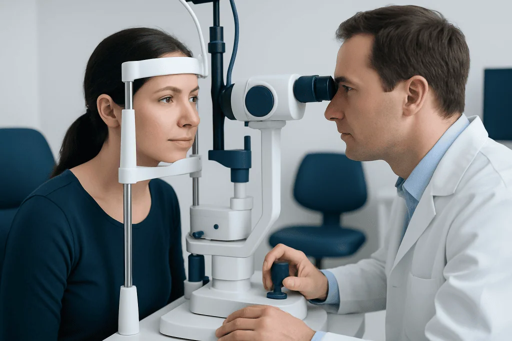
Assessment generally begins with a detailed eye history and examination. Tonometry measures intraocular pressure, while pachymetry measures corneal thickness. The ophthalmologist examines the optic nerve, often using dilated eye examination and retinal imaging, and may perform visual field testing to check for subtle functional changes. Gonioscopy evaluates the drainage angle and helps rule out angle-closure risk.
If observation is appropriate, follow-up visits are scheduled at intervals based on risk. At these appointments, pressure is rechecked and the optic nerve and visual field are reviewed over time. A pattern of change is more informative than a single measurement, so keeping scheduled appointments is an important part of care.
For eye-drop treatment, the clinician explains which drop to use, how often to use it and how to apply it safely. Usually, hands are washed first, one drop is placed in the lower eyelid pocket without touching the bottle tip to the eye or lashes, and the eye is gently closed. Light pressure at the inner corner of the eyelid for a short period may reduce drainage into the nose and limit absorption into the body.
For suitable patients, laser trabeculoplasty is typically performed as an outpatient procedure. Numbing drops are applied, a special contact lens may be placed on the eye, and laser spots are applied to the drainage tissue. The procedure is usually brief. Pressure may be checked afterwards, and anti-inflammatory drops may be prescribed depending on the clinician’s protocol.
Benefits, Risks and Recovery Timeline
The main benefit of treatment is a lower intraocular pressure and a reduced likelihood of pressure-related optic nerve damage over time. Eye drops can be effective when used regularly, while laser trabeculoplasty may reduce or delay the need for drops for some people. Results differ between individuals, and treatment response is assessed with repeat pressure measurements and optic nerve monitoring.
Eye drops can cause local effects such as stinging, redness, dry-eye symptoms, blurred vision shortly after use or changes around the eyelids, depending on the medication class. Some drops can also have effects elsewhere in the body, particularly in people with certain heart, lung or other medical conditions. It is important to tell the prescribing clinician about all medicines, allergies and medical history.
After laser trabeculoplasty, many people return to usual non-strenuous activities the same day or the following day. Temporary blurred vision, light sensitivity, mild irritation or redness may occur. The pressure-lowering effect can take several weeks to become fully apparent, and some patients still need eye drops. Rare complications include a temporary pressure rise, inflammation or insufficient response.
Surgery is generally reserved for situations where eye pressure remains too high despite less invasive options or where glaucoma risk is particularly high. Recovery after surgery is longer and requires close postoperative visits. The ophthalmologist explains the specific expected benefits and risks before any procedure.
What to Avoid and Everyday Self-Care
What to avoid if you have ocular hypertension? A person should not miss follow-up eye appointments, stop prescribed drops suddenly, share eye medication, or use someone else’s drops. They should also avoid starting steroid eye drops, creams near the eyes, inhalers or oral steroids without telling the clinician who monitors their eye pressure, as corticosteroids can raise pressure in susceptible people.
General health habits support overall eye health, although they are not a substitute for prescribed care. Regular physical activity, balanced nutrition, not smoking and management of conditions such as high blood pressure or diabetes can be helpful for general vascular health. Large amounts of caffeine at one time may cause a short-lived rise in pressure in some people, so moderation is sensible.
Anyone using eye drops should ask about practical techniques if treatment is difficult because of arthritis, tremor, poor vision or travel. A family member, pharmacist or eye-care professional may be able to help with drop-administration aids. Contact lens wearers should also ask whether lenses need to be removed before using their particular medicine.
Acibadem International’s multidisciplinary specialists at JCI-accredited hospitals evaluate raised eye pressure and provide individualized monitoring and treatment options for international patients.
When to Seek Medical Care
Arrange a routine eye-care appointment if a screening test suggests elevated eye pressure, if there is a family history of glaucoma, or if an eye-care professional recommends monitoring. People taking corticosteroids for extended periods should ask whether their eye pressure needs checking, especially if they have other glaucoma risk factors.
Seek urgent eye assessment for sudden severe eye pain, a red eye with blurred vision, colored halos around lights, sudden vision loss, severe headache with nausea or vomiting, or symptoms after an eye injury. These symptoms may be caused by conditions other than ocular hypertension, but prompt evaluation is important because some eye emergencies can threaten vision.
New or worsening side effects after starting pressure-lowering eye drops should also be discussed promptly with the prescribing clinician. Treatment can often be adjusted rather than abandoned, helping the person find an approach that is both effective and manageable.
Frequently asked questions
What is the usual first treatment for ocular hypertension?
The first step may be regular monitoring rather than immediate medication when a person’s overall glaucoma risk is low. When treatment is recommended, prescription eye drops and laser trabeculoplasty are common options. The best choice depends on pressure level, optic nerve findings, eye anatomy, medical history and personal preferences.
Can ocular hypertension cause blindness?
Ocular hypertension itself means there is raised pressure without confirmed glaucomatous damage. If it progresses to glaucoma and remains untreated or inadequately controlled, optic nerve damage can lead to permanent vision loss. Regular monitoring and treatment when indicated can reduce this risk.
Will eye drops cure ocular hypertension?
Eye drops can lower eye pressure effectively, but they do not usually provide a permanent cure for the underlying tendency toward raised pressure. They generally work only while they are used as prescribed. Follow-up testing determines whether the treatment remains effective.
Can stress raise eye pressure?
Stress may affect overall health and can be associated with short-term changes in some body functions, but it is not usually considered the main cause of persistent ocular hypertension. A raised pressure reading should be assessed clinically rather than attributed to stress alone. Ongoing monitoring helps distinguish a temporary variation from a sustained issue.
Can exercise help lower eye pressure?
Regular moderate exercise may have a modest beneficial effect on eye pressure and supports general health. It should not replace prescribed drops, laser treatment or scheduled monitoring. People with glaucoma or other eye conditions should ask their eye specialist about activities that may need individual guidance.
How often should ocular hypertension be checked?
The follow-up schedule varies according to pressure level and glaucoma risk. Some people need checks every few months, while others with lower risk may be reviewed less often. An ophthalmologist sets the interval after considering eye pressure, corneal thickness, optic nerve tests and visual fields.
References
- American Academy of Ophthalmology
- National Eye Institute
- Glaucoma Research Foundation
- National Health Service
- Mayo Clinic
This article is for general information only and is not a substitute for professional medical advice. Please consult a qualified doctor about your individual situation.
Hypertension in Turkey — costs, top hospitals & a free quote
JCI-accredited · board-certified surgeons · reply within 24h
Add us as a Preferred Source to see more of our trusted health content across Google Search, AI Overviews and Discover.