Orbital Lesions Treatment in Gilbert: How It Works, Results and What to Expect

Orbital lesions are abnormal areas in the eye socket and can be benign, inflammatory, vascular or cancerous. New eye bulging, vision changes, double vision, pain or a growing eyelid or eye-socket mass should be assessed promptly.
Key Takeaways
- Orbital lesions are abnormal areas in the eye socket and can be benign, inflammatory, vascular or cancerous.
- New eye bulging, vision changes, double vision, pain or a growing eyelid or eye-socket mass should be assessed promptly.
- Imaging and, in selected cases, biopsy help specialists determine whether monitoring, medical treatment or surgery is appropriate.
- Orbitotomy is a specialized operation that provides access to the eye socket while aiming to preserve vision, eye movement and appearance.
- Recovery and outlook depend on the lesion’s type, size, location and the treatment required.
Orbital lesions treatment in Gilbert begins with confirming the type, location and behavior of the lesion through an eye examination and imaging. Treatment may range from observation to medication, surgery, radiation therapy or systemic cancer treatment, depending on the diagnosis and its effect on vision and surrounding structures.
Overview: How Orbital Lesions Treatment in Gilbert Works
Orbital lesions treatment in Gilbert is tailored to the cause of an abnormal mass, swelling or tissue change within the orbit, the bony space around the eye. The first goal is to identify whether the lesion is harmless, inflammatory, vascular, infectious or cancerous, then to determine whether it threatens vision, eye movement, comfort or nearby structures.
Some orbital lesions do not need immediate intervention and can be followed with repeat examinations and imaging. Others require medication, a biopsy, surgical removal, radiation therapy or systemic treatment. Care is often coordinated by ophthalmologists with expertise in the orbit, radiologists, pathologists, neurologists, neurosurgeons, ear, nose and throat surgeons, and oncology specialists when appropriate.
“Orbital lesion” is a descriptive term rather than one diagnosis. Lesions may arise from the eye socket’s fat, muscles, nerves, blood vessels, tear gland, bone or tissues that have spread from another part of the body. A personalized plan is important because the safest and most effective approach depends on the exact diagnosis.
Symptoms and When to Seek Medical Care
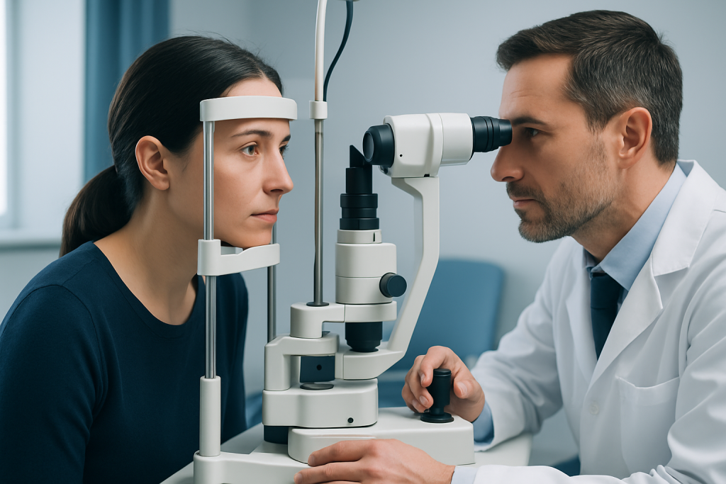
Symptoms vary with a lesion’s location and rate of growth. Some lesions are discovered incidentally on imaging performed for another reason, while others gradually cause one eye to appear more prominent, displaced or swollen. Not every symptom indicates a tumor, but an eye-care professional should assess persistent or unexplained changes.
- Bulging or forward displacement of one eye
- Double vision or reduced eye movement
- Blurred, reduced or changing vision
- Eye pain, pressure, redness or persistent swelling
- A new eyelid lump, drooping eyelid or visible change in eye position
- Numbness around the forehead, cheek or upper face
When to seek medical care: A person should arrange prompt medical assessment for new or progressive eye bulging, double vision, pain, vision changes or a noticeable orbital mass. Sudden vision loss, severe eye pain, rapidly increasing swelling, fever with eye swelling, or difficulty moving the eye requires urgent medical evaluation.
How Fast Do Orbital Tumors Grow?
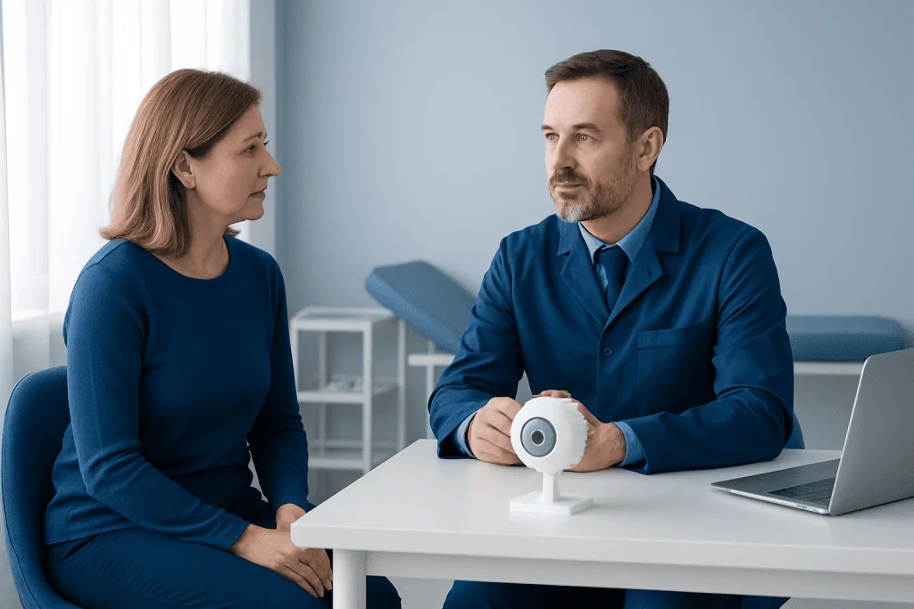
Orbital tumors do not all grow at the same speed. Many benign lesions grow slowly over months or years and may cause subtle changes before they are noticed. Certain vascular lesions can enlarge intermittently, while inflammatory conditions may develop over days to weeks.
Some cancers and infections can progress more quickly, sometimes over weeks. However, growth rate alone cannot reliably identify whether a lesion is benign or malignant. Imaging findings, symptoms, age, medical history and, when needed, tissue testing are used together to establish a diagnosis.
A perceived rapid change should not be ignored, especially when it occurs with pain, impaired eye movement or reduced vision. Early assessment helps clinicians protect sight and determine whether time-sensitive treatment is needed.
Diagnosis and Candidacy for Treatment
Evaluation usually starts with a detailed history and eye examination. The clinician checks vision, pupils, eye pressure, color vision, visual fields, eye movements and the position of each eye. Medical history is also important, including thyroid disease, autoimmune conditions, previous cancers, recent infection, trauma and medications.
CT and MRI scans are commonly used to show the lesion’s size, density, blood supply, relationship to the optic nerve and involvement of bone or nearby sinuses. Ultrasound, blood tests or specialized imaging may be useful in selected cases. If imaging cannot establish the cause, a biopsy may be recommended to examine cells under a microscope.
A person may be a candidate for active treatment when a lesion affects vision, compresses the optic nerve, causes significant double vision or pain, grows on serial imaging, has uncertain features, or is suspected to be cancerous. In contrast, stable lesions with reassuring features may be monitored safely through planned follow-up.
What Are the Treatment Options for Orbital Lesions?
Treatment options for orbital lesions include observation, medicines, minimally invasive diagnostic procedures, surgery, radiation therapy and systemic cancer treatment. Observation can be appropriate for a small, stable lesion that is not affecting vision or eye movement. Follow-up visits may include repeat eye examinations and imaging at intervals chosen by the treating team.
Medication may be used for inflammatory, autoimmune or infectious causes. Depending on the diagnosis, this can include anti-inflammatory medicines, immune-modifying treatment or antimicrobial therapy. These treatments should only be used under medical supervision because they are not suitable for every orbital condition.
Surgery may be advised to obtain a diagnosis, relieve pressure, remove a lesion, improve eye position or preserve vision. When cancer is confirmed or strongly suspected, treatment may also involve radiation therapy, chemotherapy, targeted therapy or immunotherapy. The approach is determined through a multidisciplinary review and is based on the specific tumor type.
For lesions requiring surgery, an experienced orbital team can discuss orbitotomy surgery and whether it is suitable for the lesion’s location. Related conditions such as thyroid eye disease may instead require medical management, orbital decompression or other targeted care.
Is Orbitotomy a Major Surgery?
Orbitotomy is a specialized surgical procedure that creates access to the orbit so a surgeon can biopsy or remove a lesion. Whether it is considered “major” depends on the lesion’s depth, size, relationship to the optic nerve and eye muscles, and whether an external incision, eyelid approach or an endoscopic route is used.
Although it is not the same as surgery inside the eye or brain surgery, orbitotomy is delicate because the orbit contains structures essential for vision and eye movement. It is typically performed under anesthesia in a hospital or surgical setting by an orbital ophthalmic surgeon, sometimes alongside neurosurgery or ENT specialists for lesions near the skull base or sinuses.
Before surgery, patients receive individualized guidance about imaging, anesthesia, medication adjustments and expected recovery. The surgeon should explain the planned approach, possible alternatives, the need for biopsy, and the specific risks associated with the lesion’s position.
Procedure Steps, Recovery Timeline, Benefits and Risks
For an orbitotomy, the team first reviews imaging and plans the safest route to the lesion. On the day of surgery, anesthesia is provided and the surgeon makes a carefully placed incision, often in a natural eyelid crease, through the conjunctiva, or by another route suited to the lesion. The lesion may be sampled, partially removed or removed more completely, depending on what can be done safely. Tissue is sent for pathology when needed.
After surgery, mild swelling, bruising, tenderness, tearing and temporary blurred or double vision can occur. Many people return home the same day or after a short hospital stay, but this varies by procedure and overall health. Initial swelling often improves over one to two weeks, while healing, final pathology results and return to usual activities may take longer. Follow-up visits monitor vision, eye movement, wound healing and the need for additional treatment.
Potential benefits include obtaining a definitive diagnosis, relieving pressure or symptoms, preserving vision and treating a lesion that is growing or concerning. Risks can include bleeding, infection, scarring, dry eye, eyelid position changes, persistent double vision, injury to eye muscles or nerves, incomplete removal, recurrence and, rarely, reduced vision. The individual risk profile should be discussed directly with the surgical team.
How Serious Is an Orbital Tumor?
An orbital tumor can range from a benign, slow-growing lesion to a condition requiring urgent treatment. Its seriousness depends on the underlying diagnosis, speed of change, location and whether it affects the optic nerve, eye muscles, blood vessels, bone or surrounding tissues. A benign tumor can still be important if it presses on structures needed for vision.
Malignant orbital tumors are less common than many noncancerous and inflammatory causes of orbital swelling, but they need timely specialist care. The diagnostic process is designed to clarify the situation rather than assume the worst. Imaging and pathology can guide treatment and help the team discuss an individualized outlook.
Acibadem International’s multidisciplinary specialists and JCI-accredited hospitals diagnose and treat orbital conditions for international patients, with care plans based on the lesion and the patient’s overall health. Ongoing follow-up remains important after treatment, particularly when a lesion may recur or requires further cancer-directed care.
Frequently asked questions
What is an orbital lesion?
An orbital lesion is an abnormal area of tissue, swelling or mass in the orbit, which is the space around the eye. It may be benign, inflammatory, vascular, infectious or cancerous, so assessment is needed to identify the cause.
Can an orbital lesion be harmless?
Yes. Some orbital lesions are benign and remain stable without threatening vision or health. Even a benign lesion may need follow-up or treatment if it grows, causes symptoms or presses on the optic nerve or eye muscles.
Will every orbital lesion need surgery?
No. Some lesions can be safely monitored, and others respond to medication or require non-surgical cancer treatment. Surgery is generally considered when a diagnosis is uncertain, symptoms are significant, vision is at risk, or removal is needed.
How long does recovery after orbitotomy take?
Early bruising and swelling commonly improve over one to two weeks, although recovery differs according to the surgical approach and lesion location. Vision, eye movement and the surgical site are checked during follow-up, and complete healing may take several weeks or longer.
Can orbital tumors cause double vision?
Yes. A lesion can affect or displace the muscles that move the eye, leading to double vision or restricted eye movement. Double vision should be assessed by an eye-care professional, particularly when it is new or worsening.
What tests are used to diagnose an orbital tumor?
An ophthalmic examination and imaging, usually MRI or CT, are central to evaluation. Blood tests, ultrasound and biopsy may also be used when they can clarify the cause or guide treatment.
References
- American Academy of Ophthalmology
- National Cancer Institute
- National Eye Institute
- Merck Manual Professional Edition
- Orbit Society
This article is for general information only and is not a substitute for professional medical advice. Please consult a qualified doctor about your individual situation.
Oncology care in Turkey — second opinion and treatment plan
JCI-accredited · board-certified surgeons · reply within 24h
Add us as a Preferred Source to see more of our trusted health content across Google Search, AI Overviews and Discover.