Osseous Dysplasia Treatment: How It Works, Results and What to Expect

Osseous dysplasia is a broad term that can include fibrous dysplasia and cemento-osseous dysplasia, which have different causes and management needs. Treatment is individualized; symptom-free, stable areas often require monitoring rather than an operation.
Key Takeaways
- Osseous dysplasia is a broad term that can include fibrous dysplasia and cemento-osseous dysplasia, which have different causes and management needs.
- Treatment is individualized; symptom-free, stable areas often require monitoring rather than an operation.
- Surgery may be considered for functional problems, substantial deformity, repeated fractures, nerve compression, or suspected infection.
- Cemento-osseous dysplasia usually has a favorable outlook, but unnecessary biopsy or dental procedures in affected areas may increase infection risk.
- New pain, swelling, drainage, numbness, difficulty walking, or a rapid change in bone shape should be assessed promptly.
Osseous dysplasia treatment depends on the specific condition, location, symptoms, and whether the affected bone is stable. Many people need observation and regular imaging only, while selected cases benefit from medication, dental treatment, corrective surgery, or fracture-prevention care.
Osseous Dysplasia Treatment: What It Involves
Osseous dysplasia treatment is guided by the exact diagnosis and by how the affected bone is behaving over time. In many cases, especially when there is no pain, infection, fracture risk, or functional limitation, the safest approach is regular clinical review and imaging rather than immediate treatment. This helps clinicians track the area while avoiding procedures that may not be necessary.
“Osseous dysplasia” may describe different disorders of bone development or bone remodeling. Two important examples are fibrous dysplasia, in which normal bone is replaced by fibrous tissue and immature bone, and cemento-osseous dysplasia, a jaw condition involving cementum-like and bone-like tissue around tooth roots. Although both may appear on imaging, they differ in their typical location, symptoms, and care plan.
When treatment is needed, its goals are to relieve symptoms, protect bone strength, preserve function, address dental infection, and improve significant deformity where appropriate. Care may involve oral and maxillofacial specialists, dentists, orthopedic surgeons, endocrinologists, radiologists, and pain or rehabilitation professionals depending on the affected area.
How Treatment Decisions Are Made: Candidacy and Assessment
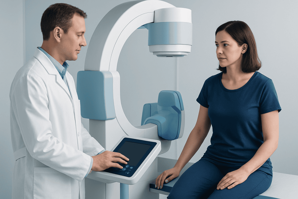
A clinician first confirms the type of dysplasia and excludes other conditions that can resemble it on an X-ray, CT scan, MRI scan, or dental radiograph. The assessment considers the site and size of the lesion, symptoms, growth pattern, fracture history, dental health, age, and whether one bone or several bones are involved. Blood tests may be used when there is concern about associated hormonal conditions or abnormal bone turnover.
People are more likely to be considered for active treatment when dysplasia causes persistent pain, progressive asymmetry, reduced movement, difficulty chewing or seeing, nerve-related symptoms, repeated fractures, or a high likelihood of structural weakening. In jaw lesions, infection, exposed bone, drainage, or a need for carefully planned dental treatment can also affect the plan.
Not every imaging change needs a biopsy. For example, cemento-osseous dysplasia can often be identified from its characteristic location and radiographic appearance in combination with a dental examination. A biopsy or surgery may be avoided when the appearance is typical because procedures in poorly vascularized dysplastic jaw bone can sometimes lead to delayed healing or infection.
- Observation may suit stable, symptom-free disease.
- Medication may help selected people with bone pain but does not remove dysplastic bone.
- Surgery is reserved for defined functional, structural, or cosmetic indications.
- Dental prevention and infection control are particularly important in cemento-osseous dysplasia.
How Osseous Dysplasia Treatment Works: Step by Step

For monitoring, the process generally starts with baseline imaging and a review of symptoms. Follow-up timing is individualized: a clinician may recommend periodic dental X-rays for jaw disease or repeat imaging of long bones, the skull, or other involved sites when fibrous dysplasia is being followed. Patients are encouraged to report changes rather than waiting for the next scheduled review.
For symptomatic fibrous dysplasia, treatment may begin with non-surgical pain management, correction of contributing factors such as low vitamin D when present, and physical therapy or protective devices when mobility is affected. Medications that influence bone turnover are sometimes considered by specialists for persistent bone pain in selected patients. They are not routinely used simply to change imaging findings, and their potential benefits and risks require individual discussion.
If an operation is appropriate, planning commonly includes detailed imaging to map the anatomy and decide whether reshaping, stabilization, correction of deformity, or removal of a problematic area is needed. During surgery, the team may contour bone, reinforce weakened bone with fixation, correct alignment, or address compression of nearby structures. In the jaw, surgery is generally limited to situations such as significant expansion, infection that does not settle with conservative care, or a diagnosis that remains uncertain.
Dental management focuses on preserving healthy teeth and preventing infection. A dentist may monitor tooth vitality, gum health, and restorations, while coordinating with an oral and maxillofacial surgeon before extractions, implants, or other invasive work in an affected region. This careful planning is an important part of fibrous dysplasia care when the facial bones or jaw are involved.
Recovery Timeline, Benefits and Possible Risks
Recovery depends greatly on the treatment used. After a monitoring visit or routine dental preventive care, normal activity usually continues immediately. Recovery after orthopedic or craniofacial surgery may take weeks to months, with the pace influenced by the bone treated, the extent of reconstruction, overall health, and the need for rehabilitation. The surgical team provides individualized guidance on activity, wound care, nutrition, and follow-up imaging.
Potential benefits of treatment include less pain, improved ability to walk or use the affected limb, correction of deformity, reduced risk of fracture in selected cases, relief of compression symptoms, and better dental comfort or function. Surgery does not always permanently eliminate fibrous dysplasia, particularly when it is performed before skeletal maturity, so long-term follow-up remains important.
All procedures have possible risks. These may include bleeding, infection, delayed healing, scarring, changes in sensation, damage to nearby teeth or nerves, incomplete symptom relief, and recurrence or continued growth of dysplastic tissue. Jaw procedures can be especially challenging when blood supply within the lesion is limited. A specialist can explain how these risks apply to the individual situation and what alternatives are available.
When complex care is required, Acibadem International’s multidisciplinary specialists at JCI-accredited hospitals can assess and treat patients with bone and jaw dysplasia, including international patients, through coordinated imaging, dental, surgical, and rehabilitation planning.
What Causes Osseous Dysplasia?
The cause depends on the form of osseous dysplasia. Fibrous dysplasia is usually caused by a genetic change that happens after conception in some body cells. It is not typically inherited from a parent and is not contagious. The timing and distribution of this cell change determine whether one bone or multiple bones are affected.
In fibrous dysplasia, the altered cells interfere with normal bone formation, leading to replacement of mature bone with fibrous tissue and irregular bone. Some people with more extensive disease also have hormone-related features as part of McCune-Albright syndrome, which requires specialist evaluation and follow-up.
The cause of cemento-osseous dysplasia is less clearly defined. It is considered a benign disorder of bone and cementum-like tissue formation in the jaws. It is not cancer, and it is usually discovered incidentally during dental imaging. Genetics, local tissue factors, and hormonal influences may contribute, but it is not caused by poor oral hygiene.
At What Age Does Fibrous Dysplasia Stop Growing?
Fibrous dysplasia is commonly diagnosed in childhood or adolescence and often becomes less active after skeletal maturity, when most bones have completed their growth. For many people, the lesions then remain relatively stable. However, stability does not mean that follow-up is unnecessary, particularly if the disease affects weight-bearing bones, the face, or the skull.
Some lesions can continue to cause symptoms or change during adulthood, especially if there are fractures, deformity, hormonal abnormalities, or involvement near critical structures. A sudden increase in pain, swelling, or growth should not automatically be attributed to fibrous dysplasia and should be medically assessed.
For children and adolescents, treatment planning aims to protect function while recognizing that further growth is expected. Surgeons may recommend monitoring or staged procedures in some situations, because reshaped or reconstructed bone can change as the skeleton develops.
What Is the Prognosis for Cemento-Osseous Dysplasia?
The prognosis for cemento-osseous dysplasia is generally very good. Most people have no symptoms, maintain normal daily function, and need only regular dental examinations and imaging follow-up. The condition is benign and does not usually transform into cancer.
The main concern is the possibility of secondary infection in areas where the tissue becomes dense and has a reduced blood supply. This is more likely after trauma, ill-fitting dentures, tooth extraction, or other invasive dental procedures, although many people never develop this complication. Good oral hygiene and early attention to dental problems can reduce avoidable risks.
If infection occurs, treatment may require a combination of dental care, antibiotics when clinically appropriate, and carefully planned surgical management. The approach should be individualized by a dental professional or oral and maxillofacial specialist familiar with the condition.
Can Cemento-Osseous Dysplasia Cause Root Resorption?
Root resorption is not a typical defining feature of cemento-osseous dysplasia. In many cases, the teeth associated with the lesions remain vital and their roots are preserved. Imaging may show changes around roots that can be mistaken for inflammatory dental disease, which is why accurate interpretation is important.
However, large or expansile lesions, secondary infection, or another coexisting jaw condition may affect nearby teeth and supporting structures. If an X-ray suggests root shortening, displacement, loss of tooth vitality, or a lesion with an unusual appearance, further assessment is appropriate rather than assuming it is due to cemento-osseous dysplasia alone.
Unnecessary root canal treatment or extraction should be avoided when teeth are healthy and the radiographic findings are characteristic of the condition. A dentist can use examination findings, vitality testing, and comparison with previous images to make a safer treatment decision.
Prevention, Self-Care and When to Seek Medical Care
Osseous dysplasia cannot always be prevented, but complications can often be reduced. People with fibrous dysplasia should attend recommended follow-up visits, protect weight-bearing bones according to medical advice, maintain adequate nutrition for bone health, avoid smoking, and seek guidance before starting high-impact activity when a bone is weakened. Physical therapy may support safe movement, strength, and balance when needed.
For cemento-osseous dysplasia, consistent oral hygiene, regular dental reviews, well-fitting dentures, and prompt treatment of gum or tooth problems are especially valuable. Patients should tell dental professionals about the diagnosis before invasive procedures are planned. This allows the team to consider alternatives and coordinate specialist input when required.
When to seek medical care: A person should arrange timely medical or dental assessment for new or worsening bone pain, swelling, redness, drainage, fever with jaw symptoms, a new limp, reduced ability to bear weight, numbness, vision or hearing changes, a sudden change in facial shape, or a suspected fracture. Urgent evaluation is appropriate after a significant injury, severe pain, or signs of infection.
Regular monitoring provides reassurance for many people, while early review of new symptoms supports timely and appropriate care. Individual decisions should be made with a qualified clinician who can interpret symptoms and imaging in the context of the specific diagnosis.
Frequently asked questions
Is osseous dysplasia cancer?
Osseous dysplasia is generally a benign condition, meaning it is not cancer. However, the exact type of lesion should be diagnosed carefully because several bone and jaw conditions can look similar on imaging. New or unusual changes should be reviewed by a qualified clinician.
Does every person with fibrous dysplasia need surgery?
No. Many people with fibrous dysplasia do not need surgery and are managed with observation, symptom control, and periodic imaging. Surgery is considered when there is a clear concern such as fracture risk, major deformity, functional impairment, compression of nearby structures, or persistent symptoms.
Can teeth be removed in cemento-osseous dysplasia?
Teeth can be removed when there is a clear dental reason, but extractions in affected jaw areas should be planned carefully. Dense dysplastic tissue may have reduced blood supply and can be more prone to delayed healing or infection. A dentist or oral and maxillofacial specialist should assess the risks and alternatives first.
Can fibrous dysplasia return after surgery?
Dysplastic bone can continue to change after surgery, and regrowth may occur, particularly when treatment is performed before skeletal maturity. This does not mean surgery is ineffective; it may still be important for function, fracture prevention, or symptom relief. Long-term follow-up helps identify changes early.
What tests are used to diagnose osseous dysplasia?
Diagnosis commonly uses clinical examination and imaging such as X-rays, CT, MRI, or dental radiographs. Blood tests may be used in selected cases, particularly when fibrous dysplasia may be associated with hormonal concerns. Biopsy is reserved for lesions with uncertain or atypical features.
Can osseous dysplasia cause pain?
It can, although some people have no symptoms. Pain may result from bone weakening, fracture, deformity, pressure on nearby structures, or secondary infection in jaw lesions. Persistent, worsening, or sudden pain should be assessed to determine its cause.
References
- National Institutes of Health
- Orphanet
- American Association of Oral and Maxillofacial Surgeons
- International Consortium for Fibrous Dysplasia and McCune-Albright Syndrome
This article is for general information only and is not a substitute for professional medical advice. Please consult a qualified doctor about your individual situation.
Joint and spine care in Turkey — expert assessment & treatment
JCI-accredited · board-certified surgeons · reply within 24h
Add us as a Preferred Source to see more of our trusted health content across Google Search, AI Overviews and Discover.