

Osteosarcoma Xray Knee: Preparation, Procedure and Results

A knee X-ray is quick, painless, and commonly used as the first imaging test for possible bone abnormalities. Osteosarcoma most often develops near the knee, typically in the lower thigh bone or upper shinbone.
Key Takeaways
- A knee X-ray is quick, painless, and commonly used as the first imaging test for possible bone abnormalities.
- Osteosarcoma most often develops near the knee, typically in the lower thigh bone or upper shinbone.
- Many common knee problems cause pain, so knee pain alone does not mean cancer.
- Suspicious X-ray findings are assessed with MRI, chest imaging, laboratory tests, and biopsy by a specialist team.
- Early evaluation of persistent or worsening symptoms helps clinicians identify the cause and plan appropriate care.
An osteosarcoma Xray knee is usually an initial imaging test when a clinician needs to assess persistent bone pain, swelling, or an unexplained lump around the knee. X-rays can identify suspicious changes in bone, but they cannot confirm osteosarcoma; further imaging and a carefully planned biopsy are needed for diagnosis.
Osteosarcoma Xray Knee: What the Test Can Show
An osteosarcoma Xray knee is a standard X-ray examination of the bones around the knee. It may be requested when someone has persistent pain, swelling, a palpable lump, reduced movement, or symptoms that do not fit a straightforward injury. The examination takes images of bone using a small amount of radiation and is often the first step in investigating a possible bone tumor.
Osteosarcoma is a cancer that begins in bone-forming cells. It most often affects the growing ends of long bones, particularly the lower end of the femur (thighbone) and the upper end of the tibia (shinbone), which are close to the knee. On an X-ray, clinicians may see an area where bone has been destroyed, newly formed abnormal bone, a change in the normal bone outline, or a reaction in the outer bone covering.
An X-ray cannot establish a diagnosis by itself. Infection, a healing fracture, benign bone tumors, and some other conditions can also create abnormal-looking bone changes. If the X-ray is concerning, the next steps commonly include MRI to define the local area, imaging of the chest to check for spread, and a biopsy planned by the orthopedic oncology team.
Who May Need a Knee X-Ray and How It Works
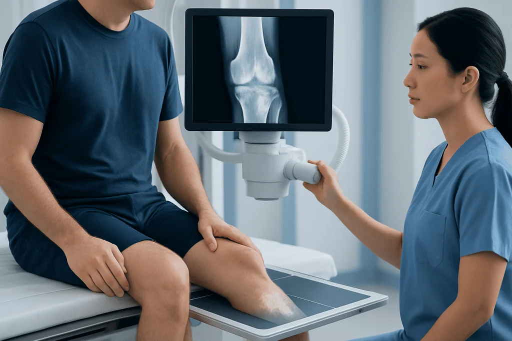
A clinician may recommend a knee X-ray for children, teenagers, or adults with symptoms that are persistent, progressive, or difficult to explain. Osteosarcoma is more common in adolescents and young adults, but it can occur at other ages. The test may also be used after an injury when symptoms or examination findings suggest that bone needs closer assessment.
The X-ray machine directs a controlled beam through the knee. Dense structures such as bone absorb more radiation and appear lighter on the resulting image, while soft tissues appear darker. Several views are usually taken, often from the front and side, so the radiologist can assess the bone from different angles.
Most people are suitable for an X-ray. Patients should tell the imaging team if they are or may be pregnant, as the team can confirm whether imaging is necessary and use appropriate safeguards. Previous X-rays, MRI scans, and medical records can be helpful for comparison, but they are not required before the examination.
Preparation and Step-by-Step Procedure

Little preparation is needed for an osteosarcoma Xray knee. The patient can usually eat, drink, and take regular medicines as normal unless a clinician gives different instructions. Comfortable clothing is helpful, and metal objects or jewelry near the knee should be removed because they can affect image quality.
At the imaging department, a radiographer explains the process and positions the leg on the X-ray table or in front of the imaging detector. The patient may be asked to sit, lie down, or stand depending on the views needed. Keeping still for a few seconds while each image is taken helps create clear results.
The radiographer usually steps behind a protective screen to operate the machine but can see and communicate with the patient throughout. The imaging itself is brief, commonly completed within minutes, although positioning can take longer if the knee is painful or movement is limited. The test does not involve injections, anesthesia, or recovery time.
Afterward, a radiologist reviews the images and sends a report to the referring clinician. If there is a suspicious finding, the clinician should explain what it may mean and arrange further assessment promptly. It is important not to interpret an X-ray report without medical guidance, because the significance depends on symptoms, examination, and other tests.
Results, Benefits, Risks, and What Happens Next
A normal knee X-ray can be reassuring, but it may not explain every cause of knee pain. Soft-tissue injuries involving cartilage, ligaments, tendons, or early bone marrow changes are often better assessed with MRI. If symptoms continue despite a normal X-ray, a clinician may recommend follow-up or additional imaging.
If a scan shows a lesion that could represent osteosarcoma, the usual next investigation is an MRI of the entire affected bone and nearby joint. MRI helps show the size of the lesion and whether nearby muscles, nerves, blood vessels, or the joint may be involved. Chest imaging is also often part of staging because the lungs are a common site of spread when osteosarcoma metastasizes.
The main benefit of X-ray is that it is fast, accessible, and very useful for evaluating bone structure. Radiation exposure from a knee X-ray is low. The test is generally safe, although avoiding unnecessary radiation remains important, particularly during pregnancy. The main limitation is that it cannot confirm whether an abnormality is cancerous.
A biopsy is needed to make a definite diagnosis. It should be planned by the team that would provide any later surgery, because the biopsy pathway must be positioned carefully. In suspected bone cancer, referral to a specialist orthopedic oncology and sarcoma service is an important part of safe, coordinated care.
How Do I Know If My Knee Pain Is Osteosarcoma?
Knee pain is common and is much more often caused by injury, overuse, arthritis, tendon problems, or growth-related pain than by osteosarcoma. Pain alone cannot identify bone cancer. Osteosarcoma-related discomfort may begin gradually and become more persistent over time, sometimes worsening at night or occurring at rest, but these features are not specific to cancer.
Other possible signs include a growing swelling or firm lump near the knee, reduced ability to use the leg, limping, or pain that does not improve as expected after rest or routine injury care. In some cases, the bone may weaken and fracture after relatively minor force. These symptoms warrant medical assessment, but they do not confirm a diagnosis.
A clinician will consider the person’s age, symptoms, injury history, physical examination, and imaging. An X-ray may reveal whether there is a bone abnormality, while MRI and biopsy clarify the diagnosis. Seeking assessment for persistent symptoms is a practical step that supports timely care without assuming the cause is cancer.
Is Bone Cancer in the Knee Curable?
Some cases of bone cancer in the knee, including localized osteosarcoma, can be treated with curative intent. The outlook depends on factors such as whether the cancer has spread, the tumor’s response to treatment, its location, and whether it can be fully removed safely. Individual prognosis should always be discussed with the treating oncology team.
Treatment for osteosarcoma commonly combines chemotherapy with surgery. Chemotherapy may be given before surgery to treat cancer cells throughout the body and help assess response, followed by surgery to remove the tumor. Whenever appropriate, surgeons aim to preserve the limb using reconstruction, although the safest operation differs from person to person.
Care is delivered by a multidisciplinary team that may include orthopedic oncologists, medical oncologists, radiologists, pathologists, rehabilitation specialists, nurses, and psychosocial support professionals. At Acibadem International, multidisciplinary specialists in JCI-accredited hospitals assess and treat osteosarcoma for international patients, with care plans tailored to clinical findings and personal needs.
How Fast Does Osteosarcoma Spread?
Osteosarcoma can grow and spread, but its pace varies between individuals and cannot be predicted from symptoms or an X-ray alone. When it spreads beyond the original bone, it most commonly travels to the lungs. This is why chest imaging is usually included when osteosarcoma is being evaluated or staged.
Doctors assess the extent of disease promptly using imaging and biopsy results. MRI evaluates the tumor around the knee and throughout the involved bone, while chest CT may provide detailed images of the lungs. Depending on the clinical situation, other scans may be used to assess other bones or areas of the body.
Prompt assessment does not mean that every painful knee is an emergency or that cancer is likely. It means that a persistent, worsening, or unexplained bone-related symptom should be reviewed by a qualified clinician. If osteosarcoma is diagnosed, treatment planning begins without unnecessary delay and is based on the full staging assessment.
What Is the Survival Rate for Sarcoma in the Knee?
There is no single survival rate for “sarcoma in the knee” because sarcoma includes many different tumor types, and outcomes vary considerably. For osteosarcoma, prognosis is influenced most strongly by whether the cancer is localized or has spread at diagnosis, as well as response to chemotherapy and the ability to remove the tumor completely.
Published survival estimates are based on groups of patients treated in earlier periods and cannot predict what will happen for one person. Advances in imaging, surgical reconstruction, chemotherapy, supportive care, and specialist team management continue to shape treatment approaches. A treating team can explain the most relevant outlook after diagnosis and staging are complete.
It can be helpful to ask the care team what type of sarcoma is present, whether it is localized, what the treatment goals are, and which factors are most relevant in the individual situation. Clear discussions can support informed decisions while avoiding conclusions based on generalized figures found online.
When to Seek Medical Care
Medical review is advisable for knee or bone pain that persists beyond a few weeks, steadily worsens, wakes someone from sleep, or does not improve as expected after a minor injury. A new or enlarging lump, visible swelling, unexplained limp, reduced movement, or repeated pain in the same area should also be assessed.
Urgent assessment is appropriate if there is sudden severe pain after minor trauma, inability to bear weight, a suspected fracture, or rapidly worsening swelling. Fever, marked redness, or feeling unwell alongside bone pain may indicate infection or another condition that also needs timely medical attention.
Most concerning symptoms will have explanations other than osteosarcoma, but evaluation provides clarity and directs treatment. A clinician may begin with examination and X-ray, then refer to an orthopedic, radiology, or oncology specialist if results require further investigation.
Frequently asked questions
Does an X-ray always show osteosarcoma in the knee?
X-rays often show bone changes that raise concern for osteosarcoma, but they cannot diagnose it with certainty. MRI, other staging scans, and a biopsy are needed to confirm the diagnosis and guide treatment planning.
Is a knee X-ray painful?
The X-ray itself is painless and does not involve needles or injections. Positioning the leg may be uncomfortable if the knee is already painful, but the radiographer can help arrange the patient as comfortably as possible.
How long does it take to receive knee X-ray results?
The timing varies by imaging center and clinical urgency. A radiologist reviews the images, and the referring clinician usually explains the report and any next steps.
Can osteosarcoma be mistaken for a sports injury?
Early osteosarcoma symptoms can resemble a sports injury because they may include pain, swelling, or limping around the knee. Pain that persists, progresses, occurs at rest or at night, or is accompanied by a lump should be evaluated by a clinician.
Why is a biopsy planned by a specialist team?
The biopsy route must be carefully selected so it does not interfere with later surgery if cancer is found. A specialist team can obtain enough tissue for accurate testing while protecting future treatment options.
Can a normal X-ray rule out every serious cause of knee pain?
No. A normal X-ray is useful for assessing many bone problems, but it may not show soft-tissue injuries or some early bone marrow changes. Continued or worsening symptoms should be discussed with the clinician, who may recommend MRI or other evaluation.
References
- National Cancer Institute
- American Cancer Society
- National Health Service
- RadiologyInfo.org
- European Society for Medical Oncology
This article is for general information only and is not a substitute for professional medical advice. Please consult a qualified doctor about your individual situation.
Joint and spine care in Turkey — expert assessment & treatment
JCI-accredited · board-certified surgeons · reply within 24h
Add us as a Preferred Source to see more of our trusted health content across Google Search, AI Overviews and Discover.

More from the Health Library



Related Specialists

Dr. Erbil Aydemir
Pediatrics
Dr. Melike Zeynep Çapoğlu
Oral & Dental Health
Dr. Cengiz Akbenlioğlu
Pediatrics
Dr. Cem Tangay
Orthopedic Surgery & Traumatology