Painless Lump in Middle of Chest Male Explained: Common Triggers and Red Flags

The pointed lower end of the breastbone, called the xiphoid process, can feel like a firm midline chest lump and is usually normal. Common non-cancerous causes include lipomas, epidermoid cysts, prominent cartilage, and changes after an injury.
Key Takeaways
- The pointed lower end of the breastbone, called the xiphoid process, can feel like a firm midline chest lump and is usually normal.
- Common non-cancerous causes include lipomas, epidermoid cysts, prominent cartilage, and changes after an injury.
- A painless lump still deserves medical assessment if it is new, growing, hard, fixed in place, or associated with skin, breast, or general health changes.
- A clinician can often identify the likely cause through examination and may use ultrasound, X-ray, CT, or other tests when needed.
- Urgent assessment is important for a lump with breathing difficulty, chest pressure, infection signs, rapid enlargement, or unexplained weight loss.
A painless lump in the middle of the chest in a male is commonly related to the normal breastbone or xiphoid process, a harmless fatty lump, or a skin cyst. However, because painless lumps can occasionally need treatment, any new, enlarging, hard, fixed, or unexplained lump should be checked by a qualified clinician.
What can cause a painless lump in the middle of the chest?
A painless lump in the middle of the chest in a male is often not dangerous. One of the most frequent explanations is a normal structure called the xiphoid process, the small section of bone and cartilage at the lower tip of the sternum (breastbone). It may feel like a firm bump below the main flat part of the breastbone, especially in people with a slim body frame or after weight loss.
Other common explanations include a lipoma (a soft, non-cancerous collection of fatty tissue), an epidermoid cyst beneath the skin, or a naturally prominent area of chest cartilage. A lump can also become more noticeable after a chest injury, exercise-related strain, inflammation, or a change in body weight. The location, texture, mobility, and speed of change provide useful clues.
Painlessness is generally reassuring, but it does not by itself confirm the cause. A lump that is genuinely new, continues to grow, feels hard and immobile, or has no clear explanation should be examined. Most chest wall lumps are non-cancerous, but careful assessment helps identify the small number that need further investigation or treatment.
Understanding the location: skin, breast tissue, chest wall, or bone
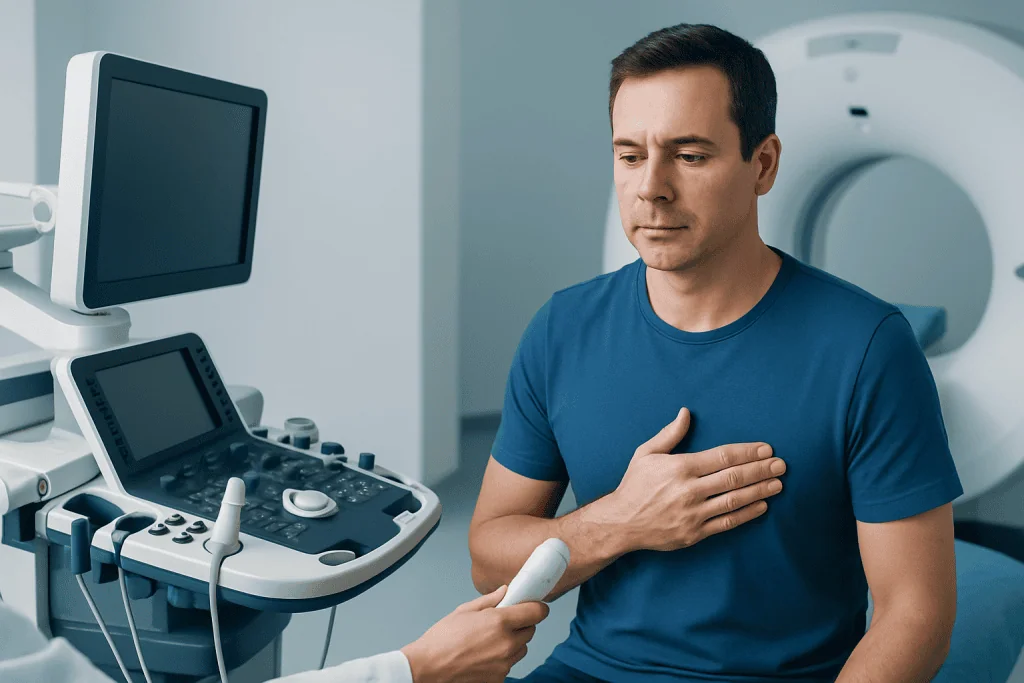
The term “middle of the chest” can describe several different areas. A lump directly in the midline may arise from the skin, the tissue beneath it, the sternum, the cartilage joining the ribs to the sternum, or the xiphoid process. A clinician will first establish whether the finding is part of the normal chest structure or a separate growth.
A skin or surface lump may move slightly with the skin and may have a visible pore, discoloration, or a smooth rounded outline. Lipomas are often soft, rubbery, and mobile under the skin. Cysts may feel rounder and firmer and can occasionally become inflamed, even if they were initially painless.
Lumps closer to one side of the chest, particularly behind or beneath the nipple, may involve breast tissue rather than the sternum. Male breast enlargement, known as gynecomastia, can create a tender or painless disc-like area under the nipple. This differs from a central bony prominence, and the distinction matters because the causes and investigations can be different.
Common triggers and non-serious causes

A prominent xiphoid process is a normal anatomical variation rather than a disease. It is usually hard, fixed, and positioned at the bottom of the breastbone. Some people first notice it after touching the area, changing weight, beginning a fitness routine, or experiencing mild local irritation. Pressing it repeatedly can make the area sore, so it is best not to keep checking or manipulating it.
Lipomas are another common cause of a painless chest lump. They develop gradually from fat cells and are usually soft and movable beneath normal-looking skin. They do not usually require treatment unless they are uncomfortable, enlarging, affecting movement, or creating uncertainty about the diagnosis. A clinician can advise whether observation, imaging, or removal is appropriate.
Epidermoid cysts can occur when skin cells become trapped beneath the surface. They are often slow-growing and painless unless inflamed or infected. Past injury may also leave a firm area during healing, particularly near a rib or the sternum. Less commonly, a bony or cartilaginous prominence may be related to a chest wall shape difference, previous fracture healing, or inflammation around a rib-sternum joint.
- Normal xiphoid process: hard, central, and fixed at the lower sternum.
- Lipoma: soft or rubbery, slow-growing, and usually mobile.
- Skin cyst: rounded lump near the surface, sometimes with a small central opening.
- Post-injury change: a firm area noticed after a blow, strain, or fracture.
Less common causes and signs that need attention
Although most painless chest lumps have a non-serious explanation, less common causes include benign growths of bone or cartilage, enlarged lymph nodes, infections, and tumors affecting skin, soft tissue, bone, breast tissue, or structures deeper in the chest. These conditions cannot be diagnosed reliably by touch alone, particularly if the lump is deep or difficult to define.
A lump deserves prompt clinical review if it is rapidly enlarging, larger than expected, very hard, irregular, or fixed firmly to deeper tissue. Changes in the overlying skin, such as redness, warmth, breakdown, dimpling, ulceration, or a persistent rash, should also be assessed. A nipple discharge, nipple inversion, or a new one-sided breast lump requires medical evaluation in men as well as women.
General symptoms can add important context. These include unexplained weight loss, persistent fever, drenching night sweats, ongoing fatigue, or a persistent cough. Such symptoms often have causes unrelated to the lump, but reporting them helps the clinician decide whether imaging or specialist assessment is needed. It is sensible to seek assessment rather than attempting to self-diagnose from online images or descriptions.
How a doctor investigates a chest lump
Assessment usually begins with a medical history and physical examination. The clinician may ask when the lump was first noticed, whether it has changed, whether there was recent injury or infection, and whether there are symptoms such as pain, fever, weight loss, or breast changes. They will examine the size, shape, consistency, mobility, tenderness, and precise location of the lump.
If the finding clearly matches a normal xiphoid process or a typical harmless skin lesion, testing may not be necessary. When the cause is uncertain, an ultrasound scan can help distinguish a cyst, lipoma, breast tissue change, or solid soft-tissue mass. An X-ray, CT scan, or MRI may be considered when a lump appears connected to bone or cartilage, lies deeper in the chest wall, or needs more detailed evaluation.
Occasionally, a doctor may recommend a biopsy, which involves taking a small tissue sample for laboratory analysis. This is not needed for every lump. It is used when imaging and examination cannot confidently identify the cause or when features raise concern. The purpose is to reach an accurate diagnosis and guide care rather than to assume the worst.
Treatment and safe self-care
Treatment depends entirely on the cause. A normal xiphoid process or stable, harmless chest wall prominence usually needs reassurance only. Avoiding repeated pressing, squeezing, or trying to “move” a lump is helpful, as this can irritate skin and nearby tissues and make the area more noticeable or tender.
Lipomas and uncomplicated cysts may be monitored if they are small, stable, and not bothersome. If a lump is painful, repeatedly inflamed, growing, or cosmetically troubling, a doctor may discuss removal or another appropriate procedure. Infected cysts or skin abscesses need clinical care; squeezing or cutting them at home can worsen infection and cause scarring.
It can be useful to note the date a lump was first noticed and whether its size or appearance changes over several weeks. A person should avoid measuring it repeatedly every day, as this may increase anxiety without providing reliable information. Instead, a brief record of changes and relevant symptoms can help during a medical appointment.
For international patients, Acibadem International’s multidisciplinary specialists at JCI-accredited hospitals can assess chest wall and breast-related symptoms, arrange appropriate imaging, and coordinate care when treatment is needed.
When to seek medical care
A non-urgent doctor’s appointment is appropriate for any new painless lump in the middle of the chest that lasts longer than a short period, is not clearly explained by normal anatomy, or causes worry. Assessment is especially important if the lump is growing, changes in shape, becomes hard or fixed, or appears after an injury that has not been medically reviewed.
More prompt medical advice is needed when there are skin changes, redness, warmth, drainage, fever, increasing pain, breast or nipple changes, or swollen glands near the collarbone or armpit. A clinician should also review a lump accompanied by unintended weight loss, persistent night sweats, or ongoing unexplained tiredness.
Emergency care is appropriate if a chest lump occurs with chest pressure or pain, shortness of breath, fainting, coughing blood, severe injury, or signs of a serious allergic reaction. These symptoms may not be caused by the lump itself, but they require urgent evaluation. In the absence of emergency symptoms, timely assessment by a primary care doctor is the safest next step.
Frequently asked questions
Is a hard lump at the bottom of the sternum normal in men?
It can be normal. The xiphoid process is a firm structure at the lower end of the breastbone and may feel like a hard central lump. If it is newly noticeable, growing, painful, or different from a fixed midline bony prominence, a clinician should assess it.
Can a painless chest lump be cancer?
Most chest lumps are not cancer, particularly when they are soft, mobile, and stable. However, some important conditions can be painless in their early stages. A new, enlarging, hard, irregular, or fixed lump should be examined rather than judged by pain alone.
What does a lipoma in the chest feel like?
A lipoma often feels soft, doughy, or rubbery and may move slightly under the skin when touched. It usually grows slowly and is not painful. A doctor can confirm whether a lump has typical lipoma features or whether imaging is needed.
Can exercise cause a lump in the middle of the chest?
Exercise does not usually create a true lump, but it can make normal chest structures more noticeable or irritate the cartilage and muscles around the breastbone. An injury during sport can also lead to swelling or a healing bony prominence. A persistent lump should still be checked, particularly after significant trauma.
Should a man see a doctor for a lump under the nipple?
Yes. A lump under the nipple may be related to gynecomastia, which is often benign, but male breast lumps should be assessed to identify the cause. Medical review is especially important for a one-sided lump, nipple discharge, skin dimpling, or a lump that is hard and fixed.
Which scan is used for a lump in the chest wall?
The most suitable scan depends on the suspected source of the lump. Ultrasound is commonly useful for superficial soft-tissue, cyst, and breast-area lumps, while X-ray, CT, or MRI may be used for bone, cartilage, or deeper chest wall findings. The examining clinician will select testing based on the physical examination and symptoms.
This article is for general information only and is not a substitute for professional medical advice. Please consult a qualified doctor about your individual situation.
Explore treatments in Turkey — costs, top hospitals & a free quote
JCI-accredited · board-certified surgeons · reply within 24h
Add us as a Preferred Source to see more of our trusted health content across Google Search, AI Overviews and Discover.

More from the Health Library

Why Do All My Teeth Hurt Suddenly? Here Is What the Evidence Says

What Do Stds Look Like? Here Is What the Evidence Says

How to Avoid Feet Sweating? Causes, Explanations, and Next Steps

Can Heat Cause Diarrhea? Here Is What the Evidence Says

Pancreatic Cancer Symptoms Explained: Common Triggers and Red Flags