

Partial Hemianopia: A Complete Medical Overview
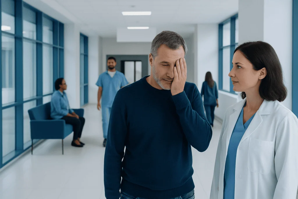
Partial hemianopia means losing part of the visual field on one side, not complete blindness in an eye. Common causes include stroke, brain injury, tumors, and other conditions affecting the visual pathways.
Key Takeaways
- Partial hemianopia means losing part of the visual field on one side, not complete blindness in an eye.
- Common causes include stroke, brain injury, tumors, and other conditions affecting the visual pathways.
- Testing often includes formal visual field exams, a full eye examination, and brain imaging such as MRI or CT.
- Treatment focuses on the underlying cause and may also include visual rehabilitation and practical coping strategies.
- Sudden visual field loss is a medical concern and should be assessed urgently.
Partial hemianopia is a type of visual field loss where a person cannot see part of one side of the world around them, even though the eyes themselves may look normal. It usually happens when the brain’s visual pathways are affected, so diagnosis focuses on both eye health and the nervous system.
Overview: what partial hemianopia means
Partial hemianopia is a loss of part of the visual field on one side. In simple terms, a person may miss objects, movement, or written words located in a particular area to the left or right, even though central sharpness of vision may remain fairly good. This differs from total blindness and also differs from blurred vision caused by common eye conditions.
The condition usually reflects a problem in the brain’s visual pathways rather than in the surface of the eye. Light may enter the eyes normally, but the signals are not fully processed because part of the route from the eyes to the brain has been injured or compressed. For this reason, partial hemianopia is often assessed by both eye specialists and neurology teams.
Partial hemianopia can affect daily life in subtle or significant ways. Some people bump into doorframes, miss cars approaching from one side, lose their place while reading, or feel unusually clumsy in crowded spaces. Others notice little at first, and the problem is only found during formal visual field testing.
How vision is affected in daily life
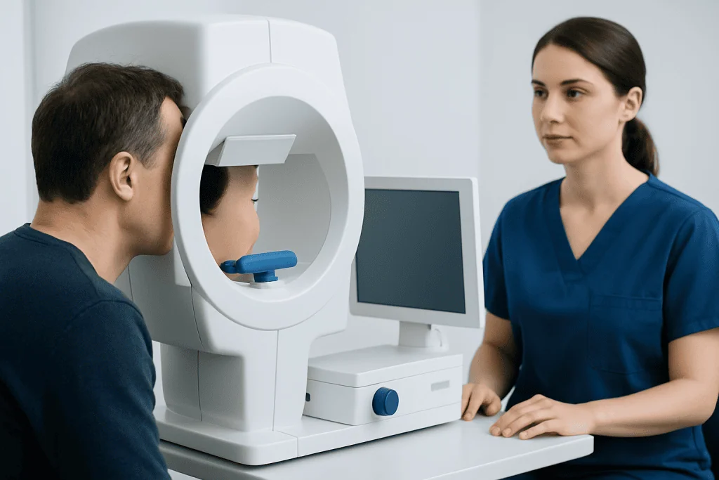
A person with partial hemianopia does not lose all vision equally. Instead, a section of the visual field is missing or reduced. The missing area may be upper or lower, more central or more peripheral, and may affect the same side in both eyes. This pattern helps doctors identify where the visual pathway may be affected.
Because the brain combines information from both eyes, visual field loss can be confusing to describe. People may say they are “not seeing well on one side” or that words disappear while reading. Some only notice symptoms when driving, walking downstairs, navigating unfamiliar places, or trying to find items on a shelf.
Common functional effects include:
- Missing objects or people on one side
- Bumping into furniture, walls, or doorframes
- Difficulty reading lines of text or scanning a page
- Trouble crossing streets or noticing vehicles from one direction
- Visual fatigue, frustration, or reduced confidence outdoors
The impact depends on how much of the field is affected, whether the change came on suddenly or gradually, and whether the brain has had time to adapt. Occupational therapy, visual scanning training, and home adjustments may help people regain confidence in everyday tasks.
Symptoms and related warning signs

The main symptom of partial hemianopia is patchy or sector-like loss of vision on one side of the visual field. It may seem as if part of the environment is missing, faded, or harder to notice. Some people mistake this for poor attention, clumsiness, or ordinary eye strain at first.
Symptoms may occur alone, but they can also appear together with signs of a neurological condition. These additional symptoms matter because they may suggest an urgent cause such as a stroke, bleeding, or a rapidly changing brain problem.
Possible associated symptoms include:
- Sudden onset of visual field loss
- Headache, especially if severe or new
- Weakness or numbness on one side of the body
- Speech difficulty or confusion
- Problems with balance, coordination, or walking
- Double vision or other new visual changes
- Seizures or episodes of altered awareness
When symptoms start suddenly, especially with weakness, trouble speaking, or a severe headache, urgent assessment is important. Visual field loss can be one sign of stroke, which requires immediate medical care.
Causes and risk factors
Partial hemianopia occurs when part of the visual pathway is disrupted. This pathway begins at the retina, travels through the optic nerves, crosses partially at the optic chiasm, and continues through deeper brain structures to the visual cortex. The exact pattern of field loss can help localize the problem.
One of the most common causes is stroke, particularly when blood flow to the occipital lobe or nearby visual pathways is interrupted. Head injury may also damage these pathways directly or through bleeding and swelling. Brain tumors, inflammation, infection, and certain neurological disorders can produce similar symptoms if they involve the same regions.
Other possible causes and risk factors include:
- Transient ischemic attack or stroke risk factors such as high blood pressure, diabetes, smoking, and heart disease
- Traumatic brain injury
- Brain tumors or masses compressing visual pathways
- Brain surgery or bleeding in the brain
- Demyelinating disease, including multiple sclerosis in some cases
- Seizure-related or migraine-related visual phenomena, which must be distinguished from persistent field loss
Some people develop visual field loss after treatment for another serious condition. In these situations, the cause may still be identifiable and manageable. A careful medical history, timing of symptoms, and examination findings are essential for determining the reason.
How partial hemianopia is diagnosed
Diagnosis begins with a detailed description of what the person is seeing and when the symptoms started. A doctor will ask whether the change was sudden or gradual, whether it affects reading or mobility, and whether there are neurological symptoms such as weakness, speech changes, or headache. This helps guide the urgency of testing.
An eye examination checks visual acuity, eye movements, pupils, and the health of the retina and optic nerve. However, a person can have partial hemianopia even when the eye examination appears largely normal. For that reason, formal visual field testing is especially important to map the exact area of loss.
Imaging of the brain is often needed because the source is commonly beyond the eye itself. Depending on the situation, doctors may request MRI scanning or CT imaging to look for stroke, bleeding, tumors, or other structural causes. Blood tests and cardiovascular evaluation may also be advised if a vascular cause is suspected.
When the pattern is complex or the diagnosis is uncertain, referral to a neuro-ophthalmology team may be helpful. In selected cases, a multidisciplinary assessment that includes neurology, neuroradiology, rehabilitation, and eye specialists gives the clearest picture of both cause and next steps.
Treatment options and visual rehabilitation
There is no single treatment for partial hemianopia because management depends on the underlying cause. If the visual field loss is due to stroke, treatment focuses on urgent stroke care and prevention of future events. If the cause is a tumor, inflammation, or another structural problem, care is directed at that condition.
Some people recover part of their visual field over time, especially in the weeks or months after an acute brain event. Others have persistent field loss and benefit most from rehabilitation. This may include visual scanning exercises, reading strategies, orientation training, and practical methods to compensate for the missing field.
Supportive treatment may include:
- Neuro-ophthalmology follow-up
- Occupational therapy for safety and daily function
- Vision rehabilitation programs
- Advice on home layout, lighting, and fall prevention
- Driving assessment according to local regulations
- Treatment of the underlying neurological or vascular condition
When a structural cause requires intervention, doctors may consider therapies such as brain tumor surgery if appropriate. Near the end of the care pathway, patients may also seek coordinated assessment at centers such as Acibadem International, where multidisciplinary specialists and JCI-accredited hospitals evaluate and treat international patients with neurological and visual disorders.
Self-care, safety, and adaptation
Living with partial hemianopia often involves learning new visual habits. Turning the head more fully toward the affected side, scanning the environment before moving, and using finger or line guides when reading can improve function. These strategies do not cure the problem, but they can make daily activities easier and safer.
At home, simple adjustments may reduce accidents. Good lighting, clear walkways, contrast markings on steps, and organized storage can help a person compensate for reduced awareness on one side. Family members may also support adaptation by approaching from the stronger visual side while encouraging regular scanning practice.
Driving, cycling, and certain work tasks may be affected. People should avoid driving until they have been medically assessed and understand the legal and safety requirements in their area. If visual field loss follows a neurological event, rehabilitation and follow-up are often important before returning to demanding activities.
Emotional adjustment matters too. Losing part of the visual field can be frustrating and tiring, especially when the problem is invisible to others. Reassurance, practical education, and specialist rehabilitation can help people regain confidence and independence over time.
When to seek medical care
Sudden partial hemianopia should be treated as a prompt medical concern. A new visual field defect may be the first sign of stroke or another serious neurological condition, especially if it appears within minutes or hours. Early evaluation improves the chance of identifying the cause and starting appropriate treatment quickly.
Urgent care is particularly important if visual field loss occurs with facial drooping, arm weakness, numbness, speech difficulty, confusion, severe headache, imbalance, or seizure. These symptoms suggest that the nervous system may be involved and should not be ignored.
Medical review is also advisable if visual field problems develop gradually, keep recurring, or begin to interfere with reading, walking, driving, or work. Even when symptoms seem mild, formal testing can confirm whether true field loss is present and whether further brain imaging is needed.
If a person has already been diagnosed, follow-up remains important. Changes in the visual field, new headaches, or additional neurological symptoms should be reported to a qualified doctor without delay.
Frequently asked questions
What is the difference between partial hemianopia and complete hemianopia?
Partial hemianopia means only part of one half of the visual field is missing. Complete hemianopia means the entire half of the visual field is lost. The distinction is important because the exact pattern may help doctors locate the problem in the visual pathway.
Can partial hemianopia affect both eyes?
Yes. Although people may describe it as vision loss on one side, the pattern usually involves the same side of the visual field in both eyes. This happens because the problem is often in the brain’s processing pathway rather than in just one eye.
Is partial hemianopia permanent?
Sometimes it improves, and sometimes it persists. Recovery depends on the cause, how quickly treatment begins, and which part of the visual pathway is affected. Even when the field loss does not fully reverse, rehabilitation can help improve day-to-day function.
Can a stroke cause partial hemianopia?
Yes, stroke is one of the most important causes of sudden visual field loss. If partial hemianopia starts suddenly, especially with weakness, speech changes, or severe headache, urgent medical assessment is needed.
How do doctors test for partial hemianopia?
Doctors usually begin with a medical history and a full eye examination. Formal visual field testing is then used to map the missing area, and brain imaging such as MRI or CT may be needed to find the cause.
Can someone drive with partial hemianopia?
That depends on the degree of field loss, local driving laws, and the results of medical assessment. Many people should stop driving until they are fully evaluated. Safety is the priority because side awareness may be reduced.
References
- American Academy of Ophthalmology
- National Eye Institute
- National Institute of Neurological Disorders and Stroke
- American Stroke Association
- Mayo Clinic
This article is for general information only and is not a substitute for professional medical advice. Please consult a qualified doctor about your individual situation.
Explore treatments in Turkey — costs, top hospitals & a free quote
JCI-accredited · board-certified surgeons · reply within 24h
Add us as a Preferred Source to see more of our trusted health content across Google Search, AI Overviews and Discover.
Check your numbers in seconds
BMI, calories, due date, blood pressure and 30+ more clinical calculators — free, instant, doctor-reviewed ranges.

More from the Health Library



Related Specialists

Dr. Süreyya Işık Coşkun
Psychiatry
Assoc. Prof. Dr. Serap Şahin Önder
Otorhinolaryngology
Dr. Hülya Yolaçan
Pulmonary Medicine
Prof. Dr. Şükrü Yazar
Aesthetic Plastic & Reconstructive Surgery