Peripheral Nerve Ultrasound: When It Is Used in Neuromuscular Diagnosis

Peripheral nerve ultrasound shows the size, shape, and course of nerves in real time. It is especially useful for suspected nerve entrapment, nerve injury, and some inflammatory or hereditary neuropathies.
Key Takeaways
- Peripheral nerve ultrasound shows the size, shape, and course of nerves in real time.
- It is especially useful for suspected nerve entrapment, nerve injury, and some inflammatory or hereditary neuropathies.
- The test is painless, does not use radiation, and can complement EMG and nerve conduction studies.
- Ultrasound can help guide treatment decisions by locating the exact site and severity of nerve involvement.
- Results are most helpful when interpreted together with symptoms, examination findings, and other tests.
Peripheral nerve ultrasound is a noninvasive imaging test that helps doctors examine nerves in real time. It is often used alongside clinical evaluation and nerve conduction studies to clarify the cause of weakness, numbness, pain, or muscle changes.
Overview
Peripheral nerve ultrasound is a specialized imaging technique used to examine nerves outside the brain and spinal cord. Using high-frequency sound waves, it creates detailed images of superficial nerves and surrounding tissues, such as muscles, tendons, blood vessels, and connective tissue. Because the test is dynamic, the doctor can assess the nerve while the patient moves a limb or changes position.
In neuromuscular diagnosis, this test has become a valuable complement to the neurological examination and electrodiagnostic testing. While nerve conduction studies and electromyography evaluate how nerves and muscles function, ultrasound shows structure. This combination can help answer an important question: whether symptoms are caused by nerve compression, inflammation, injury, or another process affecting the nerve’s shape or pathway.
Peripheral nerve ultrasound is commonly used when a person has numbness, tingling, pain, weakness, muscle wasting, or unexplained sensory changes. It may be particularly helpful in conditions involving trapped or enlarged nerves, such as carpal tunnel syndrome, ulnar neuropathy at the elbow, traumatic nerve injury, and selected polyneuropathies.
When It Is Used
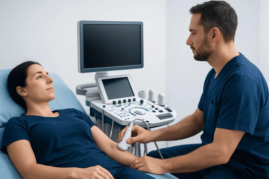
Doctors may recommend peripheral nerve ultrasound when symptoms suggest a focal nerve problem. Common examples include hand numbness at night, tingling in the ring and little fingers, foot drop, wrist weakness, or pain radiating along a specific nerve distribution. In these situations, ultrasound can help locate the exact point where a nerve is compressed, stretched, scarred, or otherwise affected.
The test is also used when physical examination and electrodiagnostic tests do not fully explain symptoms. For instance, a person may have pain or weakness that points to a nerve problem, but the source may be difficult to define. Ultrasound can reveal nerve swelling, flattening, displacement, or abnormal mobility that may not be obvious from symptoms alone.
In some neuromuscular disorders, ultrasound helps assess multiple nerves and muscles in a short time. This can support the evaluation of inherited neuropathies, inflammatory neuropathies, or muscle disease patterns. It may also be useful after injury or surgery to check nerve continuity, identify neuromas, or guide decisions about peripheral nerve surgery.
What Ultrasound Can Show
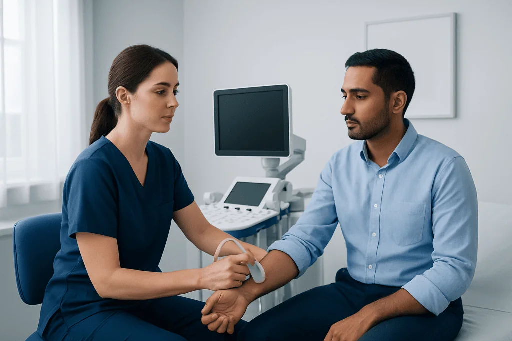
Peripheral nerve ultrasound is especially good at showing structural changes. A healthy nerve usually has a recognizable internal pattern and follows a predictable course. When disease is present, the nerve may look enlarged, flattened, darker than usual, more vascular, or irregular in outline. Comparing the affected side with the unaffected side often adds useful information.
Ultrasound can identify several important findings:
- Focal nerve enlargement at a compression site
- Flattening of the nerve as it passes through a tight tunnel
- Changes in the nerve’s internal fascicular pattern
- Nerve displacement, subluxation, or abnormal movement
- Scar tissue, masses, cysts, or surrounding inflammation
- Muscle changes that suggest chronic denervation
These details may help distinguish among common causes of symptoms. For example, in entrapment neuropathies, ultrasound can show exactly where the nerve is pinched. In traumatic injuries, it can help determine whether the nerve is intact, partially torn, or surrounded by scar tissue. In broader neuropathies, enlargement affecting several nerves can suggest a generalized process rather than a single local compression.
Conditions Commonly Evaluated
One of the most frequent uses of peripheral nerve ultrasound is the evaluation of entrapment neuropathies. These happen when a nerve is compressed as it passes through a narrow space. Examples include median nerve compression at the wrist, ulnar neuropathy at the elbow, and peroneal nerve compression near the knee. Ultrasound can confirm the site of compression and identify contributing structures such as thickened tissue, ganglion cysts, or anatomical variations.
The test is also useful in nerve injury. After trauma, stretch injury, laceration, fracture, or surgery, ultrasound can assess the continuity of the nerve and any swelling above or below the injury. This information can be important when planning rehabilitation, follow-up, or procedures such as EMG and nerve conduction studies or surgical repair.
In selected people with peripheral neuropathies, ultrasound provides additional clues about whether the problem may be inflammatory, hereditary, or multifocal. It can also be used in some muscle disorders to look for muscle wasting or changes in muscle texture, although it is not a replacement for other imaging or laboratory tests. When symptoms involve the neck or spine as well as the limbs, doctors may need to combine ultrasound findings with broader evaluations, including MRI.
How the Test Is Performed and What to Expect
Peripheral nerve ultrasound is usually performed in an outpatient setting by a radiologist, neurologist, or another clinician trained in neuromuscular imaging. The patient sits or lies in a comfortable position, and a clear gel is placed on the skin. A small handheld probe is moved along the course of the nerve to obtain images from different angles.
The test is generally painless and does not involve radiation, injections, or sedation. The clinician may ask the patient to bend the wrist, elbow, knee, or ankle during the scan. This helps assess whether the nerve glides normally or becomes compressed or unstable during movement. In many cases, both sides of the body are examined for comparison.
Preparation is simple. Patients may be asked to wear loose clothing that allows easy access to the arm or leg being examined. Depending on the clinical question, the ultrasound may be done before or after an electrodiagnostic study. When both are needed, together they can provide a more complete picture of nerve structure and function.
How It Helps With Diagnosis and Treatment Planning
Peripheral nerve ultrasound rarely works as a stand-alone diagnosis. Instead, it helps doctors bring together several pieces of information: symptoms, neurological examination, laboratory results, electrodiagnostic tests, and imaging findings. This combined approach is particularly valuable because similar symptoms can arise from different causes, such as a trapped nerve, a root problem in the spine, a generalized neuropathy, or a muscle disorder.
By showing the location and extent of structural change, ultrasound can help determine whether treatment should focus on conservative care, further testing, or a procedure. For example, if a nerve is mildly enlarged at a common entrapment site, treatment may involve splinting, activity changes, physical therapy, or medication. If a mass, severe compression, or loss of nerve continuity is found, referral to a surgeon may be considered.
Ultrasound can also assist with follow-up. Repeated imaging may be used to monitor recovery after injury, assess postoperative changes, or document the progression of a nerve disorder. Near the end of the care pathway, patients seeking comprehensive evaluation may be seen at centers such as Acibadem International, where multidisciplinary specialists in JCI-accredited hospitals diagnose and treat neuromuscular and peripheral nerve conditions for international patients.
Limitations, Self-Care, and When to Seek Medical Advice
Although peripheral nerve ultrasound is very useful, it has limits. Deep nerves and structures covered by bone can be harder to visualize, and the quality of the examination depends on the equipment used and the experience of the clinician. In some cases, normal ultrasound findings do not rule out a nerve problem, especially if symptoms are early, intermittent, or related more to function than structure.
Because of these limits, patients should not assume that one test alone provides the full answer. Persistent numbness, tingling, weakness, hand clumsiness, muscle wasting, burning pain, or balance problems deserve medical evaluation. Sudden weakness, rapidly worsening symptoms, loss of bladder or bowel control, or symptoms after major trauma need urgent assessment.
Self-care depends on the cause. General measures may include avoiding repetitive strain, adjusting workstation posture, using splints when advised, controlling diabetes, limiting alcohol, and following a rehabilitation plan. Early assessment can improve the chance of identifying treatable causes and preventing long-term nerve damage.
Frequently asked questions
What is peripheral nerve ultrasound used for?
It is used to look at the structure of nerves outside the brain and spinal cord. Doctors often use it to evaluate nerve compression, nerve injury, unexplained numbness or weakness, and some types of peripheral neuropathy.
Is peripheral nerve ultrasound painful?
No, the test is usually painless. It involves moving an ultrasound probe over the skin with gel, and it does not use needles or radiation.
How is it different from EMG or nerve conduction studies?
Ultrasound shows what the nerve looks like, including swelling, compression, or structural damage. EMG and nerve conduction studies show how well the nerve and muscles are working, so the tests often complement each other.
Can ultrasound diagnose carpal tunnel syndrome?
It can strongly support the diagnosis by showing swelling or compression of the median nerve at the wrist. However, doctors usually interpret the findings together with symptoms, examination results, and sometimes nerve conduction testing.
Do patients need any preparation before the test?
Preparation is usually minimal. Wearing comfortable clothing that allows access to the arm or leg being examined is often enough, and patients should follow any specific instructions from their doctor.
Can a normal ultrasound rule out a nerve problem?
Not always. Some nerve conditions affect function more than structure, and some changes may be too subtle or too deep to see clearly, so additional tests may still be needed.
References
- American Association of Neuromuscular & Electrodiagnostic Medicine
- Radiological Society of North America
- National Institute of Neurological Disorders and Stroke
- American Academy of Neurology
- European Academy of Neurology
This article is for general information only and is not a substitute for professional medical advice. Please consult a qualified doctor about your individual situation.
Peripheral Nerve in Turkey — costs, top hospitals & a free quote
JCI-accredited · board-certified surgeons · reply within 24h
Add us as a Preferred Source to see more of our trusted health content across Google Search, AI Overviews and Discover.

Check-up & Preventive Medicine
Comprehensive health screenings designed for early detection and prevention.
3 specialists in this unit