PET-CT Scan in Cancer Care: Why It Is Used and What to Expect
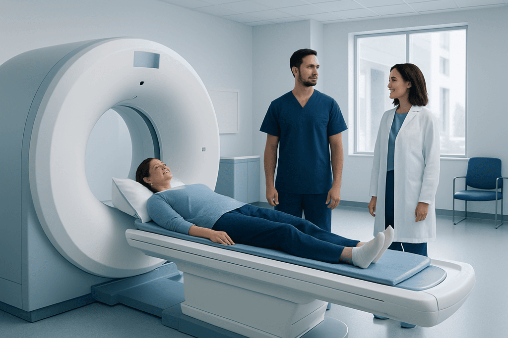
A PET-CT scan combines PET imaging, which shows cellular activity, with CT imaging, which shows detailed body structures. In cancer care, PET-CT may help with diagnosis, staging, treatment planning, and checking whether treatment is working.
Key Takeaways
- A PET-CT scan combines PET imaging, which shows cellular activity, with CT imaging, which shows detailed body structures.
- In cancer care, PET-CT may help with diagnosis, staging, treatment planning, and checking whether treatment is working.
- Most PET-CT scans use a small amount of a radioactive glucose tracer, commonly FDG, which is given through a vein.
- Patients are usually asked to avoid food for several hours before the scan and to follow specific instructions about diabetes medicines if relevant.
- PET-CT is generally safe, but it is not suitable for every situation and results must be interpreted by experienced specialists.
A PET-CT scan combines metabolic and anatomical imaging to help doctors detect, stage, and monitor many types of cancer. Knowing how the test works and what to expect can make the experience clearer and less stressful for patients.
Overview: What Is a PET-CT Scan?
A PET-CT scan is an imaging test that combines two technologies in one examination. PET stands for positron emission tomography, which shows how active cells and tissues are. CT stands for computed tomography, which uses X-rays to create detailed cross-sectional images of the body. When these two types of information are combined, doctors can see both where an abnormal area is located and how it is behaving.
In cancer care, PET-CT is often used because many cancer cells use more glucose than normal cells. For this reason, the most common PET tracer is a mildly radioactive form of glucose called FDG. After FDG is injected into a vein, areas with higher glucose use may appear brighter on the scan. This does not automatically mean cancer, but it gives doctors important information to interpret together with medical history, physical examination, laboratory tests, biopsy results, and other imaging.
A PET-CT scan is not a treatment and it does not remove tissue. It is a diagnostic and monitoring tool. The scan may help a medical team make more informed decisions about whether cancer is present, how far it has spread, which treatment approach may be appropriate, and how well treatment is working.
Why PET-CT Is Used in Cancer Care
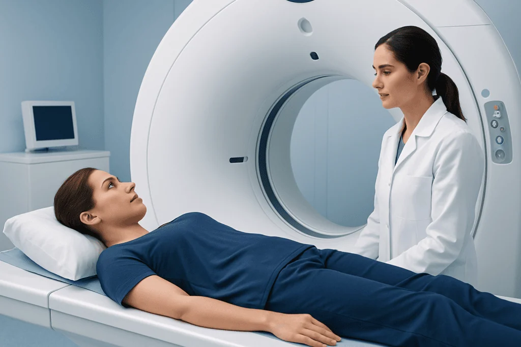
PET-CT can be helpful at different stages of a cancer journey. It may be requested when doctors need more information after an abnormal CT, MRI, ultrasound, or biopsy. It can also help determine whether cancer is limited to one area or has spread to lymph nodes or other organs. This process is called staging, and it is essential for choosing the most appropriate treatment plan.
In some cancers, PET-CT is used before surgery or radiotherapy to help define the treatment area. It may also be performed after chemotherapy, immunotherapy, radiotherapy, or surgery to evaluate response. If a tumor becomes less active on PET imaging, this may suggest that treatment is having an effect, although interpretation depends on the cancer type and timing of the scan.
Common reasons for requesting PET-CT in oncology include:
- Helping diagnose a suspicious mass when additional information is needed
- Staging cancers such as lymphoma, lung cancer, colorectal cancer, melanoma, head and neck cancer, and certain gynecologic cancers
- Planning radiotherapy or surgery in selected cases
- Checking whether treatment has reduced cancer activity
- Looking for recurrence when symptoms, tumor markers, or other tests raise concern
Not every cancer is best evaluated with PET-CT. Some tumors do not absorb FDG strongly, and some small lesions may be below the scanner’s detection limits. For this reason, the decision to use PET-CT is individualized by the oncology and nuclear medicine team.
How to Prepare for a PET-CT Scan
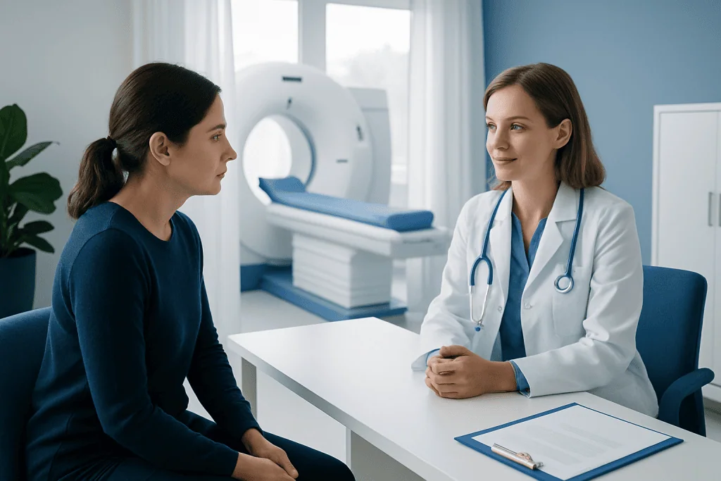
Preparation is important because the most common PET tracer, FDG, behaves like glucose in the body. Patients are typically asked not to eat for several hours before the scan, although drinking plain water is usually encouraged. Food, sugary drinks, and some forms of exercise can affect how the tracer is distributed and may make the images harder to interpret.
Patients should tell the imaging team if they have diabetes, are pregnant, may be pregnant, or are breastfeeding. Diabetes requires special planning because blood glucose levels can influence scan quality. The medical team may provide individualized instructions about meals, insulin, tablets, and scan timing. Patients should not stop prescribed medicines unless their doctor or imaging center specifically advises them to do so.
Before the appointment, patients may be asked about allergies, kidney problems, recent infections, recent surgery, and previous imaging tests. Comfortable clothing without metal parts is helpful. Jewelry, watches, removable dental appliances, and some clothing items may need to be removed. Bringing previous scan reports and images can help the radiology or nuclear medicine physician compare changes over time.
What Happens During the Procedure
On arrival, the patient is usually checked in and asked to confirm medical details. A small intravenous line is placed, often in the arm or hand. The tracer is then injected through the vein. The injection usually feels similar to a routine blood test or IV placement. After injection, there is a waiting period, often spent resting quietly in a comfortable room so the tracer can circulate and be taken up by body tissues.
During this waiting time, patients are usually asked to remain still, avoid talking too much, and stay warm. Muscle activity can increase tracer uptake and may create confusing findings. When it is time for the scan, the patient lies on a narrow table that moves slowly through a large, ring-shaped scanner. The scanner does not usually enclose the body as tightly as some MRI machines, and the test is painless.
The PET and CT images are acquired either together or in sequence during the same session. The CT part may be performed with or without contrast material, depending on the clinical question and the center’s protocol. If contrast is used, patients may feel a brief warm sensation or metallic taste. The imaging staff can communicate with the patient throughout the scan and will provide instructions about keeping still or holding the breath for short moments if needed.
The full appointment may take a few hours because of preparation and tracer uptake time, although the actual scanning time is often shorter. After the scan, most patients can return to normal activities unless they received sedation, contrast instructions, or specific advice from the care team.
Understanding PET-CT Results
PET-CT results are reviewed by physicians trained in nuclear medicine and radiology. They assess areas of tracer uptake, compare them with CT anatomy, and consider whether the pattern is typical for cancer, inflammation, infection, healing tissue, or normal body activity. Some organs, such as the brain, heart, kidneys, and bladder, may normally show FDG activity, so expert interpretation is essential.
The report may describe the location, size, and metabolic activity of abnormal areas. A commonly used measurement is called SUV, or standardized uptake value, which reflects how much tracer is present in a specific area. SUV can be useful, but it is only one part of interpretation. It should not be read in isolation because many factors can influence it, including blood glucose level, timing of imaging, scanner technique, and the type of tissue involved.
After the report is complete, the treating doctor explains what the findings mean in the context of the patient’s diagnosis and treatment plan. Sometimes PET-CT confirms the next step. In other situations, it may lead to additional tests, such as MRI, endoscopy, follow-up imaging, or biopsy. A suspicious PET-CT finding does not always mean cancer, and a normal scan does not always exclude cancer, especially if a lesion is very small or a tumor type has low FDG uptake.
Safety, Radiation, and Limitations
A PET-CT scan involves exposure to a small amount of radiation from both the PET tracer and the CT scan. In medical imaging, radiation is used only when the expected benefit of the information outweighs the potential risk. The tracer loses radioactivity over time and is removed from the body mainly through urine. Drinking water after the scan, if allowed, can help clear the tracer more quickly.
Patients who are pregnant or may be pregnant should inform the medical team before the scan. PET-CT during pregnancy is considered only when clearly necessary and when the expected benefit is significant. Breastfeeding patients should also ask for specific instructions, as temporary precautions may be recommended depending on the tracer and local protocol.
PET-CT has limitations. Inflammation, infection, recent surgery, radiation changes, or healing wounds can sometimes show increased FDG uptake and mimic cancer. Conversely, very small tumors or cancers with low metabolic activity may not be clearly visible. Blood glucose levels, body movement, and timing after treatment can also affect image quality. For these reasons, PET-CT is most useful when interpreted as part of a complete medical assessment rather than as a standalone answer.
After the Scan and When to Speak With a Doctor
After a PET-CT scan, patients are usually advised to drink fluids and empty the bladder regularly, unless they have been told to limit fluids for another medical reason. Most people can eat normally and resume usual activities. If contrast material was used, the imaging team may provide additional instructions, especially for patients with kidney disease, diabetes, or previous contrast reactions.
Patients should contact their doctor or imaging center if they develop unusual symptoms after the test, such as a rash, breathing difficulty, persistent dizziness, or swelling at the injection site. These reactions are uncommon, but it is appropriate to seek advice if something feels unexpected. Patients should also speak with their oncology team if they are unsure about preparation instructions, medication timing, or how the results will affect treatment decisions.
The results are usually sent to the referring doctor, who discusses them with the patient and, when appropriate, with a multidisciplinary cancer team. Acibadem International’s multidisciplinary specialists and JCI-accredited hospitals provide diagnostic imaging and cancer care for international patients, including PET-CT when clinically indicated. As with any imaging test, the best next step should be decided with a qualified physician who understands the patient’s full medical situation.
Frequently asked questions
Is a PET-CT scan painful?
The scan itself is not painful. The main discomfort is usually the small needle used to place the intravenous line for the tracer injection. During the scan, the patient needs to lie still on the table, which may feel uncomfortable for some people but is generally well tolerated.
How long does a PET-CT appointment take?
The total appointment commonly takes a few hours because there is a waiting period after the tracer injection. The actual imaging portion is usually shorter than the full visit. Timing can vary depending on the type of scan, whether contrast is used, and the center's protocol.
Can PET-CT detect all cancers?
No imaging test can detect every cancer in every situation. PET-CT is very useful for many cancer types, but some tumors do not absorb the tracer strongly, and very small lesions may be difficult to see. Doctors choose PET-CT when it is likely to provide helpful information for a specific clinical question.
Why is fasting needed before a PET-CT scan?
Fasting helps reduce the effect of recent food intake on blood glucose and tracer distribution. Since the common tracer FDG behaves like glucose, eating before the scan may affect image quality. Patients should follow the exact fasting and medication instructions provided by the imaging center.
Is the radiation from PET-CT dangerous?
PET-CT uses radiation, but the dose is carefully controlled and the test is recommended only when the expected medical benefit is greater than the potential risk. The radioactive tracer naturally decays and leaves the body, mainly through urine. Patients who are pregnant, may be pregnant, or are breastfeeding should always inform the medical team before the scan.
What happens if the PET-CT shows a bright area?
A bright area means increased tracer uptake, but it does not always mean cancer. Infection, inflammation, healing tissue, and normal organ activity can also cause increased uptake. The doctor will interpret the finding together with the patient's history, examination, laboratory tests, previous scans, and sometimes biopsy results.
References
- American Cancer Society
- National Cancer Institute
- European Association of Nuclear Medicine
- Radiological Society of North America
- Society of Nuclear Medicine and Molecular Imaging
This article is for general information only and is not a substitute for professional medical advice. Please consult a qualified doctor about your individual situation.
Oncology care in Turkey — second opinion and treatment plan
JCI-accredited · board-certified surgeons · reply within 24h
Add us as a Preferred Source to see more of our trusted health content across Google Search, AI Overviews and Discover.

Medical Oncology Department
Medical treatment of cancer with chemotherapy, immunotherapy and targeted therapies under a multidisciplinary tumor board.
60 specialists in this unit