Pet Scan vs MRI: Key Differences and How Doctors Tell Them Apart
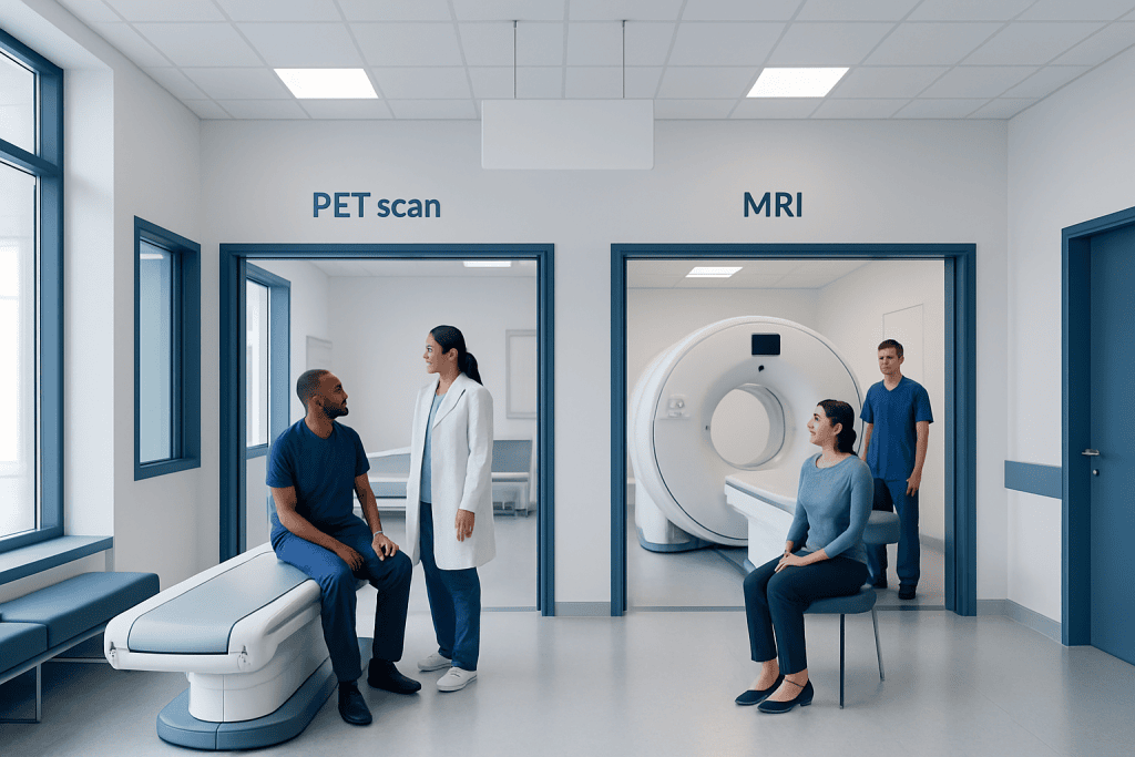
PET scans show how actively cells and tissues are working, often using a small amount of radioactive tracer. MRI uses powerful magnets and radio waves to create detailed images of soft tissues and body structures.
Key Takeaways
- PET scans show how actively cells and tissues are working, often using a small amount of radioactive tracer.
- MRI uses powerful magnets and radio waves to create detailed images of soft tissues and body structures.
- PET is commonly used in cancer evaluation, some heart conditions, and selected brain disorders.
- MRI is especially useful for the brain, spine, joints, muscles, pelvic organs, and many soft-tissue conditions.
- The best scan depends on the medical question; neither test is universally better than the other.
PET scans and MRI scans provide different types of medical information: PET identifies changes in tissue activity, while MRI produces detailed pictures of organs, soft tissues, and structures. A doctor selects one, or sometimes both, according to the symptoms, suspected condition, and part of the body being examined.
Pet Scan vs MRI: A Side-by-Side Comparison
When comparing a PET scan vs MRI, the key difference is what each examination measures. A positron emission tomography (PET) scan highlights metabolic activity, meaning how tissues use energy or process a tracer. Magnetic resonance imaging (MRI) creates highly detailed pictures of anatomy, including soft tissues, organs, nerves, blood vessels, and bones.
Both scans can be valuable, and they are sometimes used together because they answer different questions. For example, MRI may show the exact location and structure of an abnormal area, while PET may help show whether it is metabolically active. The choice is based on the person’s symptoms, medical history, previous test results, and the reason for imaging.
| Feature | PET scan | MRI scan |
|---|---|---|
| Main purpose | Shows metabolic or functional activity in tissues | Shows detailed anatomy and soft-tissue structure |
| How it works | Uses a radiotracer detected by a special camera | Uses magnetic fields and radio waves |
| Radiation exposure | Yes, from the radiotracer; PET is often combined with CT, which also uses radiation | No ionizing radiation |
| Common uses | Cancer staging, treatment response, infection or inflammation evaluation, selected brain and heart conditions | Brain, spine, joints, muscles, heart, pelvic organs, liver, and soft-tissue assessment |
| Typical preparation | May require fasting and blood glucose checks, depending on the tracer | Metal screening; contrast instructions may be provided when needed |
| Scan experience | Usually includes an injection, a waiting period, then imaging while lying still | May be noisy and enclosed; requires lying still for detailed image sequences |
What a PET Scan Shows
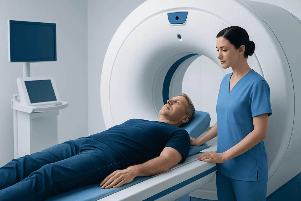
A PET scan uses a very small amount of a radiotracer, usually given through a vein. The tracer travels through the body and collects differently in tissues according to their activity. A scanner detects this distribution and produces images that help clinicians assess areas of unusually high or low activity.
The most widely used PET tracer is related to glucose, the body’s main energy source. Because many cancer cells use glucose more actively than surrounding tissues, PET can help identify areas that may need further assessment. However, increased uptake is not always cancer. Healing tissues, infection, inflammation, and some normal organs can also show increased tracer uptake, so results must always be interpreted in clinical context.
PET is frequently performed as a combined PET/CT examination. CT supplies anatomic detail and helps localize the PET findings precisely. In selected situations, PET may also be paired with MRI, particularly when detailed soft-tissue imaging is useful, such as in parts of the brain, head and neck, pelvis, or certain pediatric assessments.
What an MRI Scan Shows
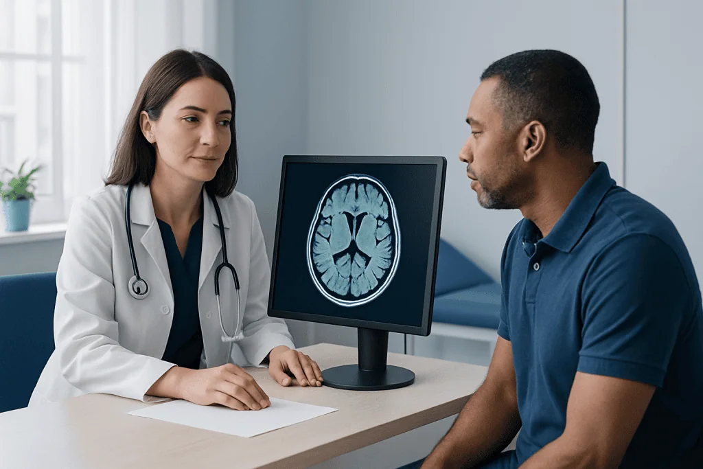
MRI does not use X-rays or radioactive tracers. Instead, it uses a strong magnetic field and radiofrequency energy to produce detailed images of the body. It is particularly effective at distinguishing among different soft tissues, such as muscle, fat, ligaments, cartilage, nerves, brain tissue, and many internal organs.
Doctors often request MRI when they need to evaluate the brain and spinal cord, suspected nerve compression, stroke-related changes, joint injuries, ligament or tendon problems, and abnormalities in the pelvis or abdomen. MRI can also assess the heart, blood vessels, breast tissue, liver, and prostate in appropriate clinical settings.
Some MRI examinations use a contrast agent, usually gadolinium-based, to make blood vessels, inflammation, scars, or certain lesions easier to see. Before contrast is given, the imaging team reviews kidney health, allergies, pregnancy status, and other relevant factors. Most people can have MRI safely, but certain implanted devices, metal fragments, or older types of clips may require special review.
How Clinicians Tell Which Scan Is Needed
Clinicians do not generally choose PET or MRI simply because one scan is more advanced. They begin with the clinical question: Is the goal to understand tissue activity, map anatomy, investigate a symptom, stage a known condition, monitor treatment, or clarify an uncertain finding? The answer guides the most suitable test.
For example, if a person has back pain with possible disc disease or nerve compression, MRI is often more informative because it clearly shows discs, nerves, vertebrae, and surrounding soft tissues. If a person has a confirmed cancer and the team needs to assess whether disease may be active elsewhere in the body, PET/CT may be considered depending on the cancer type and treatment plan.
Medical history also matters. A recent surgery, infection, vaccination, inflammatory disease, diabetes, kidney impairment, pregnancy, claustrophobia, or an implanted medical device can affect preparation or test selection. Previous scans are equally important because comparing images over time can help determine whether a finding is stable, improving, or changing.
What to Expect Before, During, and After Each Scan
Preparation for a PET scan commonly includes instructions about eating, drinking, exercise, and diabetes medicines. For many FDG-PET scans, fasting for a specified period is necessary because blood glucose levels can influence tracer distribution. Patients should follow the imaging center’s exact instructions and tell the team about diabetes, pregnancy, breastfeeding, medicines, and recent illnesses or procedures.
During a PET scan, the radiotracer is administered, followed by a quiet waiting period while it circulates. The imaging itself involves lying still on a table that moves through the scanner. The appointment may take longer than the scanning time because of preparation and tracer uptake. Afterward, patients may usually return to normal activities, although they may receive temporary guidance about hydration and close contact with infants or pregnant people.
For MRI, patients complete a safety screening form before entering the scan room. Jewelry, hearing aids, removable dental items, and other metal objects must be removed. The machine makes loud tapping or knocking sounds, so ear protection is provided. People who feel anxious in enclosed spaces should mention this in advance; the care team can discuss practical support, scan breaks, or other options where appropriate.
Benefits, Limitations, and Safety Considerations
PET can detect changes in tissue activity before structural changes are obvious on an anatomic scan. This can be useful for evaluating some cancers, monitoring response to treatment, or investigating selected inflammatory, neurologic, and cardiac conditions. Its limitation is that PET findings can be nonspecific: active inflammation and infection may resemble some cancer-related patterns, and some tumors do not take up a particular tracer strongly.
MRI offers excellent detail without ionizing radiation, which can be especially helpful when repeated imaging is needed. However, it may not be the best first test for every concern. MRI is less suited to some urgent situations, may take longer than CT, and can be affected by movement. It also does not directly measure metabolism in the same way as PET.
Both tests are performed by trained imaging professionals with safety procedures in place. Patients should provide complete information about allergies, medicines, kidney disease, implanted devices, possible pregnancy, and prior imaging. An imaging result is only one part of diagnosis; doctors combine it with symptoms, examination findings, laboratory results, pathology when needed, and other tests.
When to Seek Medical Care
New or persistent symptoms should be discussed with a qualified doctor rather than self-diagnosed through imaging information. Medical review is particularly important for unexplained weight loss, a persistent new lump, coughing up blood, ongoing severe pain, seizures, new weakness, changes in speech or vision, or symptoms that steadily worsen. A clinician can determine whether imaging is needed and which test is most appropriate.
Urgent medical care is needed for symptoms that may signal an emergency, including sudden weakness on one side of the body, facial drooping, difficulty speaking, sudden severe headache, chest pain, severe shortness of breath, fainting, or sudden loss of vision. These symptoms require immediate assessment and should not wait for an outpatient PET scan or MRI appointment.
People who have been advised to undergo PET or MRI should ask why the scan is being recommended, whether contrast or a tracer will be used, how to prepare, and when results will be available. Acibadem International’s multidisciplinary specialists and JCI-accredited hospitals support international patients who need diagnostic evaluation and treatment planning for a range of conditions.
Frequently asked questions
Is a PET scan better than an MRI?
Neither scan is better in every situation. PET is designed to show metabolic activity, while MRI is designed to show detailed body structure. The most appropriate examination depends on the suspected condition and the clinical question.
Can a PET scan detect cancer better than MRI?
PET can be very useful for evaluating many cancers because it may identify metabolically active areas throughout the body. However, not every cancer is best assessed with PET, and inflammation can also cause increased uptake. MRI may provide more detailed information for cancers affecting certain organs or soft tissues.
Does MRI have radiation?
No. MRI uses magnetic fields and radio waves rather than ionizing radiation. Some MRI examinations use contrast material, but this is different from radiation exposure.
Is a PET scan painful?
A PET scan is usually not painful, apart from the brief needle insertion used to administer the tracer. The person must lie still during imaging, which may be uncomfortable for some people. The care team can help with positioning and explain each step.
Why might a doctor order both PET and MRI?
The scans provide complementary information. MRI can define the size, location, and structure of an abnormality, while PET can show whether an area has altered metabolic activity. Using both may help clinicians make a more complete assessment in selected cases.
Can people with metal implants have an MRI?
Many people with modern implants can safely undergo MRI, but every device must be reviewed before the scan. Some pacemakers, neurostimulators, aneurysm clips, cochlear implants, or retained metal fragments require specific safety checks. Patients should never assume an implant is MRI-safe without confirmation.
This article is for general information only and is not a substitute for professional medical advice. Please consult a qualified doctor about your individual situation.
Explore treatments in Turkey — costs, top hospitals & a free quote
JCI-accredited · board-certified surgeons · reply within 24h
Add us as a Preferred Source to see more of our trusted health content across Google Search, AI Overviews and Discover.

More from the Health Library

Wegovy Pill vs Injection: Key Differences and How Doctors Tell Them Apart

Implantation Symptoms: Common Causes, Related Conditions, and When to See a Doctor

Is E Coli Contagious? A Doctor-Reviewed Answer

Back Ache Upper Right Explained: Common Triggers and Red Flags

What Does Running Do for Your Body? A Doctor-Reviewed Answer