Placenta Abruption Ultrasound: Preparation, Procedure and Results

Ultrasound can support the diagnosis of placental abruption but cannot exclude it on its own. Vaginal bleeding, abdominal pain, back pain, contractions and reduced fetal movement need prompt medical assessment during pregnancy.
Key Takeaways
- Ultrasound can support the diagnosis of placental abruption but cannot exclude it on its own.
- Vaginal bleeding, abdominal pain, back pain, contractions and reduced fetal movement need prompt medical assessment during pregnancy.
- A transabdominal ultrasound is painless, uses no radiation and usually requires no recovery time.
- Care depends on the severity of bleeding, the baby’s condition and how far the pregnancy has progressed.
- Placental abruption is an obstetric emergency when bleeding is heavy, pain is severe or fetal movement changes.
A placenta abruption ultrasound is a safe imaging examination used to assess the placenta, baby, amniotic fluid and possible bleeding behind the placenta. It may show signs of placental separation, but because some abruptions are not visible on a scan, doctors also rely on symptoms, examination findings and fetal monitoring.
Placenta abruption ultrasound: how it helps
Placenta abruption ultrasound is usually performed when a pregnant person has vaginal bleeding, abdominal pain, uterine tenderness, frequent contractions or a change in fetal movement. Placental abruption means that the placenta begins to separate from the inner wall of the uterus before birth. The placenta provides oxygen and nutrients to the baby, so a suspected abruption requires timely clinical assessment.
Ultrasound helps the care team look for bleeding or a collection of blood behind the placenta, assess the placenta’s location and review the baby’s growth, fluid around the baby and heartbeat. However, not all abruptions can be seen on ultrasound, especially when bleeding is small, recent or concealed behind the placenta. For this reason, a normal ultrasound result does not always rule out an abruption.
Doctors interpret imaging alongside the person’s symptoms, vital signs, abdominal examination, blood tests when needed and fetal heart-rate monitoring. This combined approach helps determine whether observation, hospital care or delivery is safest.
Who may need this examination and how to prepare
An ultrasound may be recommended at any stage of pregnancy when placental abruption is suspected. It may also be used after abdominal trauma, such as a fall or vehicle collision, or when there is unexplained bleeding in later pregnancy. Risk factors can include high blood pressure, smoking, cocaine use, previous placental abruption, abdominal injury and certain pregnancy complications, although an abruption can occur without identifiable risk factors.
Preparation is usually simple. For an abdominal pregnancy ultrasound, patients can generally eat and drink as usual unless their healthcare team gives different instructions. A full bladder may occasionally help early in pregnancy, but it is commonly not necessary later in pregnancy. Comfortable two-piece clothing can make the examination easier.
Patients should not delay urgent assessment to prepare for a scan. Heavy bleeding, severe or persistent abdominal pain, fainting, a rigid or very tender abdomen, or reduced fetal movement should be assessed immediately in an emergency maternity setting.
What happens during a placenta abruption ultrasound

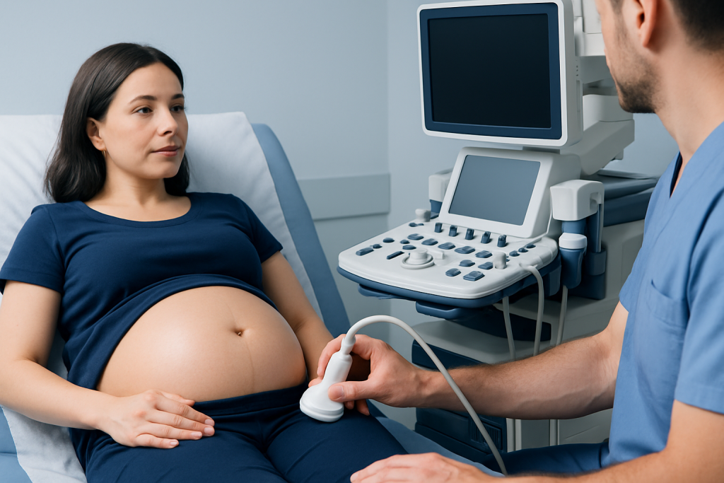
The examination commonly starts with a transabdominal ultrasound. The patient lies on an examination couch, and a sonographer applies gel to the abdomen. A handheld probe is moved over the skin to produce real-time images using sound waves. The procedure does not use ionizing radiation and is not known to harm the baby when medically indicated.
The sonographer evaluates the placental position and appearance, checks for possible blood collections, measures amniotic fluid and reviews fetal movement and cardiac activity. Doppler techniques may sometimes be used to assess blood flow in the umbilical cord or fetal circulation when there are concerns about fetal wellbeing or growth.
In selected circumstances, a clinician may recommend a transvaginal ultrasound for a clearer view of the cervix or lower part of the uterus. This involves gently placing a covered ultrasound probe into the vagina. It does not cause placental abruption and is generally safe when performed by trained professionals, but the team will explain why it is needed before proceeding.
The scan often takes about 20 to 40 minutes, although timing varies with the clinical situation. In an emergency, fetal monitoring and other assessments may take place at the same time rather than waiting for imaging alone.
What are the ultrasound findings of placental abruption?
Possible ultrasound findings of placental abruption include a blood collection between the placenta and uterine wall, sometimes called a retroplacental hematoma. A collection may appear darker or brighter than surrounding tissue depending on when the bleeding occurred and how the blood has changed over time.
Other possible findings include a thickened placenta, an irregular placental surface, blood within the amniotic fluid, or separation at the edge of the placenta. Ultrasound may also identify findings that affect management, such as low amniotic fluid, poor fetal growth or changes in blood-flow studies. These features are not specific to abruption and must be interpreted by an obstetric specialist.
Importantly, an ultrasound may look normal despite a genuine abruption. Concealed bleeding may not be easy to distinguish from placental tissue, and a small separation can be difficult to visualize. Clinical concern therefore remains important even when imaging does not show a hematoma.
Can they see a placental abruption on ultrasound?
Sometimes, but not always. Ultrasound can detect some cases when there is a visible collection of blood or an area where the placenta appears separated from the uterine wall. It is particularly useful for checking the baby’s condition and looking for other causes of bleeding, including placenta previa, in which the placenta lies near or over the cervix.
Ultrasound has limited sensitivity for placental abruption, meaning a negative result cannot reliably exclude the condition. Doctors may diagnose a suspected abruption based mainly on symptoms such as bleeding and pain, uterine findings, fetal heart-rate changes and the overall clinical picture.
When an abruption is suspected, repeated ultrasound may be appropriate in some stable pregnancies to monitor the placenta, fetal growth and amniotic fluid. The timing and frequency of follow-up depend on individual circumstances and should be guided by an obstetric team.
Results, benefits, risks and recovery after the scan
Results may be available immediately in urgent settings, while a formal report may follow after review by a radiologist, sonographer or obstetric specialist. The clinician will explain whether the scan shows a possible hematoma, whether the placenta is appropriately located and whether additional monitoring is needed. A scan alone does not determine the full severity of an abruption.
The principal benefits are that ultrasound is noninvasive, widely available and able to provide useful information without radiation. It can help guide urgent decisions and establish a baseline for follow-up. Its main limitation is that it may miss an abruption, so it should never be used to dismiss ongoing concerning symptoms.
There is no physical recovery period after a routine ultrasound. The gel is wiped away, and most patients can return to usual activities unless their care team recommends rest, observation or hospital admission because of symptoms. If bleeding or pain continues, the patient should follow the individualized plan provided by their maternity team.
Management of confirmed or suspected abruption can involve observation, IV fluids, blood tests, fetal monitoring, treatment for complications and, when necessary, delivery. Decisions depend on maternal stability, bleeding, gestational age and fetal wellbeing. In complex situations, coordinated high-risk pregnancy care can bring obstetric, maternal-fetal medicine, neonatal and anesthesia expertise together.
Are there warning signs before a placental abruption?
Warning signs can include vaginal bleeding, sudden or persistent abdominal pain, lower back pain, painful or frequent contractions, uterine tenderness, and a feeling that the abdomen is unusually tight. Some people may notice reduced or changed fetal movement. Bleeding may be light, heavy or absent because blood can remain trapped behind the placenta.
Symptoms vary, and their severity does not always reflect the amount of placental separation. For example, concealed bleeding can cause significant pain or uterine tenderness without obvious vaginal bleeding. Any bleeding or new abdominal pain in pregnancy deserves contact with a qualified maternity clinician for advice.
While risk cannot always be prevented, attending prenatal appointments and managing conditions such as high blood pressure can support a healthier pregnancy. Avoiding smoking, alcohol and illicit drugs is also important. People who have experienced a previous abruption may benefit from early discussion with an obstetrician about monitoring in a future pregnancy.
Can a baby be saved if the placenta detaches?
Many babies can be supported and delivered safely when placental abruption is recognized and treated quickly, but outcomes depend on how much of the placenta has separated, how quickly the separation occurred, the baby’s gestational age and the condition of both parent and baby. A small, stable abruption may sometimes be monitored carefully, particularly when the baby is preterm and fetal testing is reassuring.
A larger or rapidly progressing abruption can reduce the baby’s oxygen supply and may require urgent delivery. If the baby is very premature, neonatal specialists may provide intensive care after birth. The maternity team focuses on protecting the health of the pregnant person while making timely decisions for fetal wellbeing.
Acibadem International’s multidisciplinary specialists and JCI-accredited hospitals provide assessment and treatment for high-risk pregnancy concerns for international patients. Individual care plans are based on examination findings, fetal monitoring and ultrasound results rather than on imaging alone.
When to seek medical care
Urgent medical care is needed during pregnancy for heavy vaginal bleeding, severe or worsening abdominal pain, repeated painful contractions, dizziness, fainting, shoulder pain after trauma, fluid leakage, or a clear reduction in fetal movement. These symptoms can have several causes, but they require prompt evaluation because placental abruption and other pregnancy complications can develop quickly.
Patients should contact their maternity unit or emergency services immediately rather than driving themselves if they feel unwell, are bleeding heavily or have severe pain. They should bring pregnancy records if available, but should not delay leaving home to find them.
For lighter bleeding, mild cramping or uncertainty about fetal movement, it is still appropriate to call a maternity care provider promptly for guidance. A clinician can advise whether same-day assessment, fetal monitoring or ultrasound is needed.
Frequently asked questions
Is placenta abruption ultrasound safe during pregnancy?
Yes. Diagnostic ultrasound uses sound waves rather than radiation and is considered safe when performed for an appropriate medical reason by trained professionals. The care team uses the examination to assess the placenta and the baby while considering the full clinical situation.
Can an ultrasound rule out placental abruption?
No. Ultrasound can identify some signs of abruption, but some cases are not visible, particularly when bleeding is small or concealed. Persistent symptoms or concerning fetal monitoring findings still need medical attention even if the scan appears normal.
What happens if an ultrasound shows blood behind the placenta?
A blood collection behind the placenta can suggest placental abruption, but the next steps depend on its size, symptoms, fetal wellbeing and gestational age. The team may recommend observation, repeat testing, hospital monitoring or delivery when safety concerns are present.
Does placental abruption always cause vaginal bleeding?
No. Some bleeding can remain behind the placenta, which is called concealed bleeding. This is why abdominal pain, uterine tenderness, contractions or reduced fetal movement should be assessed even when there is no visible bleeding.
How quickly are placenta abruption ultrasound results available?
In urgent maternity care, preliminary findings are often reviewed immediately with the treating team. A formal imaging report may be completed later, but urgent clinical decisions are based on the scan, symptoms, examination and fetal monitoring together.
Can placental abruption happen again in a future pregnancy?
A previous placental abruption increases the chance of recurrence, although many later pregnancies progress without it. People with this history should discuss pre-pregnancy planning and individualized monitoring with an obstetrician or maternal-fetal medicine specialist.
References
- American College of Obstetricians and Gynecologists
- Royal College of Obstetricians and Gynaecologists
- Society for Maternal-Fetal Medicine
- Merck Manual Professional Edition
This article is for general information only and is not a substitute for professional medical advice. Please consult a qualified doctor about your individual situation.
Ultrasound in Turkey — costs, top hospitals & a free quote
JCI-accredited · board-certified surgeons · reply within 24h
Add us as a Preferred Source to see more of our trusted health content across Google Search, AI Overviews and Discover.
Check your numbers in seconds
BMI, calories, due date, blood pressure and 30+ more clinical calculators — free, instant, doctor-reviewed ranges.

Check-up & Preventive Medicine
Comprehensive health screenings designed for early detection and prevention.
2 specialists in this unitMore from the Health Library



Related Specialists

Assoc. Prof. Dr. Hatice Gamze Demirdağ
Dermatology
Assoc. Prof. Dr. Mustafa Çorum
Physical Medicine & Rehabilitation
Dr. Gökan Güler
Emergency Service
Dt. İpek Mehmetoğlu Torun
Oral & Dental Health