Posterior Vitreous Detachment Treatment: How It Works, Results and What to Expect

Most posterior vitreous detachments do not need surgery or medication. A dilated retinal examination is important, especially when symptoms first begin.
Key Takeaways
- Most posterior vitreous detachments do not need surgery or medication.
- A dilated retinal examination is important, especially when symptoms first begin.
- Retinal tears or detachments are uncommon but need prompt treatment to protect vision.
- Floaters and flashes commonly become less noticeable over weeks to months.
- Posterior vitreous detachment cannot be reversed, but related retinal complications can often be treated.
Posterior vitreous detachment treatment is most often careful observation and follow-up eye examinations, because the vitreous usually separates from the retina naturally without needing a procedure. New flashes, a sudden increase in floaters, or a curtain-like shadow require urgent assessment to rule out a retinal tear or retinal detachment.
Overview: What posterior vitreous detachment treatment involves
Posterior vitreous detachment treatment usually means observation, education about warning symptoms, and scheduled retinal examinations rather than an operation. A posterior vitreous detachment (PVD) happens when the vitreous gel inside the eye naturally becomes more liquid and separates from the retina, the light-sensitive tissue lining the back of the eye. It is common with aging and often occurs without harm to vision.
The main purpose of care is to identify the small number of people who develop a retinal tear, bleeding in the vitreous, or retinal detachment while the vitreous is pulling away. If the retina remains intact, symptoms such as floaters and flashes often settle with time. If a tear is found, prompt retinal detachment surgery and retinal repair may be recommended to prevent more serious vision loss.
PVD may feel unsettling, particularly when symptoms appear suddenly. An ophthalmologist can examine the eye after dilating the pupil, explain the findings, and advise when follow-up is needed. Most people can continue their usual daily activities after they have been assessed, while remaining alert for changes in their vision.
How posterior vitreous detachment develops and who may need treatment

The vitreous is a clear, gel-like substance that helps the eye keep its rounded shape. Over time, it naturally shrinks and becomes more watery. Eventually, it can pull away from the retina; this is called a posterior vitreous detachment. It is not the same as retinal detachment, although the two conditions can be related.
PVD becomes more likely with older age. It can also occur earlier in people with significant short-sightedness, after eye injury, following eye surgery such as cataract surgery, or in association with inflammation inside the eye. Having PVD in one eye may also make it more likely to occur in the other eye later.
Most people are candidates for monitoring rather than an active procedure. Treatment is considered when the examination shows a retinal tear, a retinal detachment, or another cause of the symptoms that needs care. A clinician may also arrange closer follow-up if there is vitreous bleeding, because it can make a retinal tear more likely or more difficult to see during the first examination.
Symptoms and the eye examination process
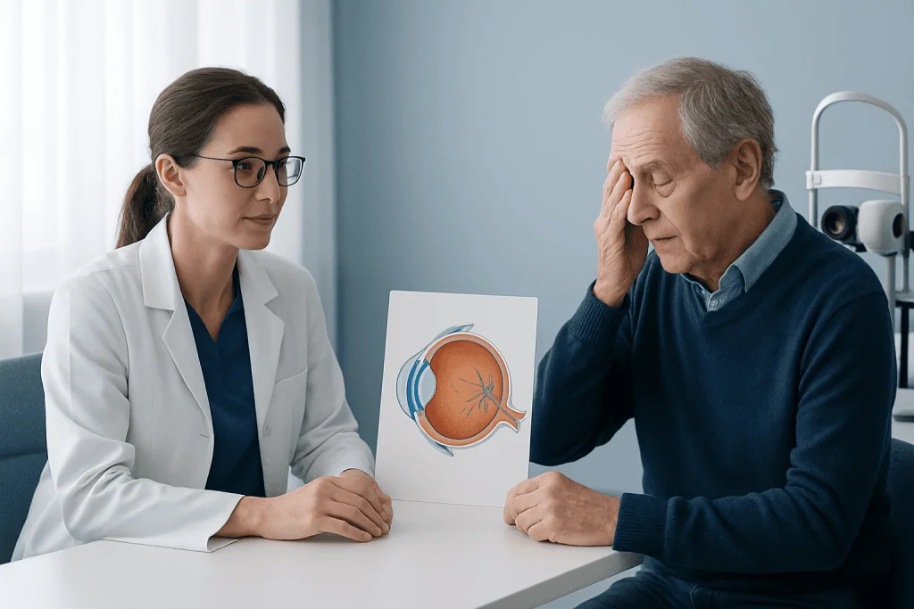
The most common symptoms are new floaters and flashes of light. Floaters may look like dots, threads, cobwebs, or small moving shapes that drift across vision. Flashes are often brief streaks or arcs of light, especially in dim surroundings or when moving the eyes. These symptoms can be noticeable even when central vision remains clear.
During diagnosis, the eye doctor asks about the timing and pattern of symptoms, relevant eye history, medicines, and any previous retinal problems. Eye drops are usually used to widen the pupils so the retina can be examined thoroughly. The doctor may use special lenses and bright light to look for retinal breaks, bleeding, or areas where the retina may be lifting.
If the retina cannot be fully viewed because of bleeding or a dense cataract, an eye ultrasound may be used. Further testing is guided by the individual findings. A normal initial examination is reassuring, but follow-up may still be advised because some retinal tears develop after symptoms first start.
- Seek urgent assessment for a new shower of floaters.
- Seek urgent assessment for persistent or increasing flashes.
- Arrange immediate care for a dark curtain, shadow, missing area of vision, or sudden reduction in vision.
How treatment works when intervention is needed
There is no treatment that reattaches the vitreous to the retina once a straightforward PVD has occurred, and this is usually not necessary. The clinical focus is protecting the retina. When no retinal complication is present, an ophthalmologist generally recommends watchful waiting, planned review when appropriate, and clear instructions about symptoms that should prompt urgent reassessment.
If a retinal tear is identified, laser photocoagulation may be used to create small, controlled scars around the tear. These scars help secure the retina to the eye wall and reduce the chance that fluid will pass through the tear and cause retinal detachment. In some cases, freezing treatment, called cryotherapy, may be used instead.
A retinal detachment requires more extensive repair. Depending on its type, location, and severity, treatment may involve laser or freezing therapy, a gas bubble placed inside the eye, a scleral buckle, or vitrectomy surgery. These procedures are tailored to the retinal findings rather than to PVD itself. Acibadem International’s multidisciplinary eye specialists in JCI-accredited hospitals evaluate retinal symptoms and provide treatment planning for international patients.
For a small number of people, persistent floaters significantly affect quality of life after the retina has been confirmed to be healthy. Options may be discussed with a retinal specialist, but procedures aimed at floaters have potential risks and are not routinely needed for typical PVD symptoms.
Step by step: monitoring, retinal repair and follow-up
For uncomplicated PVD, the first step is a comprehensive dilated eye examination. The ophthalmologist confirms whether the vitreous has detached and checks carefully for retinal tears or detachment. The patient receives practical advice about warning signs and may be asked to return for another examination, particularly during the early weeks after symptoms begin.
If laser treatment is needed for a retinal tear, it is commonly performed as an outpatient procedure. Numbing drops are used, and the patient sits at a laser machine while the specialist applies laser spots around the affected area. Vision can be blurred temporarily after dilation, so arranging transport home is sensible. The specialist explains individual aftercare and whether activity adjustments are needed.
When surgery is required for retinal detachment, the process is more individualized. The surgeon discusses the recommended technique, anesthesia, positioning requirements if a gas bubble is used, and follow-up appointments. Recovery instructions can differ considerably, so patients should follow the retinal team’s advice rather than relying on general guidance.
Follow-up examinations are an important part of treatment because the retina can change after the initial assessment. People should not wait for their planned appointment if they develop a sudden change in floaters, flashes, visual field, or clarity of vision.
Recovery timeline, benefits and possible risks
How long symptoms last varies. Flashes often reduce over several weeks as the pulling forces on the retina settle. Floaters may remain visible for months, but the brain often adapts so they become much less distracting. Some floaters can persist long term without indicating ongoing damage.
The benefit of monitoring is early recognition of a retinal complication while avoiding unnecessary procedures in people with an uncomplicated PVD. A normal retinal examination is reassuring, though it does not replace the need to seek help if symptoms change. Follow-up allows the eye team to reassess the retina during the period when a delayed tear could occasionally appear.
Laser treatment for a retinal tear is intended to lower the risk of retinal detachment, but it cannot guarantee that no further tear or detachment will occur. Temporary discomfort, blurred vision, inflammation, and visual changes can occur after treatment. Retinal surgery has additional risks, including infection, bleeding, raised eye pressure, cataract development, recurrent detachment, and incomplete visual recovery; the specialist discusses these in relation to the expected benefit.
Any sudden loss of vision, a growing dark shadow, significant eye pain, or marked redness after a procedure should be reported promptly. These symptoms need individual evaluation and should not be managed at home.
What should be avoided, and what makes symptoms worse?
After a PVD has been assessed and the retina is intact, most routine activities are usually safe. However, people should follow any specific restrictions given by their ophthalmologist, especially if a retinal tear has been treated or surgery has been performed. Activities with a risk of eye injury should be avoided, and protective eyewear is sensible for work, sport, or tasks involving flying particles.
There is no reliable evidence that reading, computer use, ordinary walking, or normal eye movements make an uncomplicated PVD worse. Stress, fatigue, bright backgrounds, and paying close attention to the visual field can make floaters feel more noticeable, but they do not cause the vitreous to detach further. Sudden new symptoms are more important than day-to-day variation in how noticeable existing floaters feel.
It is wise to avoid rubbing or pressing on the eye, particularly after an examination or retinal procedure. People who have been prescribed drops or given positioning instructions after treatment should use them exactly as directed. They should also tell the eye team about new eye trauma, severe headache with vision changes, or symptoms in the other eye.
When to seek medical care
New flashes or floaters should be checked promptly by an eye professional, ideally with a dilated retinal examination. This is particularly important if symptoms are sudden, numerous, or occurring for the first time. Although PVD is often harmless, only an examination can determine whether the retina has been affected.
Emergency assessment is needed for a curtain-like shadow, a new blind area, rapidly worsening vision, a sudden large increase in floaters, or repeated flashes. These can be warning signs of retinal tear or detachment, where timely care is important. A person should seek urgent help rather than waiting to see whether symptoms settle.
People with previous retinal tears or detachment, high short-sightedness, recent eye surgery, eye trauma, or inflammatory eye disease should have a lower threshold for contacting their ophthalmologist. Regular eye care and quick reporting of new symptoms support early detection of retinal problems.
Frequently asked questions
How long does it take for posterior vitreous detachment to get better?
Flashes often improve over a few weeks as the vitreous finishes separating and traction on the retina decreases. Floaters may remain noticeable for several months and may not disappear completely, but many people find that they fade or become easier to ignore over time. A follow-up examination may be recommended during the early period after symptoms begin.
What should I avoid doing if I have a posterior vitreous detachment?
After an eye examination confirms an uncomplicated PVD, most everyday activities are generally acceptable. Avoid eye injury, rubbing or pressing on the eye, and any activity restrictions specifically given by the ophthalmologist after laser treatment or surgery. New or worsening symptoms should prompt urgent eye assessment rather than changes to activity alone.
What makes posterior vitreous detachment worse?
A PVD is a natural structural change and is not usually worsened by reading, screens, or normal exercise. Floaters may seem more prominent against bright backgrounds or when a person is tired, but this does not necessarily mean the condition has progressed. A sudden increase in floaters, flashes, shadowing, or reduced vision may signal a retinal complication and needs prompt review.
Can you reverse posterior vitreous detachment?
Posterior vitreous detachment cannot be reversed because the vitreous does not normally reattach to the retina. In most cases, reversal is not needed because PVD itself is harmless. Care focuses on detecting and treating complications such as retinal tears or retinal detachment if they occur.
Does posterior vitreous detachment require surgery?
Most people with PVD do not require surgery. Surgery or laser treatment is considered when the examination finds a retinal tear, retinal detachment, or another significant complication. Procedures solely to remove troublesome floaters are considered only selectively because they have potential risks.
Can posterior vitreous detachment cause blindness?
An uncomplicated PVD does not usually cause blindness. However, in some people, traction during the detachment can lead to a retinal tear, which may progress to retinal detachment without treatment. Prompt assessment of new flashes, floaters, or a curtain-like shadow helps protect vision.
References
- American Academy of Ophthalmology
- National Eye Institute
- Royal College of Ophthalmologists
- Mayo Clinic
This article is for general information only and is not a substitute for professional medical advice. Please consult a qualified doctor about your individual situation.
Explore treatments in Turkey — costs, top hospitals & a free quote
JCI-accredited · board-certified surgeons · reply within 24h
Add us as a Preferred Source to see more of our trusted health content across Google Search, AI Overviews and Discover.

More from the Health Library



Related Specialists

Dt. İletişim | 444 55 44
Oral & Dental Health
Assoc. Prof. Dr. Nurcan Kızılcık
Anesthesiology
Fzt. Mehmet Sabah Ateş
Physical Medicine & Rehabilitation
Assoc. Prof. Dr. Özgür Timurkaynak
Dermatology