Prenatal Screening Spina Bifida: How It Works, Results and What to Expect
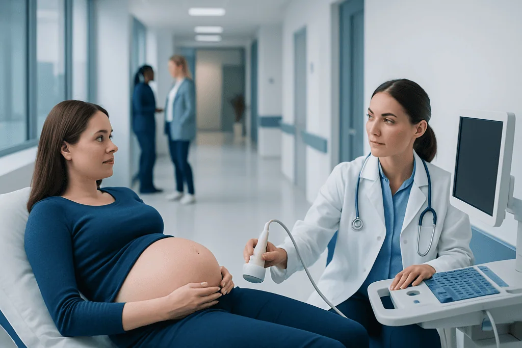
A detailed ultrasound around 18 to 22 weeks is the main prenatal test used to look for spina bifida. Maternal serum AFP screening can identify pregnancies with a higher chance of an open neural tube defect, but it cannot confirm a diagnosis.
Key Takeaways
- A detailed ultrasound around 18 to 22 weeks is the main prenatal test used to look for spina bifida.
- Maternal serum AFP screening can identify pregnancies with a higher chance of an open neural tube defect, but it cannot confirm a diagnosis.
- Most screening tests are safe and non-invasive; amniocentesis is diagnostic but has a small procedure-related risk.
- An abnormal screening result does not always mean the baby has spina bifida and should be reviewed with an experienced maternity team.
- Early specialist assessment helps families understand findings, pregnancy care options and planning for birth and newborn treatment.
Prenatal screening for spina bifida usually combines a detailed pregnancy ultrasound with, in some settings, a maternal blood test that measures alpha-fetoprotein (AFP). Screening estimates the chance of an open neural tube defect; diagnostic testing may be offered when a result or scan finding needs clarification.
Prenatal screening spina bifida: how it works
Prenatal screening spina bifida refers to tests during pregnancy that look for signs suggesting an open neural tube defect. The neural tube is an early fetal structure that develops into the brain and spinal cord. In spina bifida, part of the spinal column does not close fully; severity varies considerably depending on the type and location of the opening.
Screening does not diagnose every type of spina bifida and cannot predict every aspect of a child’s health. It is designed to identify pregnancies that may benefit from a more detailed scan, specialist review or diagnostic testing. A normal result is reassuring, but it does not completely exclude all spinal or neurological differences.
The two main approaches are maternal serum AFP testing and ultrasound. AFP is a protein made by the developing baby. Higher-than-expected AFP in the pregnant person’s blood may occur with an open neural tube defect, although it can also have other explanations, such as inaccurate pregnancy dating or a multiple pregnancy.
Who may be offered screening and when
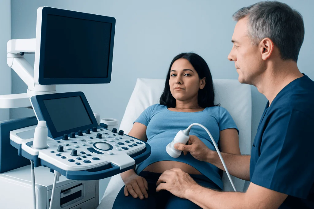
Screening policies differ between countries, hospitals and individual pregnancies. Many people are offered a routine detailed fetal anatomy ultrasound in the second trimester, whether or not they have known risk factors. Maternal serum AFP screening may be offered during the second trimester, often between about 15 and 20 weeks of pregnancy, depending on local practice.
A clinician may recommend earlier counselling or targeted imaging when there is a previous pregnancy affected by a neural tube defect, a family history, certain anti-seizure medicines, pre-existing diabetes, obesity, or concerns about nutrition and folate intake. Having a risk factor does not mean that a baby will have spina bifida; it simply helps the maternity team tailor screening and support.
People planning pregnancy are generally advised to discuss folic acid supplementation with a healthcare professional. Folic acid taken before conception and in early pregnancy lowers the chance of neural tube defects, but it cannot prevent every case. Those with a previous affected pregnancy or certain medical circumstances may need individual advice.
What happens during the tests

For maternal serum AFP screening, a healthcare professional takes a routine blood sample from a vein in the arm. The laboratory interprets the AFP level using factors such as gestational age, maternal weight and whether there is more than one fetus. The blood draw itself is brief, and normal activities can usually continue straight away.
The detailed anatomy ultrasound is usually performed between 18 and 22 weeks. A sonographer places gel on the abdomen and moves a handheld probe over the skin. The examination evaluates the spine and looks carefully at the brain, skull, legs and other organs. In many cases of open spina bifida, changes in the fetal brain can help specialists recognize the condition even when the spinal opening is difficult to view directly.
If screening raises concern, the next step may be a targeted ultrasound performed by a fetal medicine specialist. Amniocentesis may also be discussed. In this procedure, ultrasound guidance is used to pass a fine needle through the abdomen into the amniotic sac to collect fluid. Testing amniotic fluid for AFP and acetylcholinesterase can help confirm an open neural tube defect. The procedure is usually brief, but the care team will explain its small risks, limitations and alternatives before consent.
How long does it take to get results from a spina bifida test?
Result timing depends on the test, laboratory and whether a specialist review is needed. Maternal serum AFP results are often available within several days to about two weeks. The maternity team should explain how and when results will be communicated, including whom to contact if questions arise while waiting.
Some ultrasound findings can be discussed on the day of the scan, particularly when the images are clear. However, a formal report or a follow-up appointment may be needed before conclusions are made. If a targeted scan or amniocentesis is arranged, the timing of final results can vary, and the specialist team can provide the most accurate estimate.
Waiting for additional testing can be stressful. It may help to write down questions, ask for a clear explanation of the next steps and bring a trusted person to appointments where possible. Support from an obstetrician, fetal medicine specialist and genetic counsellor can help families interpret information in context.
Can you tell if a baby has spina bifida in the womb?
Yes, many cases of open spina bifida can be identified before birth, most commonly through a detailed second-trimester ultrasound. Detection depends on the type of spina bifida, the position of the baby, scan quality, gestational age and the experience of the imaging team. Open forms, including myelomeningocele, are more likely to be found before birth than closed forms.
When a scan suggests spina bifida, a fetal medicine team may use high-resolution ultrasound and sometimes fetal MRI to understand the spinal and brain findings more clearly. These tests can provide important information for pregnancy monitoring and delivery planning, but they cannot predict every future effect on movement, bladder or bowel function, learning, or independence.
Further assessment may also look for associated differences, including hydrocephalus or Chiari II malformation. Families may be referred to specialists in maternal-fetal medicine, neonatology, pediatric neurosurgery, rehabilitation and urology. This coordinated approach supports informed, individualized planning before delivery.
Is spina bifida picked up at a 12 week scan?
A 12-week scan is valuable for confirming the pregnancy location, estimating gestational age, checking early fetal development and, where offered, assessing markers for certain chromosome conditions. Some major neural tube abnormalities may be suspected at this stage, but a 12-week scan is not the main or most reliable assessment for spina bifida.
The detailed anatomy ultrasound at around 18 to 22 weeks is the usual key examination for evaluating the fetal spine and related brain features. If there are early concerns, known risk factors or limited images, a clinician may arrange earlier follow-up and a targeted scan at the appropriate gestational age.
It is important not to interpret an early scan result alone as confirmation or exclusion of spina bifida. The pregnancy care team can explain what was visible at that point, what remains uncertain and whether further imaging is recommended.
What do the results of a spina bifida test indicate?
A screen-negative or low-risk result means the test did not identify features associated with a higher chance of an open neural tube defect. It is reassuring, but it is not an absolute guarantee. Routine pregnancy care and scheduled ultrasound assessments should still continue.
A screen-positive or high-risk AFP result means the AFP level was higher than expected for that pregnancy. It does not confirm spina bifida. Reasons may include incorrect pregnancy dating, twins or other fetal, placental or pregnancy-related factors. Usually, the first step is to confirm gestational age and arrange a detailed ultrasound.
If a targeted ultrasound and, when chosen, amniocentesis confirm an open neural tube defect, the team will discuss the specific findings and available care pathways. This may include continued monitoring, delivery at a center with neonatal and surgical expertise, and discussion of postnatal surgery or, in carefully selected circumstances, fetal surgery. Decisions are personal and should be supported by clear, non-directive counselling.
Benefits, limitations and when to seek medical care
The main benefit of prenatal screening is information. Finding a possible concern before birth can allow time for confirmatory testing, specialist consultations and planning for the safest place and team for delivery. Screening is non-invasive when it involves blood testing and ultrasound. Amniocentesis can provide more definitive information about open neural tube defects, but it is invasive and carries a small risk of complications, including fluid leakage, infection, bleeding and pregnancy loss.
All prenatal tests have limitations. A raised AFP level is not specific to spina bifida, ultrasound may not detect every spinal defect, and even confirmed findings cannot fully predict a child’s future needs. Asking about the purpose of each test, the possible outcomes and what each result would mean can help families decide whether testing is right for them.
Contact a maternity clinician promptly for vaginal bleeding, fluid leakage, severe or persistent abdominal pain, fever, fainting, or concerning symptoms after an invasive procedure. Routine prenatal care should also be sought if there are questions about screening results or a missed follow-up appointment. Acibadem International’s multidisciplinary specialists and JCI-accredited hospitals can support international patients who need prenatal assessment and coordinated care planning.
Frequently asked questions
Is the AFP blood test a diagnosis of spina bifida?
No. AFP testing is a screening test that estimates whether there may be an increased chance of an open neural tube defect. A high result needs follow-up, usually with a detailed ultrasound and occasionally diagnostic testing such as amniocentesis.
Does a normal ultrasound rule out all forms of spina bifida?
A normal detailed ultrasound is reassuring and can detect many open spinal defects. However, some closed forms of spina bifida can be difficult to identify before birth, so no single test excludes every possible spinal difference.
Is amniocentesis always needed after a high AFP result?
No. A detailed ultrasound is often the first follow-up test, and it may provide enough information. Amniocentesis is considered individually after discussion of what it can clarify, its limitations and its small procedure-related risks.
Can non-invasive prenatal testing (NIPT) detect spina bifida?
Standard NIPT primarily screens for certain chromosome conditions using cell-free DNA in maternal blood. It is not a screening test for open neural tube defects such as spina bifida, so ultrasound and, where offered, AFP screening remain important.
What happens if spina bifida is confirmed before birth?
A specialist team will explain the type and location of the defect, any associated findings and what is known and uncertain about likely care needs. Families can discuss pregnancy monitoring, delivery planning, newborn surgery and, for selected pregnancies in specialist centers, possible fetal surgery.
Can folic acid prevent spina bifida?
Folic acid before conception and during early pregnancy reduces the chance of neural tube defects. It does not prevent every case, and the right supplement plan should be discussed with a clinician, especially for people with a previous affected pregnancy or certain medical conditions.
References
- World Health Organization
- Centers for Disease Control and Prevention
- American College of Obstetricians and Gynecologists
- International Society of Ultrasound in Obstetrics and Gynecology
- National Institute for Health and Care Excellence
This article is for general information only and is not a substitute for professional medical advice. Please consult a qualified doctor about your individual situation.
Joint and spine care in Turkey — expert assessment & treatment
JCI-accredited · board-certified surgeons · reply within 24h
Add us as a Preferred Source to see more of our trusted health content across Google Search, AI Overviews and Discover.