Proliferative Diabetic Retinopathy Treatment: How It Works, Results and What to Expect
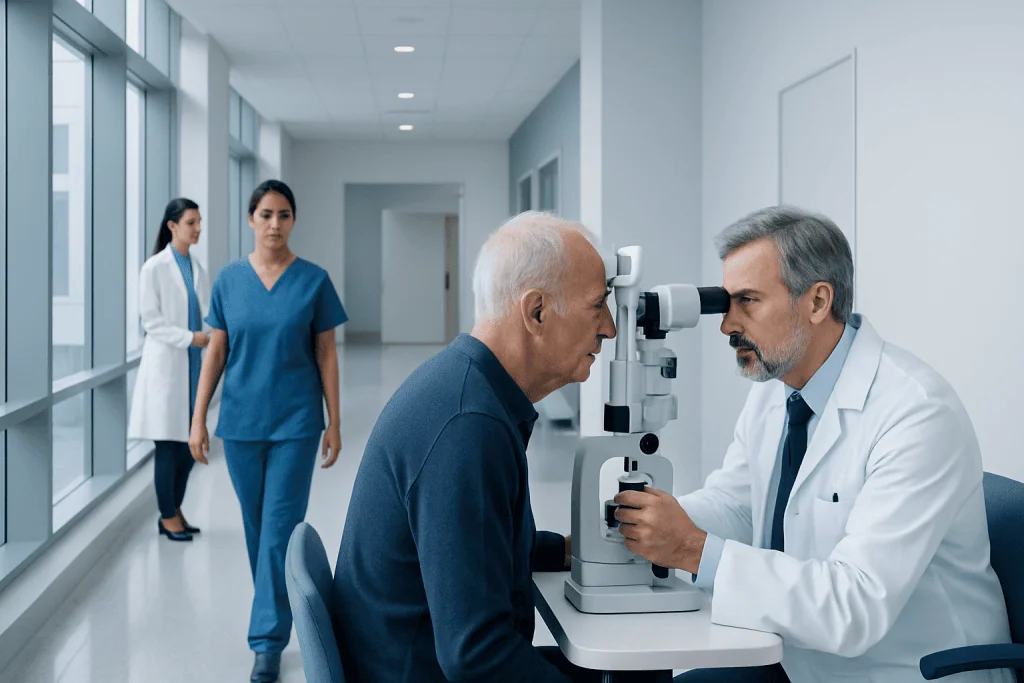
Proliferative diabetic retinopathy is an advanced form of diabetic eye disease that needs prompt assessment by an ophthalmologist. Anti-VEGF medicines and panretinal laser treatment can cause abnormal blood vessels to shrink or become less active.
Key Takeaways
- Proliferative diabetic retinopathy is an advanced form of diabetic eye disease that needs prompt assessment by an ophthalmologist.
- Anti-VEGF medicines and panretinal laser treatment can cause abnormal blood vessels to shrink or become less active.
- Vitrectomy may be recommended when bleeding, scar tissue or retinal detachment affects the vitreous or retina.
- Treatment often stabilizes vision and reduces the risk of severe vision loss, but it cannot always restore vision already lost.
- Regular eye examinations and management of blood glucose, blood pressure and cholesterol remain essential after treatment.
Proliferative diabetic retinopathy treatment aims to prevent further vision loss from fragile abnormal blood vessels that grow on the retina. Depending on the eye findings, care may include anti-VEGF injections, laser treatment, vitrectomy surgery and careful diabetes management.
Overview: How proliferative diabetic retinopathy treatment works
Proliferative diabetic retinopathy treatment works by addressing abnormal new blood vessels that develop when diabetes-related damage reduces oxygen delivery to the retina, the light-sensitive tissue at the back of the eye. These vessels are fragile and may bleed into the eye, form scar tissue or pull on the retina. Treatment is designed to make the abnormal vessels regress, reduce bleeding and lower the chance of retinal detachment and serious vision loss.
The most suitable approach depends on whether there is active vessel growth, diabetic macular edema, vitreous hemorrhage, retinal traction or detachment. Common options are injections of medicines that block vascular endothelial growth factor (anti-VEGF), panretinal photocoagulation laser therapy and vitrectomy surgery. An ophthalmologist, often a retina specialist, chooses and sequences treatment according to detailed retinal imaging and the person’s vision needs.
Diabetes care is also part of eye treatment. Keeping blood glucose, blood pressure and cholesterol within individualized targets can help slow further retinal damage. Diabetic retinopathy may progress without obvious symptoms, so scheduled dilated eye examinations remain important even when vision seems unchanged.
Who may need treatment and how the eye is assessed

People may be candidates for treatment when an eye specialist finds neovascularization, meaning abnormal new blood vessels on the retina or optic nerve. Treatment may also be needed if these vessels are associated with bleeding into the vitreous gel, recurrent hemorrhage, progressive scar tissue, macular swelling or traction on the retina. Some people have few symptoms at first, while others notice floaters, blurred or fluctuating vision, dark areas or a sudden decrease in sight.
Assessment usually includes a dilated eye examination and retinal photographs. Optical coherence tomography (OCT) provides detailed cross-sectional images of the retina and helps identify macular edema or traction. Fluorescein angiography may be used to show areas of poor blood flow, leakage and new-vessel growth. These tests help the specialist map the condition and discuss whether observation, injections, laser treatment, surgery or a combination is appropriate.
General health factors are reviewed as well. The eye team may ask about diabetes duration and control, blood pressure, kidney disease, pregnancy, smoking, medicines and previous eye procedures. Coordinated care with diabetes clinicians, primary care professionals and kidney or cardiovascular specialists can support the eye plan and overall health.
Treatment options and the step-by-step experience

Anti-VEGF injections are commonly used to reduce the signals that drive abnormal vessel growth. The eye is numbed with anesthetic drops, cleaned carefully and the medicine is injected into the white part of the eye using a very fine needle. The procedure is usually brief. Several injections over time may be needed, and regular follow-up is essential because the effect can lessen if treatment is delayed.
Panretinal photocoagulation, also called scatter laser treatment, treats selected areas of the peripheral retina. It reduces the retina’s oxygen demand and helps abnormal vessels become less active over time. The pupil is dilated and numbing drops are used; a contact lens may be placed on the eye to focus the laser. Laser eye treatment is generally performed as an outpatient procedure, sometimes over more than one session. It may be used alone or together with injections.
Vitrectomy is an operation considered when dense or persistent vitreous hemorrhage blocks vision, scar tissue pulls on the retina, or tractional retinal detachment threatens central vision. During vitrectomy surgery, a surgeon removes the cloudy vitreous gel and blood, treats the retina with laser if needed, and may address scar tissue. A gas bubble or silicone oil may occasionally be used to support the retina, which can affect recovery instructions.
Each option has a different role. Injections can be especially useful when macular edema is present, while laser provides a durable way to reduce the risk from widespread proliferative disease. Surgery is usually reserved for complications that cannot be adequately managed with injections or laser alone.
Benefits, limitations and possible risks
The main benefit of treatment is reducing the risk of further severe vision loss. Anti-VEGF therapy can rapidly reduce new-vessel activity, while laser can lower the risk of major bleeding and retinal detachment over the longer term. Vitrectomy can clear blood from the eye and relieve traction in selected situations. Many people achieve stabilization, and some experience improved vision when treatable swelling or bleeding resolves.
However, treatment cannot always reverse retinal injury or vision loss that has already occurred. Results depend on the degree of retinal damage, whether the macula or optic nerve has been affected, the presence of retinal detachment, treatment timing and ongoing diabetes control. Continued monitoring is needed because proliferative disease can recur or become active again.
Possible short-term effects of injections include temporary irritation, redness, tearing, floaters or blurred vision. Rare but important complications include infection inside the eye, retinal tear or detachment, increased eye pressure and bleeding. Laser can cause temporary blurred vision and may reduce peripheral vision, night vision or color perception in some people. Vitrectomy carries surgical risks such as infection, bleeding, raised eye pressure, retinal detachment and cataract progression. The treating ophthalmologist explains individual risks and warning symptoms before treatment.
Recovery and day-to-day care after treatment
Recovery differs by treatment type. After an injection, mild discomfort or a gritty feeling may last for a day or two, and vision can be temporarily blurry. Most people can return to usual gentle activities soon afterward, but should follow the clinic’s instructions about driving and eye drops. Increasing pain, worsening redness, sensitivity to light or a marked fall in vision should be reported urgently.
After laser treatment, vision may remain blurred or light-sensitive for several hours to several days. It can take weeks for abnormal vessels to become less active, and follow-up imaging helps determine whether further laser or injections are needed. People should arrange transport home if dilation or visual blurring makes driving unsafe.
Vitrectomy recovery is usually longer. The eye may feel irritated and vision may be limited initially by swelling, residual blood or a gas bubble. If a gas bubble is used, specific head positioning may be required, and air travel or high-altitude travel must be avoided until the bubble has completely gone. Follow-up visits are important to monitor healing, eye pressure and retinal attachment.
Long-term self-care includes taking diabetes medicines as prescribed, attending diabetes and eye appointments, avoiding smoking and seeking individualized support for nutrition, activity and blood pressure management. These measures do not replace eye treatment, but they can reduce risks to the retina and other organs affected by diabetes.
Does proliferative diabetic retinopathy go away?
Proliferative diabetic retinopathy may become inactive after treatment, meaning the abnormal blood vessels shrink or stop leaking and bleeding. Anti-VEGF injections and panretinal laser treatment can control active neovascularization, and surgery can address certain complications such as non-clearing blood in the vitreous or retinal traction.
However, diabetes-related retinal damage is not usually considered permanently cured. New vessels can return, especially if follow-up treatment is interrupted or diabetes and other vascular risk factors remain difficult to control. Ongoing dilated examinations and retinal imaging allow the specialist to detect reactivation early.
Even after successful treatment, vision may not fully return if the retina has sustained permanent damage. Early detection and timely treatment offer the best opportunity to preserve useful vision.
What is the success rate of diabetic retinopathy treatment?
There is no single success rate for diabetic retinopathy treatment because outcomes vary considerably by disease stage, the presence of macular edema, the amount of bleeding or scar tissue, and the person’s ability to attend monitoring and repeat treatment. In clinical practice, treatment is often judged successful when it makes abnormal vessels inactive, prevents major additional vision loss, controls retinal swelling or preserves retinal attachment.
Anti-VEGF injections and laser treatment have strong evidence for reducing the risk of vision-threatening complications in appropriately selected people. Vitrectomy can be effective for clearing vision-obscuring hemorrhage and managing tractional complications, although visual recovery depends on the condition of the macula and retina before surgery.
The eye specialist can provide a more individualized outlook after examining both eyes and reviewing scans. Keeping follow-up appointments is particularly important, as missed monitoring can allow active proliferative disease to progress without early symptoms.
How long does it take to recover from diabetic retinopathy laser treatment?
Most people recover from the immediate effects of diabetic retinopathy laser treatment within a few days. Blurred vision, glare, light sensitivity or mild discomfort may occur on the day of treatment and can persist briefly afterward. The dilating drops themselves may affect vision for several hours.
The biological effect of panretinal laser treatment develops more gradually. Abnormal vessels may take several weeks to regress, and the specialist may schedule a review to assess whether additional laser or injections are needed. Some people need treatment in stages, particularly when there is extensive proliferative disease.
Vision changes can be individual. A person should contact their eye clinic promptly if there is severe pain, a sudden curtain-like shadow, rapidly worsening vision, or a large increase in floaters or flashing lights.
What is the prognosis for proliferative diabetic retinopathy?
The prognosis for proliferative diabetic retinopathy is often better when it is identified and treated before major bleeding, scar formation, macular damage or retinal detachment occurs. Modern injection therapy, laser treatment and vitreoretinal surgery can substantially reduce the risk of profound vision loss for many people. Nevertheless, it remains a serious eye complication that requires long-term surveillance.
Outlook is influenced by retinal findings, treatment response, attendance at follow-up visits and management of diabetes, blood pressure and cholesterol. Kidney disease, smoking and pregnancy can also affect retinal risk in some people. A retina specialist can explain how these factors apply to an individual situation.
Acibadem International’s multidisciplinary specialists and JCI-accredited hospitals diagnose and treat diabetic eye conditions for international patients, with care plans guided by ophthalmic examination and retinal imaging.
When to seek medical care
Anyone with diabetes should have regular comprehensive dilated eye examinations at intervals advised by their eye specialist, even if they have no visual symptoms. People with known diabetic retinopathy should not postpone follow-up appointments, particularly when they are receiving injections or have recently had laser or surgery.
Urgent ophthalmic assessment is needed for sudden vision loss, a new shower of floaters, flashes of light, a dark curtain or shadow across vision, or rapidly increasing blur. These symptoms can have several causes, including bleeding or retinal detachment, and prompt evaluation can protect vision.
New eye pain, marked redness, light sensitivity or worsening vision after an injection or eye surgery also requires urgent contact with the treating eye team. For general diabetes management, people should seek medical advice if blood glucose levels are persistently difficult to manage or if other health concerns develop.
Frequently asked questions
Is proliferative diabetic retinopathy an emergency?
It requires prompt assessment by an ophthalmologist because abnormal blood vessels can bleed or cause retinal traction. Sudden loss of vision, a curtain-like shadow, flashes or many new floaters should be treated as urgent eye symptoms.
Can anti-VEGF injections replace laser treatment?
For some people, anti-VEGF injections may control proliferative disease effectively, particularly when diabetic macular edema is also present. Laser may still be recommended depending on retinal findings, ability to attend frequent visits and the risk of disease reactivation.
Is laser treatment painful?
Numbing eye drops are used, so most people feel little pain. Some may notice pressure, bright flashes of light or brief discomfort during the procedure, followed by temporary irritation or blurred vision.
Will vision improve after treatment?
Vision may improve when bleeding clears or retinal swelling decreases, but this cannot be guaranteed. A major goal of treatment is to prevent additional vision loss and preserve remaining vision.
How often are follow-up appointments needed?
The schedule varies with the treatment used and how active the retinopathy is. People receiving injections may need regular, sometimes frequent monitoring, while laser and surgery also require planned reviews to check the retina and eye pressure.
Can blood sugar control reverse diabetic retinopathy?
Improved blood glucose management can slow progression and supports the effectiveness of eye care, but it does not reliably reverse established proliferative disease. It should be combined with recommended ophthalmic treatment and follow-up.
References
- American Academy of Ophthalmology
- National Eye Institute
- American Diabetes Association
- National Institute of Diabetes and Digestive and Kidney Diseases
- International Council of Ophthalmology
This article is for general information only and is not a substitute for professional medical advice. Please consult a qualified doctor about your individual situation.
Diabetic Retinopathy in Turkey — costs, top hospitals & a free quote
JCI-accredited · board-certified surgeons · reply within 24h
Add us as a Preferred Source to see more of our trusted health content across Google Search, AI Overviews and Discover.
Check your numbers in seconds
BMI, calories, due date, blood pressure and 30+ more clinical calculators — free, instant, doctor-reviewed ranges.

Ophthalmology
Comprehensive eye care from laser vision correction to retina, cornea and cataract surgery.
75 specialists in this unit