Prostate Gland Anatomy Ultrasound: Preparation, Procedure and Results

Prostate ultrasound provides real-time images without radiation. Transrectal ultrasound usually gives the most detailed view of prostate anatomy.
Key Takeaways
- Prostate ultrasound provides real-time images without radiation.
- Transrectal ultrasound usually gives the most detailed view of prostate anatomy.
- Preparation varies by scan type and may include a comfortably full bladder or bowel preparation.
- The examination is usually brief and causes pressure or mild discomfort rather than significant pain.
- Ultrasound findings need to be interpreted alongside symptoms, examination findings and laboratory tests.
A prostate gland anatomy ultrasound is an imaging examination that uses sound waves to show the prostate’s size, shape and internal appearance. It may be performed through the abdomen or rectum, depending on the clinical question, and can help assess urinary symptoms, prostate enlargement and other prostate concerns.
Prostate Gland Anatomy Ultrasound: Overview
A prostate gland anatomy ultrasound is a scan that uses high-frequency sound waves to create real-time images of the prostate gland. It helps a clinician assess the gland’s size, outline, volume and internal texture, as well as nearby structures such as the bladder, seminal vesicles and rectum. Because ultrasound does not use ionizing radiation, it is commonly used when doctors need a safe, practical view of the urinary and reproductive system.
The prostate is a walnut-sized gland located below the bladder and in front of the rectum. It surrounds the urethra, the tube that carries urine out of the body, and produces fluid that contributes to semen. As the prostate enlarges or changes with age, it can affect urine flow. Ultrasound may help evaluate symptoms such as a weak stream, frequent urination, night-time urination, urgency or a feeling that the bladder does not empty fully.
There are two main approaches. A transabdominal ultrasound is performed over the lower abdomen and may assess the bladder and estimate prostate size. A transrectal ultrasound, often called TRUS, involves a slim lubricated probe placed gently into the rectum. It generally provides clearer, more detailed images of the prostate and may also be used to guide a biopsy when this is clinically appropriate.
How a Prostate Ultrasound Works and Who May Need One
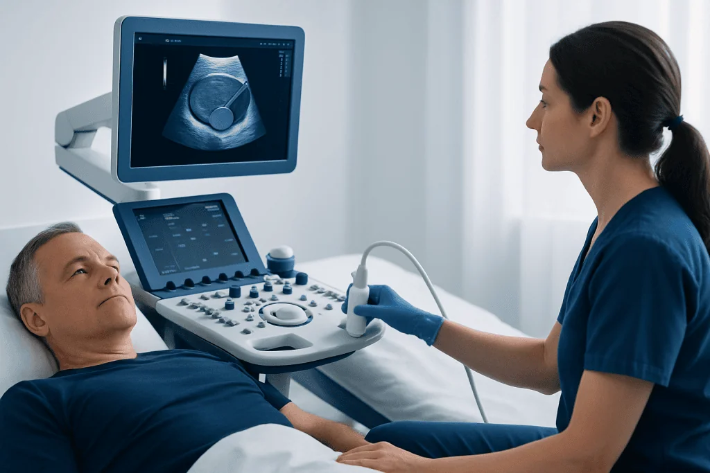
Ultrasound equipment sends sound waves into the body and detects returning echoes. A computer converts these echoes into moving images. Different tissues reflect sound differently, allowing the radiologist or urologist to evaluate the prostate’s boundaries and identify features such as enlargement, cysts, calcifications or uneven areas. However, ultrasound alone cannot reliably determine whether a finding is cancerous.
A clinician may recommend prostate ultrasound for persistent lower urinary tract symptoms, suspected prostate enlargement, recurrent urinary retention, abnormal findings during a digital rectal examination, or as part of planning for selected procedures. It can also measure post-void residual urine, meaning the amount of urine left in the bladder after urination. This can help clarify whether bladder emptying is reduced.
Transrectal ultrasound may be considered when more detailed imaging is needed or when a prostate biopsy is planned. The choice of test depends on symptoms, medical history, examination findings, prostate-specific antigen (PSA) results and previous imaging. Men with urinary symptoms may also be assessed for conditions such as benign prostatic hyperplasia, a common non-cancerous enlargement of the prostate.
How Should I Prepare for a Prostate Gland Ultrasound?
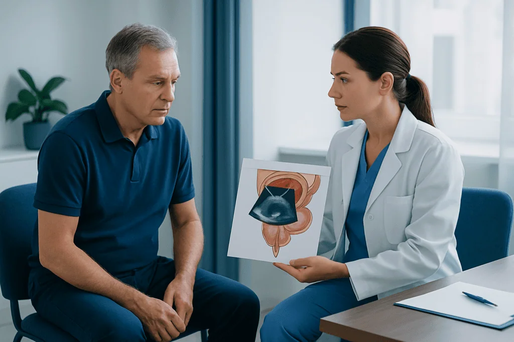
Preparation for a prostate gland anatomy ultrasound depends on whether the examination is transabdominal or transrectal. The imaging centre or urology team should provide specific instructions in advance, and these instructions should take priority because requirements can differ between hospitals and according to the reason for the test.
For a transabdominal scan, patients are often asked to arrive with a comfortably full bladder. This may involve drinking water before the appointment and avoiding urination until the first part of the examination is complete. A full bladder can improve the view of the bladder, the prostate region and the amount of urine remaining after voiding. The sonographer may then ask the patient to empty the bladder so a second scan can be performed.
For a transrectal ultrasound, some centres recommend a small enema beforehand to clear the lower bowel. Patients should tell the clinical team about blood-thinning medicines, bleeding disorders, latex allergy, heart valve disease, artificial joints, diabetes, recent rectal surgery, hemorrhoids or active rectal symptoms. They should not stop prescribed medicines, including anticoagulants, unless the clinician who manages those medicines has advised them to do so.
It can be helpful to wear comfortable clothing, bring relevant test results, and ask whether a companion is needed. For an ultrasound alone, people usually return home independently. If sedation is planned for a related procedure, such as a biopsy, the team may advise arranging transport home.
What If I Can't Poop Before Transrectal Ultrasound?
Not having a bowel movement before a transrectal ultrasound is usually not an emergency and does not always mean the appointment must be cancelled. The purpose of bowel preparation, when requested, is to reduce stool in the rectum so the probe can be inserted more comfortably and the images are less affected by gas or fecal material.
Patients should not use extra laxatives, enemas or other bowel products without first checking with the imaging centre or clinician, especially if they have kidney disease, inflammatory bowel disease, recent bowel surgery, rectal bleeding or a condition that affects fluid and electrolyte balance. Overusing bowel preparations can cause discomfort and may not improve the examination.
If the recommended preparation was not completed, the best approach is to contact the department before travelling or inform staff on arrival. The team can decide whether the scan can proceed, whether a simple preparation is appropriate, or whether rescheduling would give a better-quality examination. Being open about constipation or difficulty with the instructions helps staff tailor care safely and respectfully.
Step-by-Step: What Happens During the Procedure?
For a transabdominal ultrasound, the patient usually lies on an examination couch with the lower abdomen exposed. A sonographer applies clear gel to the skin and moves a handheld transducer across the area. The gel may feel cool, and gentle pressure is sometimes needed to obtain images. If bladder emptying is being assessed, the patient may be asked to urinate and return for repeat images.
For a transrectal ultrasound, the patient commonly lies on their side with knees bent toward the chest. A covered, lubricated ultrasound probe is inserted slowly a short distance into the rectum. The clinician obtains images while adjusting the probe position carefully. The scan itself often takes around 10 to 20 minutes, although check-in, preparation and discussion can make the overall visit longer.
If ultrasound is being used to guide a biopsy, the experience is different from an imaging-only examination. Local anesthetic, infection-prevention measures and additional instructions may be involved. A clinician should explain why a biopsy is being considered, the alternatives, possible complications and what results may mean before consent is requested. For patients who need tissue sampling, prostate biopsy services may include image-guided assessment and pathology review.
After the scan, a radiologist or urologist reviews the images in context. Some facilities provide preliminary information on the day, but a formal report may take longer. The referring clinician usually discusses the findings and any next steps.
How Uncomfortable Is a Prostate Ultrasound?
A transabdominal prostate ultrasound is typically painless. Patients may feel pressure from the probe, especially when the bladder is full, but the sensation is generally short-lived. Letting the sonographer know about tenderness or discomfort can help them adjust their technique.
A transrectal ultrasound may cause a feeling of fullness, pressure or an urge to have a bowel movement. Most people describe it as uncomfortable rather than painful. Anxiety can increase pelvic muscle tension, so slow breathing, asking questions and telling the clinician about previous rectal pain or trauma may make the process easier.
Significant pain is not expected during an imaging-only transrectal ultrasound. If pain occurs, the patient should tell the clinician immediately. The examination can be paused, repositioning can be tried, and the team can decide whether it is appropriate to continue. A biopsy can cause more discomfort than ultrasound alone, but local anesthetic is commonly used when needed.
Do I Need to Fast Before a Prostate Ultrasound?
Fasting is usually not required for a prostate ultrasound. Patients can generally eat and drink normally unless their healthcare team provides different instructions. In fact, drinking water may be important before a transabdominal scan if a full bladder is needed.
There may be exceptions when prostate imaging is combined with another examination, when sedation is planned, or when a biopsy or procedure is scheduled at the same visit. In those situations, the department may give fasting instructions to reduce risks related to sedation or to support the planned procedure. These instructions should be followed exactly.
People with diabetes, kidney disease or conditions that require regular food and fluid intake should ask for individualized guidance if fasting has been recommended. They should also confirm how to take routine medicines on the day of the appointment. Clear preparation instructions help the examination proceed efficiently and safely.
Results, Benefits, Risks and When to Seek Medical Care
A report may describe prostate volume, symmetry, echotexture and any visible focal changes. An enlarged prostate may support a diagnosis such as benign prostatic hyperplasia, but symptoms and treatment decisions depend on more than size alone. PSA testing, urine tests, physical examination, magnetic resonance imaging (MRI) and biopsy may be considered when clinically indicated. Ultrasound can identify some abnormalities, but it does not rule out all prostate diseases or replace appropriate follow-up.
The main benefits are that ultrasound is radiation-free, widely available and able to provide real-time information. Risks from imaging alone are low. Transrectal scanning can cause temporary rectal pressure or minor irritation; serious complications are uncommon. Risks are higher when a biopsy is performed and can include bleeding, infection and temporary urinary difficulties, which should be discussed with the treating team.
Medical care should be sought promptly for inability to pass urine, fever or chills with urinary symptoms, visible blood in urine that is heavy or persistent, severe pelvic pain, or new confusion or weakness in an older adult with possible infection. A routine medical appointment is appropriate for ongoing changes in urination, recurrent urinary tract infections, pelvic discomfort, or concerns about PSA or prostate examination results.
Acibadem International’s multidisciplinary specialists and JCI-accredited hospitals support international patients who need investigation and management of prostate conditions. Depending on the findings, care may include medical treatment, monitoring or discussion of options such as prostate cancer treatment when a diagnosis has been confirmed.
Frequently asked questions
What does a prostate gland anatomy ultrasound show?
It shows the prostate’s size, shape, volume and internal appearance, along with nearby structures such as the bladder. It may help assess prostate enlargement, incomplete bladder emptying, cysts or calcifications. Results are interpreted with symptoms, examination findings and tests such as PSA.
Is transrectal ultrasound the same as a prostate biopsy?
No. Transrectal ultrasound is an imaging examination, while a biopsy removes small tissue samples for laboratory analysis. Ultrasound may be used to guide biopsy needle placement, but an imaging-only scan does not involve taking tissue.
How long does a prostate ultrasound take?
The scanning portion often takes about 10 to 20 minutes. The complete visit may take longer because of registration, preparation, bladder filling or emptying, and discussion of next steps. A biopsy appointment generally takes longer than ultrasound alone.
Can a prostate ultrasound diagnose cancer?
Ultrasound can identify features that need further assessment, but it cannot confirm or exclude prostate cancer on its own. If there is concern based on PSA, examination or imaging, a clinician may recommend MRI, repeat testing or biopsy.
Can I drive after a prostate ultrasound?
Most people can drive after an ultrasound-only appointment because no sedating medicine is used. If sedation, anesthesia or a related procedure has been planned, driving may not be safe. Patients should follow the instructions provided by their care team.
What should I do if I have bleeding or fever after a prostate biopsy?
Mild temporary bleeding can occur after a biopsy, but fever, chills, worsening pain, inability to urinate, heavy bleeding or feeling very unwell require urgent medical advice. These symptoms can indicate an infection or another complication that needs prompt assessment.
References
- American Urological Association
- European Association of Urology
- National Institute of Diabetes and Digestive and Kidney Diseases
- Radiological Society of North America
- National Cancer Institute
This article is for general information only and is not a substitute for professional medical advice. Please consult a qualified doctor about your individual situation.
Ultrasound in Turkey — costs, top hospitals & a free quote
JCI-accredited · board-certified surgeons · reply within 24h
Add us as a Preferred Source to see more of our trusted health content across Google Search, AI Overviews and Discover.

Check-up & Preventive Medicine
Comprehensive health screenings designed for early detection and prevention.
3 specialists in this unitMore from the Health Library



Related Specialists

Dr. Sebiha Nihal Yılmaz
Oral Dental & Maxillofacial Surgery
Prof. Dr. Cemal Üstün
Clinical Laboratory
Prof. Dr. Özge Gümüşay
Medical Oncology
Prof. Dr. Emel Güngör
Dermatology