Pseudoaneurysm: An Evidence-Based Guide for Patients
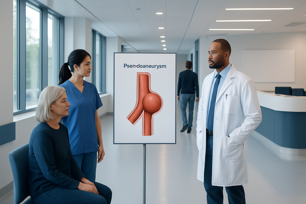
A pseudoaneurysm is different from a true aneurysm because the artery wall itself is not fully forming the bulge. It often develops after a catheter-based procedure, injury, surgery, or infection affecting an artery.
Key Takeaways
- A pseudoaneurysm is different from a true aneurysm because the artery wall itself is not fully forming the bulge.
- It often develops after a catheter-based procedure, injury, surgery, or infection affecting an artery.
- Common signs include a painful or tender lump, swelling, bruising, and sometimes a pulsing mass near the affected area.
- Ultrasound is commonly used to confirm the diagnosis and guide treatment decisions.
- Treatment may include observation, ultrasound-guided procedures, compression, or surgery depending on the situation.
- Urgent medical care is important for rapid swelling, severe pain, skin changes, numbness, weakness, or heavy bleeding.
A pseudoaneurysm is a collection of blood that forms outside an artery after the vessel wall is injured, while still communicating with the artery through a small channel. Many pseudoaneurysms are treatable, but they should be assessed by a doctor because the best approach depends on their size, location, symptoms, and cause.
Overview: what a pseudoaneurysm is
A pseudoaneurysm is a blood-filled sac that forms when blood leaks through a damaged artery wall and collects in nearby tissue. Unlike a true aneurysm, which involves widening of the artery wall itself, a pseudoaneurysm is contained by surrounding tissues or the outer layers around the vessel rather than by all normal layers of the artery.
This distinction matters because a pseudoaneurysm usually reflects a recent injury to the artery. The injury may happen after a medical procedure such as catheter insertion, after surgery, or after trauma. In some cases, infection or inflammation can weaken the vessel area and contribute as well.
Pseudoaneurysms can occur in different parts of the body, but they are especially recognized in the groin after catheter-based procedures involving the femoral artery. Smaller pseudoaneurysms may remain stable or close on their own, while others can enlarge, cause pain, press on nearby structures, or bleed. That is why medical evaluation is important even when symptoms seem mild.
Symptoms and how it may feel
The symptoms of a pseudoaneurysm depend on where it is located and how large it becomes. A person may notice a new lump near the site of a recent procedure, injection, surgery, or injury. The area can be tender, swollen, warm, or bruised, and some people describe a throbbing sensation.
Because the blood inside the sac remains connected to the artery, the lump may seem to pulsate. A clinician may hear a whooshing sound over the area with a stethoscope. In the groin, which is a common site, walking or bending the hip may make discomfort more noticeable.
Not every pseudoaneurysm causes obvious symptoms. Some are found during follow-up imaging after vascular procedures. Others may produce symptoms by pressing on nearby nerves or veins, which can lead to numbness, tingling, leg swelling, or a feeling of pressure.
- Pain or tenderness near an artery
- Visible or palpable swelling
- Bruising around the area
- A pulsating lump
- Numbness, tingling, or weakness if nearby nerves are compressed
- Rarely, active bleeding or sudden enlargement
Causes and risk factors

The most common cause of pseudoaneurysm is injury to an artery. This often happens after procedures that require a catheter to enter an artery, such as coronary angiography or other vascular interventions. In these situations, the small puncture in the artery may not seal fully, allowing blood to pass into surrounding tissue and form a sac.
Other possible causes include surgery, accidental trauma, penetrating injuries, repeated needle punctures, and infection involving a blood vessel. Less commonly, inflammation of the vessel wall or neighboring tissues may contribute. In some locations, a pseudoaneurysm can occur as a complication of vascular aneurysm disease or after treatment of another artery problem.
Several factors may make pseudoaneurysm more likely. These include the use of blood-thinning medicines, larger catheter sizes, difficult arterial access, high blood pressure, obesity, older age, and reduced ability of the puncture site to clot effectively. The exact risk varies by the procedure, the artery involved, and the person’s overall health.
Although the condition can sound alarming, many cases are recognized early and managed successfully. The key is to report new swelling, pain, or a lump after an arterial procedure rather than assuming it is only routine bruising.
How doctors diagnose pseudoaneurysm
Diagnosis begins with a medical history and physical examination. The doctor will ask about recent procedures, injuries, surgeries, symptoms, and medicines such as anticoagulants or antiplatelet drugs. Exam findings like a tender mass, pulsation, skin bruising, or a bruit can raise suspicion.
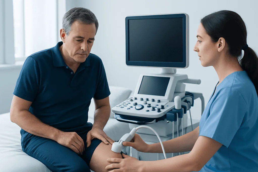
The main test used to confirm pseudoaneurysm is duplex ultrasound. This test shows blood flow in real time and can identify the sac and the narrow connecting channel, sometimes called the neck. Ultrasound is noninvasive, widely available, and also helps guide treatment when a minimally invasive approach is planned.
In some situations, doctors may use CT angiography, MR angiography, or catheter angiography to understand the anatomy more clearly, especially if the pseudoaneurysm is deep, in an unusual location, or associated with more complex artery disease. These tests may also be considered when planning angiography-based evaluation or repair.
Because several conditions can resemble a pseudoaneurysm, imaging is important for an accurate diagnosis. A hematoma, abscess, enlarged lymph node, or other soft tissue mass may look similar from the outside, but they are managed differently.
Treatment options and what determines the plan
Treatment depends on the pseudoaneurysm’s size, location, symptoms, cause, and risk of complications. Small, stable pseudoaneurysms without significant symptoms may sometimes be observed with repeat ultrasound, especially if the doctor believes they may close on their own. During this period, activity modification and careful follow-up are important.
When treatment is needed, one common option is ultrasound-guided thrombin injection. In this procedure, a doctor injects a clot-promoting medicine directly into the pseudoaneurysm sac so that it seals off. This is often effective for suitable femoral pseudoaneurysms and can avoid open surgery. In selected cases, ultrasound-guided compression may also be used, although it may be less comfortable and is not appropriate for everyone.
If the pseudoaneurysm is infected, rapidly enlarging, causing nerve or skin problems, associated with major bleeding, or not suitable for less invasive treatment, surgery may be necessary. Depending on the artery and anatomy, care may involve open repair or endovascular techniques such as endovascular treatment. If the problem affects blood flow in the limb or is linked to another serious vessel issue, doctors may also assess for related peripheral artery disease.
After treatment, follow-up imaging may be recommended to confirm closure and check for recurrence. Care is individualized, and the safest approach is the one that addresses both the pseudoaneurysm and its underlying cause.
Prevention and self-care after vascular procedures
Not every pseudoaneurysm can be prevented, but the risk can often be reduced. Careful technique during arterial access, proper closure of the puncture site, and appropriate monitoring after procedures are important clinical steps. Patients also play a role by following discharge instructions closely.
After a catheter-based procedure, the puncture site should be observed for swelling, persistent bleeding, increasing bruising, or a new lump. Strenuous lifting and vigorous activity may need to be limited for a short time, depending on the doctor’s advice. If blood-thinning medicines are prescribed, they should only be changed under medical guidance.
General self-care supports recovery but does not replace assessment. It may help to keep the area clean, avoid unnecessary pressure or massage over the site, and attend recommended follow-up appointments. Some people with vascular disease may benefit from broader cardiovascular care, including blood pressure management and smoking cessation. In complex cases, doctors may coordinate care with services such as cardiovascular surgery if structural vessel repair is needed.
Most importantly, a patient should not try to diagnose a new pulsing lump at home. Even when the problem turns out to be minor, a clinical review helps confirm whether it is a simple bruise, a hematoma, or a pseudoaneurysm.
When to seek medical care
Medical review is advisable whenever a new painful or pulsating lump appears after an arterial procedure, surgery, or injury. The same is true for swelling that is increasing instead of gradually improving. Early assessment can help prevent complications and may allow simpler treatment.
Urgent care is especially important if there is rapid enlargement, severe or worsening pain, active bleeding, faintness, skin color change, numbness, weakness, or a cold limb. These signs do not always mean a serious emergency, but they can indicate impaired circulation or ongoing bleeding and should not be ignored.
People with fever, redness, or drainage near the site should also be checked because infection can change the treatment plan. If symptoms occur after a recent hospital procedure, contacting the treating team is often the best first step.
For patients seeking coordinated vascular evaluation, Acibadem International’s multidisciplinary specialists and JCI-accredited hospitals diagnose and treat pseudoaneurysm for international patients, with treatment plans tailored to the artery involved and the person’s overall health.
Frequently asked questions
Is a pseudoaneurysm the same as an aneurysm?
No. A true aneurysm is an enlargement of an artery involving the vessel wall itself, while a pseudoaneurysm happens when blood leaks through an arterial injury and is contained by surrounding tissue. Both need medical assessment, but they are different conditions.
Can a pseudoaneurysm go away on its own?
Some small pseudoaneurysms can close spontaneously, especially if they are stable and not causing significant symptoms. However, this should only be determined by a doctor with appropriate imaging and follow-up. Self-monitoring without medical advice is not recommended.
What is the most common test for pseudoaneurysm?
Duplex ultrasound is the most commonly used test. It can show the sac, the connecting neck to the artery, and the blood-flow pattern that helps confirm the diagnosis. It is also often used during minimally invasive treatment.
Is pseudoaneurysm an emergency?
Not every pseudoaneurysm is an emergency, but some require urgent treatment. Rapid swelling, severe pain, active bleeding, skin changes, or symptoms suggesting poor blood flow should be treated urgently. A recent procedure plus a new pulsating lump should always be reported promptly.
How is a femoral pseudoaneurysm treated?
Treatment depends on size, symptoms, and anatomy. Options may include observation with repeat ultrasound, ultrasound-guided thrombin injection, compression, or surgery. The doctor chooses the safest approach based on the individual case.
Can blood thinners increase the risk of pseudoaneurysm?
Yes, blood-thinning medicines can make it harder for a puncture site to seal after an arterial procedure. This can increase the chance that a pseudoaneurysm will form or continue to enlarge. Patients should never stop these medicines on their own but should discuss concerns with their doctor.
This article is for general information only and is not a substitute for professional medical advice. Please consult a qualified doctor about your individual situation.
Explore treatments in Turkey — costs, top hospitals & a free quote
JCI-accredited · board-certified surgeons · reply within 24h
Add us as a Preferred Source to see more of our trusted health content across Google Search, AI Overviews and Discover.
Check your numbers in seconds
BMI, calories, due date, blood pressure and 30+ more clinical calculators — free, instant, doctor-reviewed ranges.

More from the Health Library

Warm Water with Lemon Honey: A Complete Medical Overview

Docosanol: What Patients Need to Know

Testosterone Pellets: What Patients Need to Know

Hemiparesis: An Evidence-Based Guide for Patients

Adenoids: A Complete Medical Overview