Pyogenic Granuloma Treatment: How It Works, Results and What to Expect

Pyogenic granulomas are benign vascular growths, despite their name; they are not pus-filled infections or cancers. Treatment may include curettage with cautery, surgical excision, laser treatment, cryotherapy or selected topical medicines.
Key Takeaways
- Pyogenic granulomas are benign vascular growths, despite their name; they are not pus-filled infections or cancers.
- Treatment may include curettage with cautery, surgical excision, laser treatment, cryotherapy or selected topical medicines.
- A removed lesion may be sent for laboratory examination, especially when the diagnosis is uncertain or the growth is atypical.
- Most treatment areas heal within a few weeks, although recurrence can occur if triggering factors remain or some lesion tissue is left behind.
- A new, fast-growing or repeatedly bleeding skin lesion should be assessed by a qualified clinician.
Pyogenic granuloma treatment usually involves removing or destroying a small, benign blood-vessel growth that bleeds easily. The best approach depends on its size, location, cause, appearance and whether tissue testing is needed to confirm the diagnosis.
Pyogenic Granuloma Treatment: How It Works
Pyogenic granuloma treatment aims to stop bleeding, remove the visible growth and reduce the chance that it returns. A pyogenic granuloma is a benign overgrowth of tiny blood vessels and inflamed tissue. It often appears as a small, bright-red bump that grows quickly and bleeds after minor contact. Despite the name, it is not caused by pus and is not a true granuloma.
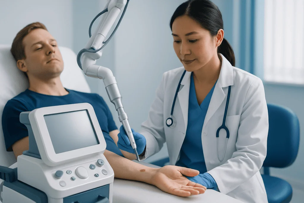
Many lesions are treated in a clinic with a local anesthetic. Common options include curettage (gently scraping away the lesion) followed by cautery to seal blood vessels, complete surgical excision, laser treatment or freezing treatment. The clinician chooses the method based on the lesion’s location, size and bleeding tendency, as well as the importance of obtaining a tissue sample.
Complete excision is often considered when a lesion is larger, repeatedly returns, occurs in an area where diagnostic certainty is important, or has an unusual appearance. Smaller lesions may be suitable for less invasive approaches. Treatment plans should be individualized after an in-person skin examination.
Why a Pyogenic Granuloma May Need Treatment
Although pyogenic granulomas are not cancerous, they can be troublesome. Their delicate surface can crack and bleed with washing, shaving, clothing friction or minor injury. Some cause soreness, crusting or concern because they change rapidly in appearance.
Treatment can provide symptom relief, reduce repeated bleeding and allow the clinician to confirm what the growth is. Several benign and more serious conditions can resemble a pyogenic granuloma, so a clinician may recommend a biopsy or examination of removed tissue when the diagnosis is not fully clear.
In selected situations, particularly for very small lesions or lesions related to a reversible trigger, observation or topical treatment may be discussed. However, a lesion should not be self-diagnosed. Repeated bleeding, persistent growth or a changing spot warrants medical assessment.
Candidacy and Assessment Before Treatment

People are usually candidates for pyogenic granuloma treatment when the lesion bleeds, is painful, grows, causes cosmetic concern, interferes with daily activities or has an uncertain diagnosis. Lesions can occur on the skin, lips, mouth, fingers and around the nails. Pregnancy-associated lesions may occur on the gums and may need coordinated dental and medical assessment.
Before treatment, the clinician asks when the growth appeared, whether there was recent trauma or irritation, and whether it has changed. They also review medicines, bleeding disorders, pregnancy status and any history of skin cancer. Examination includes the lesion’s color, shape, size, location and pattern of bleeding.
A dermatoscope, a handheld lighted magnifier, may help examine the skin. If removal is planned, tissue may be sent to pathology. This is particularly useful when a growth is darkly colored, unusually shaped, ulcerated, recurrent or not behaving as expected.
- Possible triggers include minor injury, chronic friction, ingrown nails, hormonal changes and certain medicines.
- Blood-thinning medicines should not be stopped without advice from the clinician who prescribed them.
- Oral lesions should be reviewed by a dentist, oral medicine specialist or surgeon as appropriate.
What Happens During Pyogenic Granuloma Removal
The procedure generally begins with cleaning the area. For many skin lesions, local anesthetic is injected or applied so the person should feel pressure or brief pulling rather than pain. Because these growths contain many small blood vessels, clinicians prepare to control bleeding during treatment.
With curettage and cautery, the clinician removes the raised tissue with a small instrument and uses heat or another method to seal the base. With surgical excision, the lesion and a small surrounding margin may be removed, then the wound may be closed with stitches. Laser treatment targets blood vessels, while cryotherapy uses controlled freezing to destroy the lesion tissue. The technique varies by the individual lesion and available expertise.
After treatment, a dressing may be placed over the site. The clinician provides instructions for wound care and explains whether stitches need removal. If a specimen is sent to the laboratory, follow-up includes discussing the pathology result and any next steps.
For people seeking coordinated care, Acibadem International’s multidisciplinary specialists and JCI-accredited hospitals diagnose and treat skin lesions for international patients, with the treatment approach based on clinical assessment and pathology when required.
Recovery Timeline, Benefits and Possible Risks
Initial tenderness, mild swelling, redness and a small amount of oozing can occur after treatment. The wound often forms a dry scab or crust over the first several days. It is important not to pick at the area, as this can restart bleeding and delay healing.
How long healing takes depends on the procedure and the body site. A small superficial treatment area may close in about one to two weeks, while a deeper excision or a lesion on the hand, foot or lower leg may take several weeks. Stitches, if used, are commonly removed according to the clinician’s planned schedule.
The expected benefits are less bleeding, removal of the growth, relief from irritation and diagnostic clarification when tissue is examined. Potential risks include short-term pain, infection, scarring, pigment change, delayed healing and recurrence. Recurrence does not necessarily mean a serious problem, but it should be reassessed because further treatment or pathology review may be needed.
Keeping the wound clean, following dressing instructions and avoiding friction, swimming or strenuous activity until advised can support healing. Sun protection after the skin has closed may help minimize long-lasting color changes in some people.
How serious is pyogenic granuloma?
Pyogenic granuloma is usually not serious in the sense that it is benign and does not spread through the body like cancer. However, it can cause frequent bleeding, discomfort, infection risk if the surface is repeatedly broken, and anxiety because it may grow quickly or look alarming.
Its main clinical importance is that other conditions can occasionally look similar. A clinician should evaluate a new skin growth that is changing, bleeding or not healing, rather than assuming it is a pyogenic granuloma. Examination and, when appropriate, biopsy provide reliable diagnosis.
Urgent assessment is appropriate if bleeding does not stop with steady, direct pressure, if there is spreading redness or fever, or if the lesion changes dramatically. Most people can be evaluated in a routine outpatient appointment when bleeding is controlled and there are no signs of infection.
What are the stages of pyogenic granuloma?
Pyogenic granuloma does not have formal medical stages in the way some cancers or chronic diseases do. It is more helpful to describe its typical course: a small spot develops, grows relatively rapidly over days to weeks, then may remain raised and bleed or crust repeatedly.
Early lesions are often red, pink or purple and may look smooth and shiny. As they mature, they may become more raised, develop a thin pale rim at the base, ulcerate from friction or appear darker because of dried blood. Some lesions persist unless treated, while others may shrink if an underlying trigger is removed.
After treatment, the usual sequence is a treated wound, formation of a crust or scab, gradual closure of the skin and fading redness. A clinician should review a growth that continues to enlarge, repeatedly returns, becomes increasingly painful or does not heal as expected.
What does a healing pyogenic granuloma look like? When to seek medical care
A healing treatment site commonly looks like a shallow wound with mild redness, a dark crust or scab, and gradual reduction in moisture or bleeding. As new skin forms, the area may look pink and sensitive before its color slowly settles. Small amounts of clear or lightly blood-stained drainage may occur early, depending on the procedure.
It is not possible to judge healing from appearance alone, particularly after self-treatment or repeated injury. Signs that should prompt contact with a clinician include worsening rather than improving pain, expanding redness, warmth, pus-like drainage, fever, persistent heavy bleeding, wound opening or a rapidly growing new bump at the same site.
Medical care should also be sought for any new lesion that bleeds easily, has irregular color or borders, changes quickly or persists. Until review, direct, continuous pressure with clean gauze can help control minor bleeding; emergency care is needed if bleeding is heavy or cannot be controlled.
Frequently asked questions
How long does it take for a pyogenic granuloma to heal after treatment?
Small, superficial treatment sites often heal in about one to two weeks. Healing can take several weeks after a deeper excision or when the lesion is on an area exposed to friction or slower circulation, such as the lower leg. The treating clinician can provide the most accurate timeline based on the procedure and wound location.
Can a pyogenic granuloma go away without treatment?
Some pyogenic granulomas may shrink if a triggering factor, such as repeated irritation, is removed. Many persist, bleed repeatedly or continue to grow, so assessment and treatment are often appropriate. A clinician should confirm the diagnosis before choosing observation.
Is pyogenic granuloma cancer?
No. A pyogenic granuloma is a benign growth of blood vessels and inflamed tissue, not cancer. Because some other skin conditions can look similar, a clinician may remove the lesion and send it for pathology testing when the diagnosis is uncertain.
Does pyogenic granuloma treatment hurt?
Most removal procedures use local anesthetic, which minimizes pain during treatment. Some brief pressure, pulling or heat sensation may still be felt. Mild soreness afterward is common and is usually managed with the wound-care advice provided by the clinician.
Can a pyogenic granuloma come back after removal?
Yes, recurrence is possible, especially if some lesion tissue remains or an underlying source of irritation continues. A recurrent lesion should be reviewed rather than treated repeatedly at home. The clinician may recommend a different method or pathology examination.
Should a pyogenic granuloma be removed at home?
No. Cutting, tying off, burning or using unverified products can cause significant bleeding, infection, scarring and delayed diagnosis of another condition. A qualified healthcare professional can safely assess and treat a bleeding or changing skin growth.
References
- American Academy of Dermatology Association
- DermNet
- Merck Manual Consumer Version
- National Health Service
- Mayo Clinic
This article is for general information only and is not a substitute for professional medical advice. Please consult a qualified doctor about your individual situation.
Explore treatments in Turkey — costs, top hospitals & a free quote
JCI-accredited · board-certified surgeons · reply within 24h
Add us as a Preferred Source to see more of our trusted health content across Google Search, AI Overviews and Discover.