Retinal Tear Treatment with Laser: How It Works, Results and What to Expect

Laser retinopexy creates a protective seal around a retinal tear and is intended to lower the chance of retinal detachment. The procedure is commonly completed in an eye clinic using dilating and numbing drops; most people return home the same day.
Key Takeaways
- Laser retinopexy creates a protective seal around a retinal tear and is intended to lower the chance of retinal detachment.
- The procedure is commonly completed in an eye clinic using dilating and numbing drops; most people return home the same day.
- The laser scars begin to form promptly but typically become more secure over about 1 to 2 weeks.
- New floaters, flashes, a curtain-like shadow or reduced side vision need urgent ophthalmic assessment, including after laser treatment.
- Follow-up examinations are important because additional tears or retinal detachment can occasionally develop elsewhere in the retina.
Retinal tear treatment with laser, also called laser retinopexy or photocoagulation, creates small controlled scars around a retinal tear to secure the retina to the eye wall and reduce the risk of retinal detachment. It is usually an outpatient procedure performed with numbing eye drops, and timely assessment is important when new flashes, floaters or a shadow in vision occur.
Overview: what retinal tear treatment with laser does
Retinal tear treatment with laser is a procedure used to treat a break in the retina, the light-sensitive tissue lining the back of the eye. The aim is to place a ring of small laser burns around the tear. As these spots heal, they form scar tissue that helps attach the retina more firmly to the tissue beneath it.
This treatment is often called laser retinopexy or laser photocoagulation. It does not remove floaters or reverse vision changes caused by an existing tear. Instead, it is designed to reduce the chance that fluid passes through the tear and lifts the retina away from the eye wall, a condition called retinal detachment.
A retinal tear is not always an emergency in the same way as a detached retina, but it needs prompt evaluation by an ophthalmologist. Whether laser is appropriate depends on the tear’s location, appearance and surrounding retinal tissue, as well as whether there is bleeding, fluid under the retina or other eye conditions.
How laser retinopexy works and who may be a candidate

The vitreous is a clear gel that fills most of the eye. With age, it commonly becomes more liquid and can separate from the retina; this is known as posterior vitreous detachment. During this process, traction from the vitreous may pull hard enough on a vulnerable area of retina to create a tear.
Laser energy is focused in a pattern around the retinal break, not directly into the opening itself. The treatment produces controlled inflammation followed by a firm chorioretinal adhesion, creating a barrier intended to prevent fluid from extending beneath the retina. This approach is generally considered when the retina remains attached or when there is only very limited fluid that the eye specialist believes can be safely contained.
People may be more likely to need assessment for a retinal tear if they are very short-sighted, have had eye surgery or an eye injury, have a family history of retinal detachment, or have had a retinal tear or detachment in the other eye. A specialist may also monitor some small, stable retinal changes rather than treat them immediately. The decision is individual and based on a detailed dilated retinal examination.
What happens during the laser procedure
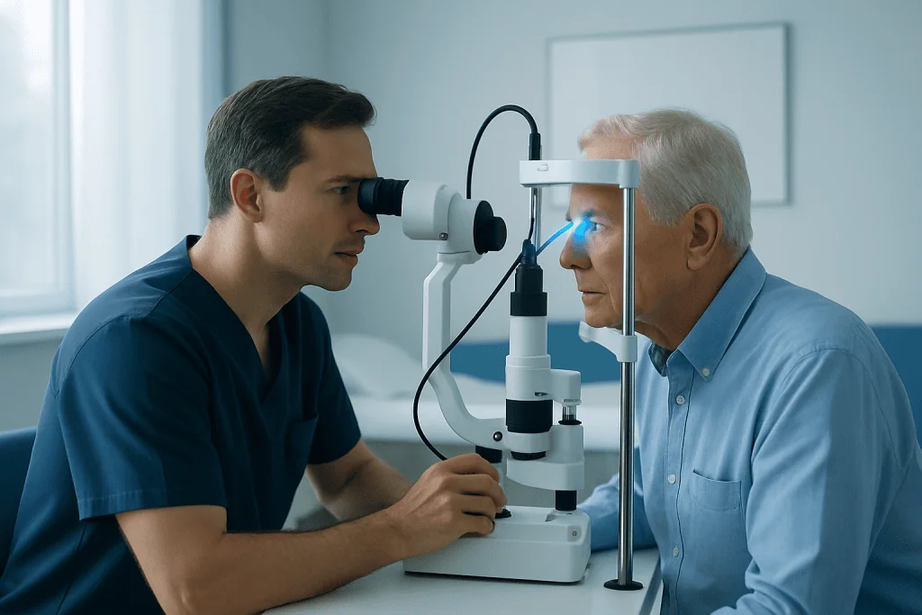
Before treatment, the eye is dilated so the specialist can view the peripheral retina. Numbing drops are used, and a contact lens may be placed gently on the eye to keep it open and allow the laser to be focused accurately. In some situations, the doctor may recommend an injection of local anaesthetic around the eye, particularly if treatment is extensive or difficult to tolerate.
The patient is seated at a laser microscope and asked to look in certain directions while the doctor applies laser spots around the tear. Bright flashes of light are expected. Some people notice brief discomfort, pressure or a stinging sensation, but severe pain is not typical and should be reported during the procedure.
Laser retinopexy often takes about 15 to 30 minutes, although the full appointment can take longer because of examination, preparation and recovery from dilating drops. Vision may remain blurred and light-sensitive for several hours. A companion or arranged transport home is helpful because driving is usually not advisable until vision has returned to normal.
Laser treatment is different from surgery for an established retinal detachment. If the retina has detached extensively, treatment may require procedures such as vitrectomy, scleral buckling or pneumatic retinopexy rather than laser alone.
Recovery timeline and everyday activity
Immediately after treatment, temporary blurred vision, sensitivity to light, mild aching and awareness of new or pre-existing floaters can occur. These symptoms commonly settle over hours to a few days, although vision can be affected longer when the tear or laser treatment is close to the central retina. The treating ophthalmologist may advise anti-inflammatory eye drops or other medicines when appropriate.
How long does it take for a retinal tear to heal after laser? The laser spots begin to produce an adhesive effect soon after treatment, but the scar barrier usually strengthens over approximately 1 to 2 weeks. During this interval, follow-up plans and activity advice should be followed carefully. Healing can vary with the size and location of the tear, the amount of laser needed and the health of the retina.
Can you walk after retinal tear laser surgery? In most cases, gentle walking is allowed after the procedure once the person feels steady and can see safely. However, vision may be blurred from dilation and glare, so it is sensible to have someone accompany the patient home and to avoid walking alone in unfamiliar or poorly lit places immediately afterward. The ophthalmologist may give different instructions when there are additional retinal concerns.
Follow-up is often arranged within days to weeks so the specialist can confirm that the laser scars are forming adequately and inspect the rest of the retina. Long-term monitoring may also be advised, particularly for people with high myopia, retinal disease or previous tears.
Benefits, limitations and possible risks
The principal benefit of laser retinopexy is prevention: when a suitable retinal tear is identified and treated before a significant detachment occurs, laser can substantially reduce the likelihood of that particular tear leading to retinal detachment. It is a targeted outpatient treatment and usually avoids incisions into the eye.
How successful is laser for retinal tears? Laser is highly effective at creating adhesion around many appropriately selected retinal tears, especially when treatment is performed before retinal detachment develops. No procedure can eliminate risk completely, however. A tear may need additional laser if the initial barrier is incomplete, and new tears can occur elsewhere in the retina later.
Possible short-term effects include temporary discomfort, light sensitivity, blurred vision and increased awareness of floaters. Less common complications include inflammation, bleeding, damage to nearby retinal tissue, changes in peripheral vision or failure of the tear to seal adequately. Retinal detachment can still occur, which is why new symptoms after treatment should never be ignored.
Laser does not treat every cause of visual symptoms. The specialist will assess for related problems, including vitreous changes and retinal detachment, and explain whether observation, laser, cryotherapy or retinal surgery is the safest approach.
What not to do after retinal tear laser
What not to do after retinal tear laser? For the first days to weeks, patients should avoid activities that could involve jolting, straining or a risk of eye injury until their ophthalmologist confirms it is safe. This may include heavy lifting, vigorous running, high-impact exercise, contact sports and activities where sudden head movements are likely. The exact restrictions vary, so the individual treatment plan takes priority over general advice.
Patients should not drive while their vision is blurred from dilating drops, glare or reduced visual clarity. They should avoid rubbing or pressing on the treated eye, and should use prescribed drops exactly as directed. Normal light household activities and gentle movement are often acceptable, but a doctor may recommend more caution if there is bleeding, fluid under the retina or a higher risk of detachment.
It is also important not to assume that all flashes and floaters after treatment are harmless. Although some symptoms can persist after a posterior vitreous detachment, a sudden increase in floaters, repeated flashes, a dark curtain or a reduction in side vision needs urgent reassessment. These changes may signal a new tear or a developing detachment.
When to seek medical care
Anyone who develops sudden flashes of light, a new shower of floaters, a cobweb-like visual disturbance, a curtain or shadow across vision, or a sudden loss of side vision should seek urgent eye care. These symptoms may occur with a retinal tear or detachment and need a dilated retinal examination as soon as possible.
After laser treatment, urgent contact with the treating eye team is appropriate if vision worsens, a new shadow develops, pain becomes severe, marked redness or discharge occurs, or there is a rapid increase in flashes or floaters. Even if symptoms improve, planned follow-up appointments remain important because the retina can change over time.
Acibadem International’s multidisciplinary specialists and JCI-accredited hospitals assess and treat retinal conditions for international patients. An ophthalmologist can explain whether laser retinopexy is appropriate and coordinate care if more complex retinal treatment is needed.
Frequently asked questions
Is retinal tear treatment with laser painful?
Numbing eye drops are generally used, so most people experience bright flashes, pressure or brief stinging rather than significant pain. The procedure team should be told if discomfort is difficult to tolerate, as they can pause and consider additional measures. Mild ache or irritation afterward can occur and usually improves over a short period.
Will laser treatment remove floaters?
Laser retinopexy is not intended to remove floaters. It treats the retinal tear by creating a scar barrier around it. Existing floaters may become less noticeable over time, but persistent or suddenly increasing floaters should be discussed with an eye specialist.
Can a retinal tear return after laser treatment?
The treated tear is expected to become sealed by scar tissue, but laser cannot prevent every future retinal problem. Another tear can develop in a different area, particularly while the vitreous is continuing to change. Regular follow-up and prompt reporting of new symptoms are therefore important.
How soon can a person return to work after retinal laser?
Many people can return to desk-based work within a day or two, depending on how quickly their vision clears and what their job involves. Work requiring driving, heavy physical effort, heights or machinery may need a longer pause. The treating ophthalmologist should provide individualized clearance.
Does laser retinopexy restore vision lost from a retinal tear?
The primary purpose of laser is to prevent progression to retinal detachment, not to restore vision that has already been affected. If central vision was not involved, vision may remain stable after successful treatment. Visual recovery depends on the underlying retinal condition and whether complications occurred.
What happens if a retinal tear is not treated?
Some retinal tears can allow fluid to pass beneath the retina and lead to retinal detachment, which can threaten vision. Not every retinal finding requires treatment, but this determination should be made by an ophthalmologist after a dilated examination. New warning symptoms should be assessed urgently.
References
- American Academy of Ophthalmology
- National Eye Institute
- Royal College of Ophthalmologists
- Mayo Clinic
This article is for general information only and is not a substitute for professional medical advice. Please consult a qualified doctor about your individual situation.
Explore treatments in Turkey — costs, top hospitals & a free quote
JCI-accredited · board-certified surgeons · reply within 24h
Add us as a Preferred Source to see more of our trusted health content across Google Search, AI Overviews and Discover.