Shoulder Dislocation: Emergency Care, Instability Risk, and Rehabilitation

A suspected shoulder dislocation should be assessed urgently; patients should not try to force the joint back into place themselves. Emergency care usually includes pain control, neurovascular assessment, imaging, and reduction by a trained clinician.
Key Takeaways
- A suspected shoulder dislocation should be assessed urgently; patients should not try to force the joint back into place themselves.
- Emergency care usually includes pain control, neurovascular assessment, imaging, and reduction by a trained clinician.
- Young athletes and people with ligament laxity have a higher risk of repeat dislocation or ongoing shoulder instability.
- Rehabilitation progresses from protection and gentle motion to strengthening, coordination, and sport- or work-specific training.
- Surgery may be recommended when there are significant associated injuries, repeated dislocations, or high-risk instability patterns.
A shoulder dislocation occurs when the upper arm bone comes out of the shoulder socket, most often after a fall, sports injury, or collision. Prompt medical care, careful follow-up, and structured rehabilitation help relieve pain, protect nerves and blood vessels, and reduce the risk of recurrent instability.
Overview
A shoulder dislocation happens when the head of the humerus, the ball at the top of the upper arm bone, is forced out of the glenoid, the shallow socket of the shoulder blade. The shoulder is the most mobile joint in the body, which allows a wide range of movement but also makes it more vulnerable to dislocation than many other joints.
Most shoulder dislocations are anterior, meaning the ball moves forward out of the socket. Less commonly, the shoulder may dislocate backward or downward. The injury can stretch or tear the capsule, ligaments, labrum, tendons, or nearby muscles, and it may occasionally be associated with a fracture or nerve irritation.
A dislocated shoulder is painful and usually requires urgent medical evaluation. With appropriate reduction, imaging, immobilization, and rehabilitation, many people regain good shoulder function. However, some patients, especially younger active people, may develop recurrent instability and need a more detailed orthopedic plan.
Symptoms and What It Feels Like
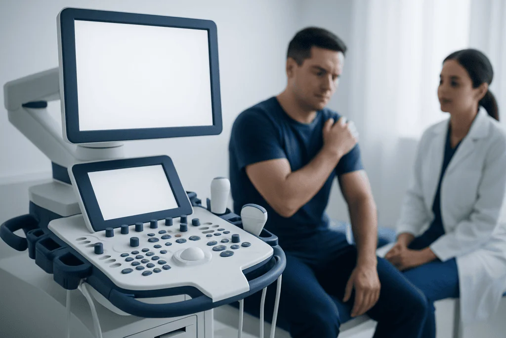
The symptoms of a shoulder dislocation are often sudden and clear. Many people feel the shoulder pop out after a fall onto an outstretched hand, a tackle, a sudden pull, or a forceful overhead movement. The arm may feel stuck in an unusual position, and moving it can be very painful.
Common signs and symptoms include:
- Severe shoulder pain immediately after injury
- Visible change in shoulder shape, such as a flattened or squared-off appearance
- Inability or reluctance to move the arm
- Swelling, bruising, or muscle spasm around the shoulder
- Numbness, tingling, or weakness in the arm or hand
- A feeling that the shoulder has slipped out or is not in the right place
Numbness over the outer shoulder or weakness when lifting the arm may suggest irritation of the axillary nerve, which can occur with dislocation. Changes in hand color, coldness, or loss of pulse are less common but require immediate attention because they may indicate blood vessel involvement.
Emergency Care and Shoulder Reduction

A suspected shoulder dislocation should be treated as an urgent injury. The safest first step is to keep the arm supported in the position that is most comfortable, avoid eating or drinking in case sedation is needed, and seek emergency medical care. Patients should not try to force the shoulder back into place themselves, because an associated fracture, trapped tissue, nerve injury, or blood vessel injury could be worsened.
In the emergency setting, clinicians examine the shoulder and check circulation, sensation, and movement in the arm and hand. X-rays are commonly used to confirm the direction of dislocation and look for fractures before and after reduction. In selected cases, especially when complex injury is suspected, CT or MRI may be used later to evaluate bone loss, labral tears, rotator cuff injury, or other soft tissue damage.
Reduction means guiding the humeral head back into the socket. This is performed by trained healthcare professionals using controlled techniques, often with pain relief, muscle relaxation, or procedural sedation when appropriate. Once the shoulder is reduced, pain usually improves significantly, but the joint still needs protection while injured tissues begin to heal.
After reduction, the arm is often placed in a sling for a short period. The exact duration depends on age, injury pattern, pain level, and physician assessment. Too little protection may irritate the injury, while prolonged immobilization can contribute to stiffness, so follow-up guidance is important.
Causes and Risk Factors for Instability
Shoulder dislocation can occur during sports, falls, road traffic accidents, seizures, electrical injuries, or work-related trauma. Contact sports, gymnastics, skiing, cycling, and activities involving overhead or extreme shoulder positions can increase risk. A previous dislocation is one of the strongest risk factors for another episode.
Instability means the shoulder does not stay securely centered in the socket. After a first dislocation, the labrum or capsule may be stretched or torn, reducing the natural stabilizing effect of these tissues. Some people develop recurrent dislocations, partial slips called subluxations, or a persistent feeling that the shoulder may give way during certain movements.
Factors that can increase the chance of recurrent instability include young age at first dislocation, participation in collision or overhead sports, ligament laxity, significant labral injury, bone loss from the socket or humeral head, and not completing rehabilitation. Older adults may be less likely to have repeated dislocations than teenagers or young adults, but they have a higher risk of associated rotator cuff tears after traumatic dislocation.
Because risk varies from person to person, follow-up with an orthopedic specialist is often helpful after a first dislocation. The goal is to identify injuries that may need closer monitoring or early treatment, and to match rehabilitation and return-to-activity decisions to the patient’s lifestyle and risk profile.
Diagnosis and Follow-Up Assessment
Diagnosis begins with the history of the injury, the position of the arm, visible deformity, pain pattern, and a careful neurovascular examination. Clinicians assess sensation, muscle strength, pulses, skin temperature, and hand function. These checks are important before and after reduction to document recovery and detect complications early.
X-rays are the standard first imaging test. They confirm that the joint is back in place after reduction and help identify fractures, such as a greater tuberosity fracture or a compression injury of the humeral head. If symptoms persist or instability is suspected, MRI can help evaluate labral tears, capsular injury, rotator cuff tears, and other soft tissue problems. CT may be preferred when detailed assessment of bone loss is needed.
Follow-up is usually arranged within days to weeks, depending on the injury. During follow-up, the physician reviews pain, range of motion, nerve function, imaging results, and the patient’s daily or athletic needs. This visit is an opportunity to plan rehabilitation, decide how long to use a sling, and discuss whether additional imaging or specialist care is needed.
Patients should mention any ongoing numbness, weakness, night pain, repeated slipping sensations, or inability to lift the arm. These symptoms do not always mean a serious complication, but they can change the treatment plan and may require further evaluation.
Treatment Options
Treatment depends on the patient’s age, activity level, direction of dislocation, first-time versus recurrent injury, imaging findings, and goals. For many first-time anterior shoulder dislocations without major associated injuries, non-surgical care is appropriate. This usually includes reduction, a short period of sling use, pain control, gradual rehabilitation, and monitored return to activity.
Medicines may be used to relieve pain and inflammation when safe for the individual patient. Ice, rest, and supported positioning can also help during the early phase. Any medication plan should consider allergies, stomach, kidney, liver, bleeding, pregnancy, and other medical factors, so patients should follow advice from a qualified clinician.
Surgical treatment may be considered when there are repeated dislocations, high-risk athletic demands, significant labral tears, major bone loss, fractures needing fixation, or persistent instability despite rehabilitation. Common procedures aim to repair or tighten the damaged soft tissues, restore stability, or address bone defects. The specific operation depends on the injury pattern and the surgeon’s assessment.
The decision between non-surgical and surgical treatment is individualized. A patient who wants to return to collision sport, for example, may have different risks and priorities than a person whose main goal is comfortable daily activity. Shared decision-making helps align treatment with safety, function, and expectations.
Rehabilitation and Return to Activity
Rehabilitation is a key part of recovery after shoulder dislocation. The early goal is to reduce pain, protect healing tissues, maintain hand, wrist, and elbow movement, and avoid stiffness. Gentle shoulder exercises are usually introduced gradually when the clinician confirms it is safe.
Rehabilitation often progresses through phases. Early exercises may focus on pain-free range of motion and posture. Later, strengthening targets the rotator cuff and shoulder blade muscles, which help keep the ball centered in the socket. Advanced rehabilitation adds coordination, balance, controlled overhead movement, and sport- or job-specific drills.
Return to sport or heavy work should not be based only on time. It usually requires near-full comfortable range of motion, good strength compared with the uninjured side, confidence during shoulder movement, and clearance from the treating clinician or physiotherapist. Returning too early may increase the risk of another dislocation, while excessive fear of movement can delay recovery.
Patients can support recovery by attending rehabilitation consistently, avoiding sudden risky positions until cleared, and reporting any slipping, catching, or sharp pain. A well-supervised program is especially important for athletes, manual workers, and people who have already had more than one instability episode.
Prevention, Self-Care, and When to See a Doctor
Not every shoulder dislocation can be prevented, but risk can be reduced. Good conditioning of the rotator cuff, shoulder blade muscles, core, and legs helps support safe movement. Athletes benefit from sport-specific technique training, appropriate protective equipment, and gradual return after injury rather than sudden full-intensity play.
After a dislocation, self-care should be guided by a healthcare professional. In general, patients should use the sling as advised, keep the elbow, wrist, and hand moving, apply cold packs safely during the early painful period, and avoid positions that make the shoulder feel unstable. They should not resume heavy lifting, contact sport, or forceful overhead activity until medically cleared.
Immediate medical care is needed for severe pain, visible deformity, numbness, tingling, weakness, coldness or color change in the hand, open wounds, or inability to move the arm after an injury. Patients should also see a doctor if the shoulder repeatedly slips, feels unreliable, remains weak, or does not regain motion as expected after reduction.
For international patients needing evaluation and treatment, Acibadem International’s multidisciplinary orthopedic, imaging, emergency, and rehabilitation teams in JCI-accredited hospitals can assess shoulder dislocation and instability and guide individualized care. As with any injury, the most appropriate plan should be made after direct examination by qualified medical professionals.
Frequently asked questions
Is a shoulder dislocation a medical emergency?
A suspected shoulder dislocation should be assessed urgently, especially if there is severe pain, deformity, numbness, weakness, or changes in hand color or temperature. Prompt care helps confirm the diagnosis, reduce the joint safely, and check for fractures or nerve and blood vessel problems.
Can someone put a dislocated shoulder back in by themselves?
It is not recommended to force a dislocated shoulder back into place without medical training. An associated fracture or soft tissue injury may be present, and improper reduction can worsen damage. The safest approach is to support the arm and seek emergency medical care.
How long does recovery take after a shoulder dislocation?
Recovery time varies depending on age, injury severity, associated tears or fractures, and activity goals. Many people resume light daily activities gradually within weeks, but return to sport or heavy work often requires longer rehabilitation and medical clearance.
Will the shoulder dislocate again?
Some people never have another dislocation, while others develop recurrent instability. The risk is higher in younger active patients, contact or overhead athletes, and people with labral injury, ligament laxity, or bone loss. Follow-up assessment and rehabilitation help define and reduce this risk.
When is surgery needed for shoulder dislocation?
Surgery may be considered for repeated dislocations, significant labral or bony injuries, fractures requiring repair, or persistent instability despite rehabilitation. It may also be discussed earlier for high-risk athletes or patients whose work requires demanding shoulder function. The decision should be individualized with an orthopedic specialist.
What exercises help after a dislocated shoulder?
Exercises usually begin with gentle, pain-free movement and progress to rotator cuff and shoulder blade strengthening. Later stages may include coordination, endurance, and sport- or work-specific training. Exercises should be prescribed or approved by a clinician or physiotherapist because doing too much too soon may increase instability.
Can an older adult have different problems after a shoulder dislocation?
Yes. Older adults may have a higher chance of rotator cuff tear or stiffness after a traumatic dislocation. Persistent weakness, night pain, or difficulty lifting the arm should be reviewed by a doctor, even if the shoulder was successfully reduced.
References
- American Academy of Orthopaedic Surgeons
- British Orthopaedic Association
- Mayo Clinic
- Cleveland Clinic
- National Institute for Health and Care Excellence
This article is for general information only and is not a substitute for professional medical advice. Please consult a qualified doctor about your individual situation.
Shoulder Dislocation in Turkey — costs, top hospitals & a free quote
JCI-accredited · board-certified surgeons · reply within 24h
Add us as a Preferred Source to see more of our trusted health content across Google Search, AI Overviews and Discover.