Skin tag on tongue: Symptoms, Causes, and Treatment — Complete Patient Guide

A true skin tag is uncommon on the tongue, and many tongue bumps are actually other benign oral lesions. Common symptoms include a small flap-like or polyp-like growth, irritation, a foreign-body sensation, or accidental biting.
Key Takeaways
- A true skin tag is uncommon on the tongue, and many tongue bumps are actually other benign oral lesions.
- Common symptoms include a small flap-like or polyp-like growth, irritation, a foreign-body sensation, or accidental biting.
- Persistent, enlarging, painful, bleeding, or ulcerated tongue growths need professional evaluation.
- Diagnosis is based on an oral exam and sometimes biopsy to confirm what the growth is.
- Treatment depends on the cause and may include observation, removing irritation, or minor surgical removal.
A skin tag on tongue usually refers to a small, soft, benign-looking growth, but true skin tags are uncommon inside the mouth. Because several harmless and less common serious conditions can look similar, a doctor or dentist should examine any tongue growth that does not go away or that changes over time.
What a skin tag on tongue usually means
A skin tag on tongue usually describes a small, soft growth that appears on the tongue and may look like a flap, tiny polyp, or raised bump. In everyday language, people often call any small hanging growth a “skin tag.” Medically, however, true skin tags are far more common on external skin than inside the mouth, so a similar-looking tongue lesion may be something else.
Many tongue growths are benign. These can include irritation fibromas caused by repeated rubbing or biting, squamous papillomas, mucosal polyps, or other noncancerous soft tissue overgrowths. Some are smooth and flesh-colored, while others are slightly white, pink, or rougher on the surface.
Because appearance alone cannot always identify the lesion with certainty, it is important not to self-diagnose. A small tongue growth that stays the same may still be harmless, but one that persists, grows, changes color, bleeds, or becomes painful should be examined by a qualified doctor or dentist.
Symptoms and how it may feel
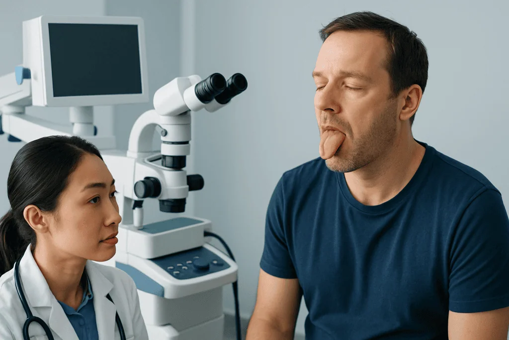
A tongue growth that is described as a skin tag may be noticed by sight, touch, or both. Some people first feel it when brushing their teeth, eating, or moving the tongue across the teeth. Others notice it in a mirror or after recurrent irritation in the same area.
Possible symptoms include:
- a small soft bump or flap on the top, side, or underside of the tongue
- a smooth or slightly rough surface
- flesh-colored, pink, or whitish appearance
- a feeling that something is “caught” on the tongue
- mild tenderness if the area is bitten or rubbed repeatedly
- annoyance when speaking, chewing, or swallowing
Many benign lesions do not cause pain. Symptoms often develop only when the growth is repeatedly traumatized by teeth, sharp dental edges, braces, or hot foods. In some cases, a lesion may become swollen after friction and then seem smaller again later.
If there is significant pain, bleeding without clear injury, a nonhealing sore, numbness, bad breath, ear pain, or difficulty swallowing, the problem may not be a simple tag-like growth. These symptoms should prompt a timely medical assessment.
Common causes and look-alike conditions

A true skin tag forms from extra folds of skin and connective tissue, but the tongue is covered by oral mucosa rather than skin. For that reason, doctors usually consider other explanations first when a patient reports a skin tag on tongue. Repeated irritation is one of the most common reasons for a benign overgrowth in the mouth.
Common possibilities include:
- Irritation fibroma: a firm or soft benign bump caused by chronic rubbing, biting, or friction.
- Squamous papilloma: a small wart-like growth that may be linked to low-risk human papillomavirus in some cases.
- Mucosal polyp or focal hyperplasia: a localized overgrowth of tissue from irritation.
- Prominent papillae or inflamed taste buds: temporary bumps that can be mistaken for a lesion.
- Mucocele: a mucus-filled cyst more often found elsewhere in the mouth but sometimes considered in the differential diagnosis.
Less commonly, a persistent tongue lesion may represent a precancerous or cancerous change, especially if it is ulcerated, hard, irregular, or associated with smoking, heavy alcohol use, or older age. This is one reason clinicians may compare it with other oral lesions such as mouth cancer during assessment, even though many tongue growths turn out to be noncancerous.
Risk factors for benign tongue lesions include accidental biting, rough teeth, dentures that rub, orthodontic appliances, tobacco exposure, and poor-fitting dental work. Not every lesion has an obvious cause, and some are only identified after removal and laboratory examination.
How doctors diagnose a tongue growth
Diagnosis usually begins with a careful history and oral examination. A doctor, dentist, or oral and maxillofacial specialist will ask how long the growth has been present, whether it has changed in size or color, and whether there are symptoms such as pain, bleeding, or trouble eating. They will also look for possible sources of friction or trauma inside the mouth.
During the exam, the clinician checks the lesion’s size, shape, color, attachment, texture, and exact location. The neck and nearby lymph nodes may also be examined if there is concern about infection or a more serious condition. In many cases, the appearance strongly suggests a benign lesion, but visual assessment alone is not always enough.
If the growth is persistent, unusual, or suspicious, a biopsy may be recommended. This means removing part or all of the tissue so it can be examined under a microscope. Biopsy is often the most reliable way to confirm whether a lesion is an irritation fibroma, papilloma, inflammatory growth, or another condition.
Some patients may be referred for oral diagnosis and biopsy or a specialist evaluation if the lesion is difficult to classify. This step helps guide treatment and provides reassurance when the lesion is benign.
Treatment options for a skin tag-like lesion on the tongue
Treatment depends on what the lesion actually is. If the growth is small, clearly benign, and not causing symptoms, a clinician may recommend observation for a short period, especially if there is a likely trigger such as recent tongue biting or irritation. Removing the source of friction can sometimes prevent recurrence or further enlargement.
When a lesion is bothersome, repeatedly traumatized, or uncertain in diagnosis, removal may be advised. Minor procedures can often be done under local anesthesia. Depending on the lesion and the treating specialist, options may include simple excision, electrosurgery, or laser-based removal such as laser treatment.
If a rough tooth edge, denture, or orthodontic appliance is contributing to the problem, correcting that issue is an important part of care. When a lesion is related to inflammation or infection, treatment focuses on the underlying cause rather than only the visible bump. Clinicians also consider whether the lesion resembles conditions that may need more specific ENT or oral surgery management, including ENT surgery in selected cases.
After removal, the tissue may be sent for pathology to confirm the diagnosis. This is a standard step for many oral lesions and helps ensure that appropriate follow-up is arranged if needed. At the end of the process, many patients learn that the growth was benign and unlikely to cause long-term problems.
Self-care, prevention, and what not to do
It is understandable to want to remove a bothersome tongue growth at home, but this is not recommended. Cutting, tying off, or applying over-the-counter wart or skin-tag products inside the mouth can cause bleeding, chemical burns, infection, and delayed diagnosis. Oral tissues are delicate and should be evaluated professionally.
Helpful self-care while awaiting assessment includes keeping the mouth clean, avoiding repeated touching of the lesion, and choosing softer foods if chewing causes irritation. It may also help to avoid very hot, spicy, or acidic foods for a few days if the area feels inflamed. Good hydration and routine oral hygiene support general healing.
Prevention is not always possible, but some steps can reduce irritation-related lesions:
- seek dental care for sharp tooth edges or broken fillings
- ensure dentures or mouth appliances fit properly
- avoid tobacco products
- limit behaviors that lead to tongue biting or rubbing
- attend regular dental checkups so oral changes are noticed early
If a person has recurring oral growths, repeated ulcers, or widespread changes in the mouth, broader evaluation may be needed. In complex cases, clinicians may consider other oral soft tissue conditions and, when relevant, compare the appearance with entities such as oral lesions.
When to seek medical care
A short-lived irritated bump on the tongue may settle on its own, especially after minor trauma. However, a growth that persists beyond about two weeks, keeps returning, or gradually enlarges should be assessed by a doctor, dentist, or oral specialist. This is particularly important if there is no clear cause such as a recent bite.
Medical care is also recommended sooner if the lesion bleeds easily, becomes painful, turns hard, develops an ulcer, changes color, or interferes with eating, speaking, or swallowing. These features do not always mean something serious, but they do mean the lesion deserves a closer look.
Urgent assessment is appropriate if there is significant swelling, fever, trouble breathing, rapid enlargement, or inability to swallow liquids. Although these situations are less common for a simple tag-like lesion, they can signal infection or another condition needing prompt treatment.
For patients seeking specialist assessment, Acibadem International’s multidisciplinary teams in JCI-accredited hospitals evaluate and treat oral and tongue lesions for international patients, with diagnosis tailored to the cause and appearance of the growth.
Frequently asked questions
Can you really get a skin tag on your tongue?
A true skin tag is uncommon on the tongue because the tongue is covered by mucosa, not regular skin. Many people use the term for any small flap-like growth, but the lesion is often another benign oral condition such as an irritation fibroma or papilloma.
Is a skin tag on tongue usually cancer?
Most small tag-like tongue growths are not cancer. Still, any lesion that persists, enlarges, bleeds, hardens, or forms an ulcer should be checked, because some serious conditions can look similar in the early stages.
Will a tongue skin tag go away on its own?
Some bumps caused by irritation or inflamed papillae may settle when the trigger is removed. A persistent flap-like growth usually should not be expected to disappear on its own and may need professional evaluation or removal.
Should a person try to remove a skin tag on the tongue at home?
No. Home removal can injure the tongue, cause bleeding or infection, and delay diagnosis of a lesion that needs medical attention. Products made for skin tags on the body should not be used inside the mouth.
Which doctor should check a growth on the tongue?
A dentist, primary care doctor, ENT specialist, or oral and maxillofacial specialist can assess a tongue lesion. Referral depends on the growth’s appearance, location, and whether biopsy or removal is likely to be needed.
How is a tongue growth removed?
Removal is usually a minor procedure done with local anesthesia. The clinician may excise the lesion with standard instruments, electrosurgery, or laser, and the tissue is often sent for laboratory examination to confirm the diagnosis.
References
- American Dental Association
- American Academy of Oral and Maxillofacial Pathology
- National Institute of Dental and Craniofacial Research
- Mayo Clinic
- World Health Organization
This article is for general information only and is not a substitute for professional medical advice. Please consult a qualified doctor about your individual situation.
Explore treatments in Turkey — costs, top hospitals & a free quote
JCI-accredited · board-certified surgeons · reply within 24h
Add us as a Preferred Source to see more of our trusted health content across Google Search, AI Overviews and Discover.