Skull Anatomy: An Evidence-Based Guide for Patients

The adult skull is usually described as 22 bones divided into cranial bones and facial bones. The skull protects the brain, houses sensory organs, and supports breathing, chewing, speaking, and swallowing.
Key Takeaways
- The adult skull is usually described as 22 bones divided into cranial bones and facial bones.
- The skull protects the brain, houses sensory organs, and supports breathing, chewing, speaking, and swallowing.
- Most skull bones are joined by fixed seams called sutures, while the lower jaw moves at the temporomandibular joints.
- Doctors use physical examination and imaging such as X-ray, CT, or MRI when skull-related problems are suspected.
- Head injury, persistent headache, facial swelling, vision changes, or fluid leaking from the nose or ear should be medically assessed.
Skull anatomy refers to the structure of the bones that protect the brain and shape the face. Understanding the skull can help patients make sense of head injuries, imaging results, dental issues, sinus problems, and some neurological symptoms.
Overview: what skull anatomy means
Skull anatomy is the study of the bones, joints, openings, and related structures of the head. In practical terms, it explains how the skull protects the brain, gives the face its shape, supports the eyes and ears, and creates spaces for the nose, mouth, and sinuses.
For patients, skull anatomy matters because many common health concerns involve these structures. Head trauma, sinus disease, jaw pain, congenital differences, dental problems, and some neurological symptoms can all relate to how the skull is built.
The adult skull is commonly described as having 22 bones: 8 cranial bones that form the protective case around the brain, and 14 facial bones that form the framework of the face. In addition, the skull contains important passages for nerves and blood vessels, along with air-filled cavities called sinuses.
Although the skull is strong, it is not a single solid piece of bone. Most skull bones meet at fibrous joints called sutures, which are fused enough to provide stability but remain visible on imaging. This design offers protection while also allowing normal growth during infancy and childhood.
How the skull is organized
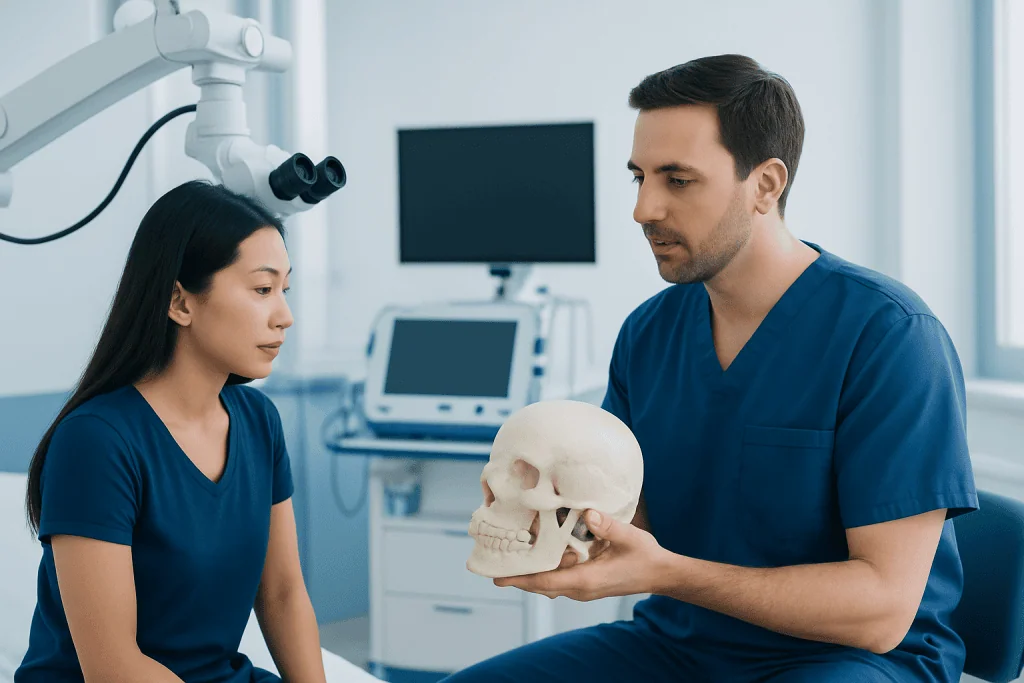
The skull is often divided into two main parts: the neurocranium and the viscerocranium. The neurocranium, also called the cranial vault and cranial base, surrounds and protects the brain. The viscerocranium forms the facial skeleton, including the eye sockets, cheekbones, nasal region, and jaws.
The 8 cranial bones are the frontal bone, two parietal bones, two temporal bones, the occipital bone, the sphenoid bone, and the ethmoid bone. These bones form the forehead, sides and roof of the skull, the skull base, and the areas that surround the ears and parts of the nasal cavity.
The 14 facial bones include the maxillae, zygomatic bones, nasal bones, lacrimal bones, palatine bones, inferior nasal conchae, vomer, and mandible. Together they support the eyes, nose, and mouth, and they help with chewing, speech, and facial appearance.
Key skull landmarks include the orbits around the eyes, the nasal cavity, the hard palate, and the foramen magnum, which is the large opening at the base of the skull where the spinal cord connects with the brain. Understanding these regions can make radiology reports and specialist consultations easier to follow.
Bones, sutures, and important openings
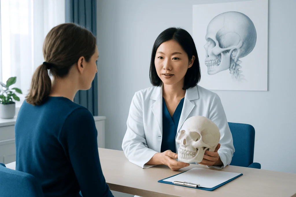
Each skull bone has a specific role. The frontal bone forms the forehead and part of the eye sockets. The parietal bones make up much of the upper sides of the skull. The temporal bones contain structures related to hearing and balance, while the occipital bone forms the back and base of the skull.
The sphenoid and ethmoid bones sit deeper within the skull and are especially important because they help form the skull base, eye sockets, and nasal cavity. The sphenoid is sometimes called the “keystone” of the skull because it connects with multiple other bones.
Sutures are the fibrous joints that connect most skull bones. Important sutures include the coronal suture between the frontal and parietal bones, the sagittal suture between the two parietal bones, the lambdoid suture between the parietal and occipital bones, and the squamous sutures between the parietal and temporal bones.
The skull also contains openings called foramina. These allow nerves and blood vessels to pass between the brain and the face, eyes, ears, and neck. Because of these pathways, a condition affecting one area of the head may sometimes cause symptoms in another, such as facial numbness, jaw pain, or visual changes.
What the skull does for the body
The skull’s best-known job is protection. It surrounds the brain and reduces the risk of injury from everyday impacts. It also protects delicate structures involved in hearing, balance, smell, and vision.
Just as important, the skull provides structural support. It creates the framework of the face, anchors muscles used for chewing and facial expression, and helps keep the airway and upper digestive tract properly shaped. The mandible, or lower jaw, is the main movable bone of the skull and is essential for speaking and eating.
The skull also contributes to normal function through its cavities and passages. The nasal cavity helps warm and filter inhaled air, while the sinuses lighten the skull and influence voice resonance. The orbits position and protect the eyes, and the temporal bone houses parts of the middle and inner ear.
Because so many structures are closely packed together in the skull, symptoms can overlap. A person may notice headache, ear symptoms, sinus pressure, jaw discomfort, or facial pain from conditions affecting nearby bones, joints, nerves, or soft tissues.
Skull development, normal variation, and related conditions
In infants, the skull is not yet fully fused. Soft spots called fontanelles and flexible sutures allow the head to pass through the birth canal and give the brain room to grow quickly during early life. Over time, these areas gradually close and harden.
There is also normal variation in skull shape between individuals. Differences in bone contour, facial proportions, sinus size, and jaw position are often part of normal human anatomy. Imaging findings may describe these variations without meaning that disease is present.
Some medical conditions can affect skull anatomy or skull function. These include fractures after trauma, sinus-related bone changes, temporomandibular joint disorders, congenital fusion of sutures, and rarely tumors or infections involving the skull. Patients may also hear related terms such as brain tumor when specialists are evaluating symptoms within the skull cavity.
Problems involving the skull base or nearby nerves can sometimes lead to complex symptoms such as hearing changes, dizziness, facial weakness, or double vision. In these situations, careful examination and imaging are important to identify whether the cause lies in the bones, joints, sinuses, nerves, or brain.
How doctors evaluate skull-related symptoms
Doctors begin with a medical history and physical examination. They ask about injury, headache, sinus symptoms, jaw pain, dental problems, hearing changes, vision changes, and any neurological symptoms such as weakness, numbness, or confusion. The examination may include checking the scalp, face, jaw movement, eye function, balance, and cranial nerves.
Imaging is often used when more detail is needed. CT scans are especially helpful for showing skull bones and are commonly used after trauma or when fractures are suspected. MRI is better for viewing the brain, nerves, soft tissues, and some skull-base problems. If a patient has severe head pain or neurological warning signs, urgent assessment may be needed to rule out conditions such as brain hemorrhage.
In selected cases, doctors may also request dental imaging, sinus evaluation, hearing tests, or specialist assessment by neurology, neurosurgery, ENT, maxillofacial surgery, or ophthalmology. The right test depends on the symptom pattern rather than on the skull alone.
When treatment planning requires detailed review of the brain or skull structures, advanced imaging may be part of the pathway, including MRI scanning or CT scanning. These tests help clarify whether symptoms relate to bone injury, sinus disease, a nerve problem, or another condition.
Treatment and self-care depend on the cause
There is no single treatment for “skull anatomy” because anatomy itself is not a disease. Care focuses on the underlying issue affecting the head or face. Minor soft-tissue injury may only need observation and symptom relief, while fractures, infections, jaw disorders, or neurological conditions may require specialist treatment.
For head trauma, doctors may recommend observation, imaging, and activity precautions. For sinus or nasal causes of facial pressure, treatment may include hydration, saline rinses, medicines, or ENT care. For jaw-related pain, approaches can include diet changes, stress reduction, dental advice, or targeted therapy for the temporomandibular joint.
If a structural problem needs more advanced management, treatment can involve several specialties working together. Depending on the diagnosis, this may include neurosurgical care or other procedures to address the brain, skull base, or nearby tissues.
General self-care supports recovery and head health. It can help to wear appropriate protective headgear during sports or work, use seat belts, manage chronic sinus or dental issues promptly, and seek medical advice rather than self-diagnosing ongoing head or facial symptoms.
When to seek medical care
Medical evaluation is important after a significant head injury, especially if there is loss of consciousness, repeated vomiting, worsening headache, confusion, seizure, weakness, unequal pupils, or fluid leaking from the nose or ears. These symptoms do not always mean a serious injury, but they should be assessed without delay.
Patients should also arrange medical care for persistent facial pain, swelling, jaw locking, unexplained vision changes, new hearing loss, frequent nosebleeds, or headaches that are unusual for them. Symptoms that continue, worsen, or affect daily life deserve a professional evaluation.
Children and infants need special attention because skull development is still ongoing. Parents should seek care for an unusual head shape, delayed closure concerns, swelling after injury, or behavior changes after a fall. A clinician can determine whether findings are part of normal development or need further tests.
For international patients who need coordinated assessment, Acibadem International’s multidisciplinary specialists and JCI-accredited hospitals diagnose and treat skull- and brain-related conditions using modern imaging and specialist care pathways.
Frequently asked questions
How many bones are in the human skull?
The adult skull is usually described as having 22 bones. These include 8 cranial bones that protect the brain and 14 facial bones that form the structure of the face.
What is the difference between cranial bones and facial bones?
Cranial bones form the protective case around the brain. Facial bones shape the face and support the eyes, nose, mouth, and jaws.
Are all skull bones fused together?
Most skull bones are joined by fixed fibrous joints called sutures. The main exception is the mandible, or lower jaw, which moves to allow chewing and speaking.
Why do doctors order a CT scan for skull problems?
CT scans show bone detail very clearly, so they are especially useful when a fracture or other bony abnormality is suspected. They are commonly used after head injury and in some sinus or skull-base evaluations.
Can skull anatomy cause headaches?
The skull itself is not usually the direct cause of common headaches, but nearby structures can contribute. Sinus disease, jaw disorders, trauma, and some neurological conditions may lead to head or facial pain.
What are fontanelles in babies?
Fontanelles are the soft spots between skull bones in infants. They are a normal part of development and allow the brain and skull to grow during early life.
When is a head injury an emergency?
Urgent medical care is needed if a head injury is followed by loss of consciousness, repeated vomiting, confusion, seizure, worsening headache, weakness, or fluid leaking from the nose or ear. These signs may suggest a more serious injury and should not be ignored.
This article is for general information only and is not a substitute for professional medical advice. Please consult a qualified doctor about your individual situation.
Explore treatments in Turkey — costs, top hospitals & a free quote
JCI-accredited · board-certified surgeons · reply within 24h
Add us as a Preferred Source to see more of our trusted health content across Google Search, AI Overviews and Discover.

More from the Health Library

Kathryn L Newton: An Evidence-Based Guide for Patients

Sciatica Pillow — Explained by Medical Evidence, Not Myths

Inspiresleep Com Cost: A Complete Medical Overview

Meat Sweats: A Complete Medical Overview

Lispro Insulin: A Complete Medical Overview