Subfalcine Herniation: A Complete Medical Overview

Subfalcine herniation happens when pressure inside the skull pushes part of one cerebral hemisphere under the falx cerebri. It is usually caused by a mass, bleeding, swelling, infection, or other condition that raises intracranial pressure.
Key Takeaways
- Subfalcine herniation happens when pressure inside the skull pushes part of one cerebral hemisphere under the falx cerebri.
- It is usually caused by a mass, bleeding, swelling, infection, or other condition that raises intracranial pressure.
- Early symptoms can be subtle, but worsening drowsiness, severe headache, vomiting, seizures, weakness, or unequal pupils need emergency assessment.
- CT scanning is commonly used urgently to identify bleeding, swelling, masses, hydrocephalus, and midline shift.
- Treatment focuses on stabilizing the person, lowering pressure in the skull, and treating the underlying cause, sometimes with emergency surgery.
Subfalcine herniation is a serious form of brain herniation in which swollen or displaced brain tissue moves beneath the falx cerebri, a firm membrane between the brain’s two hemispheres. It is a medical emergency because the shift can compress vital blood vessels and may progress to more dangerous forms of herniation without prompt treatment.
Overview: What Is Subfalcine Herniation?
Subfalcine herniation is a type of brain herniation, meaning that brain tissue has shifted from its usual position because pressure has increased inside the rigid skull. In this form, the cingulate gyrus, located on the inner surface of a cerebral hemisphere, is pushed beneath the falx cerebri. The falx is a strong fold of tissue that separates the right and left sides of the brain.
The condition is also called cingulate herniation. It commonly occurs when a problem on one side of the brain creates a pressure difference and pushes tissue toward the opposite side. Imaging reports may describe related findings such as midline shift, which means that central brain structures have been displaced from their normal position.
Subfalcine herniation is not a diagnosis on its own. It is a complication of another brain condition, such as bleeding, severe swelling, a tumor, or infection. Because ongoing pressure can reduce blood flow to brain tissue and can lead to further herniation, it requires urgent assessment in a hospital.
Why a Brain Shift Can Be Dangerous
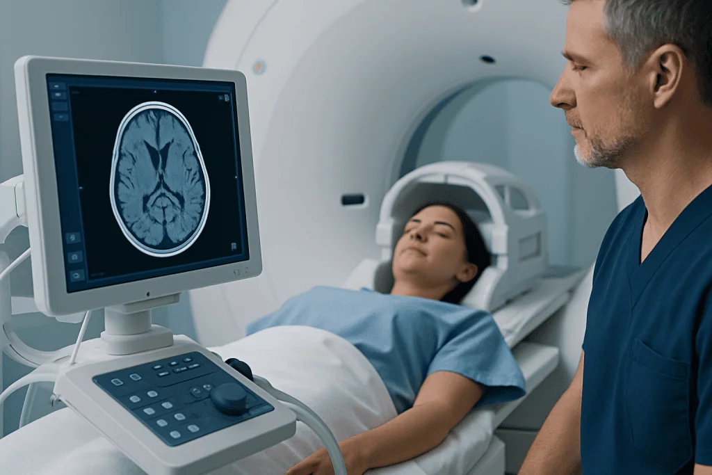
The skull provides strong protection for the brain, but it cannot expand when its contents increase in volume. The brain, blood, and cerebrospinal fluid normally coexist within this limited space. A bleed, mass, swelling, or blockage of fluid flow can disturb this balance and increase intracranial pressure.
As pressure rises, brain tissue may move across natural internal boundaries. In subfalcine herniation, the displaced tissue may compress the anterior cerebral artery or its branches. This can reduce blood supply to areas of the brain that help control leg movement, sensation, attention, and other functions.
The seriousness depends on the cause, the speed of pressure change, the amount of displacement, and whether other areas of the brain are affected. Some people have signs related mainly to the original illness, while others deteriorate rapidly as pressure rises. Careful neurological monitoring and repeat imaging may be needed because the situation can change over hours or even minutes.
Symptoms and Warning Signs

Symptoms of subfalcine herniation vary widely because they are often caused by both the underlying problem and the increased pressure in the skull. A person may initially have headache, nausea, vomiting, confusion, unusual sleepiness, irritability, slowed thinking, or a change in behavior. Following a head injury, any decline after an apparently stable period should be taken seriously.
Neurological symptoms can include weakness or numbness, often affecting the leg more than the arm, difficulty walking, problems speaking, loss of coordination, seizures, or reduced alertness. In a more severely unwell person, clinicians may find abnormal reflexes, changes in pupil response, or signs of impaired brain function during a neurological examination.
Not every person with a headache, vomiting, or confusion has increased intracranial pressure. However, a sudden severe headache, a new seizure, fainting, progressive drowsiness, repeated vomiting, new weakness, or confusion after head trauma should never be managed with home observation alone. Emergency evaluation is especially important when symptoms are worsening.
Causes and Risk Factors
Any condition that adds volume within the skull or causes extensive brain swelling can lead to subfalcine herniation. A major cause is intracranial bleeding, including bleeding after a traumatic head injury. Different types of hemorrhage, such as subdural, epidural, intracerebral, or subarachnoid bleeding, can produce pressure depending on their size and location.
Other causes include a large ischemic stroke with swelling, a brain tumor, a brain abscess, severe infection affecting the brain, and hydrocephalus, in which cerebrospinal fluid accumulates. Brain tumors can cause symptoms gradually as they enlarge, whereas bleeding or some strokes may cause pressure to rise much more quickly.
Risk factors depend on the underlying cause. Falls, road traffic injuries, contact sports injuries, blood-thinning medicines, uncontrolled high blood pressure, vascular abnormalities, cancer, and immune suppression may increase the likelihood of conditions that can raise intracranial pressure. These factors do not mean herniation will occur, but they can influence how urgently clinicians investigate concerning symptoms.
- Head trauma with a change in consciousness or repeated vomiting
- Use of anticoagulant or antiplatelet medicines after a head injury
- Known brain lesion with new neurological symptoms
- Recent stroke symptoms followed by worsening drowsiness or weakness
- Fever, severe headache, and confusion, particularly in people with weakened immunity
How Doctors Diagnose It
Subfalcine herniation is diagnosed through urgent clinical assessment and brain imaging. Clinicians first focus on airway, breathing, circulation, level of consciousness, pupil responses, limb strength, and other neurological signs. They also ask about the timing of symptoms, recent injury, medicines, prior stroke, cancer history, infections, and seizure activity when this is possible.
A non-contrast CT scan of the head is usually the first imaging test in an emergency because it is fast and can identify many forms of bleeding, swelling, fluid buildup, fractures, and major brain shift. CT findings may show compression of spaces around the brain, displacement of the midline, and the lesion responsible for the pressure.
MRI may provide more detailed information once the person is stable or when the cause is less clear. Blood tests can help identify clotting problems, infection, metabolic disturbances, and medication-related risks. Imaging and examinations are repeated when needed to assess whether swelling or bleeding is improving, stable, or progressing.
Treatment Options and Hospital Care
Treatment is urgent and is tailored to the cause of raised intracranial pressure. The immediate priorities are to support oxygenation and blood pressure, prevent further brain injury, manage seizures if present, and closely monitor neurological status. People with suspected or confirmed herniation generally need emergency care in a hospital, often with intensive care and neurosurgical involvement.
Doctors may use carefully selected measures to reduce intracranial pressure, such as elevating the head of the bed, providing appropriate pain control and sedation when required, and giving medicines that draw fluid out of swollen brain tissue. If hydrocephalus is contributing to pressure, a neurosurgeon may place a drain to remove cerebrospinal fluid in selected situations. The exact approach depends on imaging findings and the person’s overall condition.
Surgery may be necessary to remove a blood clot, drain an abscess, remove or reduce a mass, or create more space for swollen brain tissue. Craniotomy may be used when surgeons need access to remove a lesion or relieve pressure. In selected cases of severe swelling, decompressive craniectomy can create temporary extra space by removing part of the skull.
Recovery depends primarily on the cause, the severity and duration of pressure, the area of the brain affected, and how quickly treatment begins. Rehabilitation may be recommended for ongoing difficulties with movement, speech, thinking, daily activities, or emotional adjustment after the acute phase.
Prevention, Follow-Up and Recovery Support
Not all causes of subfalcine herniation can be prevented, particularly sudden bleeding or rapidly evolving swelling. However, reducing the likelihood of head injury is an important step. Seat belts, appropriate helmets, fall-prevention measures, and safe work and sports practices can lower the risk of traumatic brain injury.
People taking blood-thinning medication should seek medical advice promptly after a significant head injury, even if they initially feel well. They should not stop prescribed medicines without guidance, but should ensure that clinicians know all medications they take. Managing blood pressure, diabetes, and other vascular risk factors can also support stroke prevention.
After hospital treatment, follow-up may involve neurosurgery, neurology, rehabilitation medicine, physiotherapy, occupational therapy, speech and language therapy, or psychology. Families can help by reporting new symptoms, attending follow-up appointments, and supporting a gradual return to usual activities according to the medical team’s recommendations.
Acibadem International’s multidisciplinary specialists and JCI-accredited hospitals evaluate and treat complex neurological and neurosurgical conditions for international patients, with care plans based on the underlying cause and clinical urgency.
When to Seek Medical Care
Call emergency services immediately for a person who becomes unconscious, has a seizure, develops sudden weakness or facial drooping, has difficulty speaking, experiences a sudden severe headache, or shows confusion and declining alertness. Urgent help is also needed after a head injury when there is repeated vomiting, worsening headache, unusual behavior, unequal pupils, weakness, or increasing sleepiness.
A person with a known brain tumor, previous stroke, recent neurosurgery, or an infection affecting the nervous system should be assessed urgently if new neurological symptoms appear or existing symptoms worsen. It is safer not to drive the person to hospital if they are drowsy, confused, or having a seizure; emergency transport allows monitoring and treatment on the way.
Subfalcine herniation cannot be confirmed or ruled out by symptoms alone. It requires prompt examination and imaging by qualified medical professionals. Early assessment is the safest response whenever raised intracranial pressure is a concern.
Frequently asked questions
Is subfalcine herniation the same as a brain tumor?
No. Subfalcine herniation is a shift of brain tissue caused by increased pressure inside the skull. A brain tumor is one possible cause of that increased pressure, but bleeding, stroke-related swelling, infection, and fluid buildup can also lead to herniation.
Can subfalcine herniation be treated?
Yes, treatment is possible, but it must be provided urgently in hospital. Doctors work to lower pressure in the skull and treat the cause, which may involve medication, drainage procedures, or surgery.
What does midline shift mean on a brain scan?
Midline shift means that central brain structures have moved away from their normal position, usually because a mass, bleed, or swelling is creating pressure on one side. It is an important imaging sign that helps doctors assess urgency and plan treatment.
Can a person be awake with subfalcine herniation?
Yes. A person may be awake, especially early in the process, and symptoms may range from headache or confusion to weakness or seizures. However, alertness can worsen as pressure increases, so urgent medical evaluation is needed.
Does a head injury always cause brain herniation?
No. Most head injuries do not cause brain herniation. However, serious head injuries can cause bleeding or swelling inside the skull, and symptoms such as worsening headache, vomiting, confusion, seizure, or drowsiness require immediate assessment.
What is the outlook after subfalcine herniation?
The outlook varies substantially and depends on the cause, speed of diagnosis, severity of brain pressure, and response to treatment. Some people recover well, while others need longer-term rehabilitation or may have lasting neurological effects. The treating team can give the most relevant information based on imaging and clinical progress.
References
- National Institute of Neurological Disorders and Stroke
- American Association of Neurological Surgeons
- Merck Manual Professional Edition
- Radiopaedia
- World Health Organization
This article is for general information only and is not a substitute for professional medical advice. Please consult a qualified doctor about your individual situation.
Joint and spine care in Turkey — expert assessment & treatment
JCI-accredited · board-certified surgeons · reply within 24h
Add us as a Preferred Source to see more of our trusted health content across Google Search, AI Overviews and Discover.
Check your numbers in seconds
BMI, calories, due date, blood pressure and 30+ more clinical calculators — free, instant, doctor-reviewed ranges.