Susac MRI: Preparation, Procedure and Results
Susac MRI usually refers to a brain MRI performed with detailed sequences and often gadolinium contrast. MRI can show lesions in the corpus callosum and other brain areas that may help distinguish Susac syndrome from similar conditions.
Key Takeaways
- Susac MRI usually refers to a brain MRI performed with detailed sequences and often gadolinium contrast.
- MRI can show lesions in the corpus callosum and other brain areas that may help distinguish Susac syndrome from similar conditions.
- Preparation is usually simple, but metal implants, kidney disease, pregnancy and prior contrast reactions should be discussed in advance.
- The scan itself commonly takes 30 to 60 minutes, while the report may require specialist review in the clinical context.
- A normal or non-specific MRI does not by itself rule Susac syndrome in or out; repeat imaging may sometimes be needed.
Susac MRI is an important imaging examination used to look for characteristic changes in the brain associated with Susac syndrome. It is painless and noninvasive, and its findings are interpreted alongside eye, hearing and laboratory assessments rather than used alone.
Overview: What Is a Susac MRI?
A Susac MRI is a magnetic resonance imaging scan of the brain performed when clinicians are assessing or monitoring Susac syndrome. Susac syndrome is a rare immune-mediated condition that affects small blood vessels, most notably in the brain, retina and inner ear. MRI uses a powerful magnet and radio waves rather than ionising radiation to create detailed images of brain tissue.
In Susac syndrome, MRI may reveal a recognisable distribution of tiny areas of inflammation or injury, including changes in the corpus callosum, the band of nerve fibres connecting the two sides of the brain. The scan is usually interpreted by a neuroradiologist together with a neurologist, since findings need to be matched with symptoms, eye testing and hearing evaluation.
The purpose of a Susac MRI may be to support an initial diagnosis, exclude other possible explanations, assess disease activity, or monitor response to treatment. It is one part of a wider assessment for Susac syndrome, not a stand-alone test.
Why MRI Is Used and Who May Need It
A doctor may request a Susac MRI for a person with new neurological symptoms, unexplained visual disturbances, hearing changes, or a combination of these concerns. Symptoms can vary considerably. Some people experience headache, confusion, slowed thinking, balance difficulties or changes in mood, while others first notice a patch of missing vision or reduced hearing in one ear.
Not everyone with Susac syndrome develops the full set of brain, eye and ear features at the same time. This is one reason why MRI is valuable: it can provide evidence of central nervous system involvement even when symptoms are incomplete or evolving. However, similar MRI changes can occur in other inflammatory, vascular or infectious conditions, so careful clinical assessment remains essential.
Brain MRI is also often repeated during follow-up. Comparing new images with earlier scans can help the clinical team judge whether there are new lesions or whether previously active changes appear to be settling. The timing of follow-up scans is individual and depends on symptoms, examination findings and the treatment plan.
How a Susac MRI Works

An MRI scanner contains a strong magnet that aligns particles in the body’s tissues. Radiofrequency pulses briefly alter this alignment, and the scanner detects the signals released as tissues return to their usual state. A computer converts these signals into highly detailed images in multiple planes.
For suspected Susac syndrome, the imaging protocol commonly includes several sequences that show fluid, inflammation, old injury and recent changes in different ways. Diffusion-weighted imaging can help identify areas of recent restricted blood flow, while fluid-sensitive sequences may show lesions in the brain’s white matter or corpus callosum. The exact protocol is selected by the radiology team and may be adapted to the person’s history.
Many examinations include intravenous gadolinium-based contrast. Contrast can highlight areas where the blood-brain barrier is disrupted or inflammation is active. It is not always necessary, but it may add useful information, especially in an initial assessment or when active disease is suspected. MRI does not use X-rays, although contrast and the magnetic field still require important safety checks.
Preparation and Step-by-Step Procedure
Before the appointment, the person should tell the imaging team about any implanted device, metal fragment, surgical clip, cochlear implant, insulin pump, medication patch, pregnancy, kidney disease or previous reaction to MRI contrast. Many implants are MRI-compatible, but they must be checked carefully. The team will provide specific instructions if fasting is needed; for many routine brain MRI scans, it is not required.
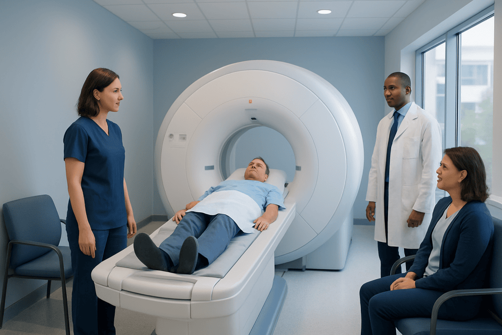
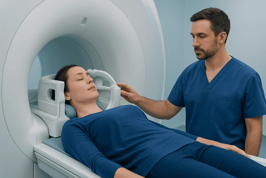
At the imaging centre, the patient completes a safety questionnaire and removes metal items such as jewellery, watches, hearing aids, hairpins and removable dental appliances. If contrast is planned, a small cannula is placed in a vein, usually in the arm. The patient lies on a padded table with a lightweight head coil positioned around the head to improve image quality.
The table then moves into the scanner. The machine makes rhythmic tapping and knocking sounds, so earplugs or headphones are provided. Remaining as still as possible is important because movement can blur images. Staff can see, hear and speak to the patient throughout the scan, and an alarm button is available if help is needed.
A typical Susac MRI takes around 30 to 60 minutes, depending on the sequences required and whether contrast is used. If contrast is given, it is usually injected partway through the examination. People with claustrophobia should mention this beforehand; the team can discuss practical comfort measures and, when appropriate, clinician-prescribed options for anxiety management.
Understanding Susac MRI Results
The radiology report describes whether there are lesions, where they are located, how they appear on different sequences and whether any lesions enhance after contrast. In Susac syndrome, radiologists may identify small round lesions in the central portion of the corpus callosum. Other changes may be present in white matter, deep grey matter or other brain structures.
These patterns can be helpful, but the report does not confirm the diagnosis by itself. The care team considers MRI results alongside an eye examination, retinal imaging or fluorescein angiography, hearing tests, neurological examination and selected blood or cerebrospinal fluid tests. This combined approach helps distinguish Susac syndrome from conditions such as multiple sclerosis, stroke-related disease, infection and other inflammatory disorders.
Results are often available after the images have been formally reviewed, which may take from the same day to several days depending on the setting. The referring clinician should explain what the findings mean for that individual. If the MRI shows no clear abnormalities but clinical suspicion remains, repeat imaging or additional specialist testing may be considered.
When imaging and other assessments support active Susac syndrome, treatment aims to control inflammation promptly and prevent further damage to the brain, eyes and hearing. Care may involve neurologists, neuro-ophthalmologists, otologists or audiologists, rheumatologists and radiologists. Neurology assessment and treatment planning can coordinate these different aspects of care.
Benefits, Risks and Recovery After the Scan
The main benefit of Susac MRI is its detailed view of brain structures without radiation exposure. It can help identify features that guide diagnosis, establish a baseline for future comparison and monitor whether disease activity may be continuing. It may also help clinicians identify alternative causes of neurological symptoms that need a different treatment approach.
MRI is generally very safe when the pre-scan safety screening is followed. The principal concerns are related to the magnetic field, which can affect certain implanted devices or move unsafe metal objects, and to contrast in selected patients. Gadolinium contrast reactions are uncommon and are usually mild, but urgent care is available if a reaction occurs. People with severely reduced kidney function require individual assessment before certain contrast agents are used.
There is no physical recovery period after a standard MRI. Most people can eat, drink, work and travel as usual immediately afterwards. If a sedative has been used for anxiety or claustrophobia, the person should follow local instructions, avoid driving and arrange an escort home.
The scan can feel challenging for people who dislike enclosed spaces or are sensitive to noise. Asking about music, communication with staff, a mirror system or an open-style scanner where available may make the experience more manageable. It is appropriate to raise any concern before the scan rather than trying to tolerate significant distress alone.
When to Seek Medical Care
Prompt medical assessment is advisable for new or worsening neurological symptoms, particularly sudden visual loss or visual field changes, new hearing loss, severe imbalance, confusion, weakness, speech difficulty or a severe unusual headache. These symptoms can have several causes, including conditions that require urgent evaluation, so they should not be assumed to be Susac syndrome.
Emergency services should be contacted immediately for symptoms that may indicate a stroke, such as sudden facial drooping, arm weakness, difficulty speaking, sudden loss of vision, severe loss of coordination or a sudden severe headache. Even if symptoms improve quickly, urgent assessment is important.
People already being monitored for Susac syndrome should contact their treating team if there are new visual, hearing or neurological changes between scheduled visits. Acibadem International’s multidisciplinary specialists and JCI-accredited hospitals can assess and treat neurological and neuro-ophthalmological conditions for international patients.
Frequently asked questions
Does a Susac MRI always require contrast?
No. A brain MRI can provide useful information without contrast, but contrast is often considered because it may show active inflammation or changes in the blood-brain barrier. The decision depends on the clinical question, prior imaging, kidney function and MRI safety assessment.
What does Susac syndrome look like on MRI?
A characteristic finding can be small, round lesions in the central fibres of the corpus callosum. There may also be lesions in other white-matter or deeper brain regions. MRI appearances vary between individuals and stages of disease, so a specialist must interpret them with other test results.
How long does a Susac MRI take?
The scan commonly takes about 30 to 60 minutes. The precise time depends on whether contrast is used, how many image sequences are needed and whether additional areas are scanned.
Can MRI diagnose Susac syndrome on its own?
No. MRI may strongly support the diagnosis, especially when characteristic features are present, but it cannot diagnose Susac syndrome in isolation. Doctors also evaluate retinal blood vessels, hearing, symptoms, examination findings and selected laboratory tests.
Is a Susac MRI painful?
MRI itself is not painful, although lying still and hearing the scanner’s loud noises can be uncomfortable. If contrast is used, there may be a brief needle prick when the intravenous line is placed. Staff provide hearing protection and can communicate throughout the examination.
Can a normal MRI rule out Susac syndrome?
A normal MRI does not always completely exclude Susac syndrome, particularly early in the condition or when symptoms mainly involve the eyes or ears. If clinical concern remains, a specialist may recommend repeat MRI or further eye and hearing assessments.
References
- National Organization for Rare Disorders
- National Institute of Neurological Disorders and Stroke
- Radiological Society of North America
- American College of Radiology
- Orphanet
This article is for general information only and is not a substitute for professional medical advice. Please consult a qualified doctor about your individual situation.
Explore treatments in Turkey — costs, top hospitals & a free quote
JCI-accredited · board-certified surgeons · reply within 24h
Add us as a Preferred Source to see more of our trusted health content across Google Search, AI Overviews and Discover.