Synovial Cyst — Explained by Medical Evidence, Not Myths
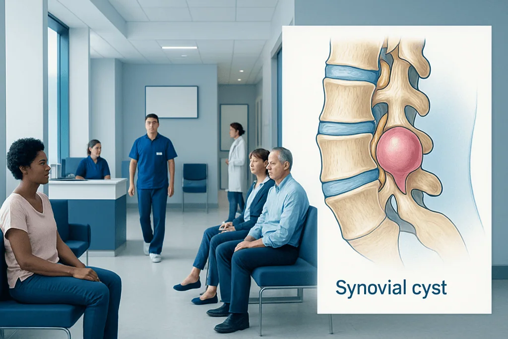
A synovial cyst usually develops from a worn or irritated spinal facet joint. Many synovial cysts do not cause symptoms and may only be found on imaging.
Key Takeaways
- A synovial cyst usually develops from a worn or irritated spinal facet joint.
- Many synovial cysts do not cause symptoms and may only be found on imaging.
- Symptoms happen when the cyst narrows space around nearby nerves.
- MRI is commonly used to confirm the diagnosis and assess nerve compression.
- Treatment ranges from observation and pain management to injections or surgery, depending on symptoms and nerve involvement.
A synovial cyst is a fluid-filled sac that forms from the lining of a joint, most often in the spine. It may cause no symptoms, but if it presses on nearby nerves, it can lead to back pain, leg pain, numbness, or weakness that often improves with appropriate evaluation and treatment.
Overview: what a synovial cyst is
A synovial cyst is a small, fluid-filled sac that forms from the lining of a joint. In everyday practice, the term most often refers to a cyst arising from the facet joints of the spine, especially in the lower back. These joints contain synovial fluid, which helps the joint move smoothly. Over time, joint wear, inflammation, or instability can allow the lining to bulge outward and form a cyst.
Medical evidence shows that a synovial cyst is usually a degenerative, not a cancerous, problem. In other words, it is commonly linked to age-related changes in the spine rather than infection or tumor. Many people have no symptoms at all, and the cyst may be discovered incidentally during imaging for another reason.
Symptoms appear when the cyst takes up space in the spinal canal or near the opening where nerves travel. This can irritate or compress nerve tissue, causing pain or neurologic symptoms. Understanding this mechanical effect helps explain why some cysts can simply be watched, while others need more active treatment.
Symptoms and how they may feel

The symptoms of a synovial cyst depend mainly on its size, location, and whether it presses on a nerve. A small cyst may cause no problems. A larger spinal synovial cyst, especially in the lumbar spine, can produce lower back pain or pain that travels into the buttock or leg.
When a nearby nerve root is compressed, symptoms may resemble sciatica. A person may notice burning pain, tingling, numbness, or weakness in one leg. Some people describe symptoms that worsen when standing or walking and improve when sitting or bending slightly forward, because body position can change pressure within the spinal canal.
Less commonly, a synovial cyst can contribute to symptoms of spinal stenosis, such as leg heaviness, cramping, or reduced walking distance. Warning signs of more urgent nerve involvement include increasing weakness, difficulty controlling the bladder or bowels, or numbness around the groin area. These symptoms need prompt medical assessment.
- Lower back pain
- Pain radiating into the buttock or leg
- Numbness or tingling
- Leg weakness
- Symptoms that worsen with standing or walking
Why synovial cysts develop: causes and risk factors

Synovial cysts are most strongly associated with degenerative changes in the spine. The facet joints can become arthritic over time, and this wear may lead to excess joint fluid and outward bulging of the synovial lining. For that reason, synovial cysts are seen more often in middle-aged and older adults.
They commonly occur in the lower lumbar spine, where motion and mechanical stress are greatest. Conditions such as facet joint arthritis, spinal instability, and degenerative spondylolisthesis may increase the likelihood of a cyst forming. These problems can exist alongside spinal stenosis or other age-related spinal changes.
It is important to separate evidence from myth. A synovial cyst is not usually caused by a single exercise session, minor movement, or poor posture alone. While strain may make existing symptoms more noticeable, the underlying process is generally chronic joint degeneration rather than one isolated event.
How doctors diagnose a synovial cyst
Diagnosis starts with a careful history and physical examination. A doctor asks where the pain is, whether it travels into the leg, and whether there are signs of numbness, weakness, balance change, or bowel and bladder problems. The examination may include checking strength, sensation, reflexes, and walking pattern.
MRI is often the most helpful imaging test because it shows soft tissues clearly, including the cyst and any pressure on nerves. It can also reveal related findings such as disc degeneration, facet joint arthritis, or narrowing of the spinal canal. In some situations, CT scans, X-rays, or other imaging may be used to evaluate bony changes or spinal alignment.
Because other conditions can cause similar symptoms, doctors may consider a differential diagnosis that includes a herniated disc, nerve root inflammation, or herniated disc. The goal is not simply to identify a cyst on a scan, but to decide whether that cyst actually explains the person’s symptoms.
Treatment options: from observation to surgery
Treatment depends on symptom severity, the degree of nerve compression, and how much symptoms affect daily life. If a synovial cyst is found incidentally and causes no symptoms, observation may be appropriate. For mild to moderate symptoms, doctors often begin with non-surgical care such as activity modification, physical therapy, and pain-relief strategies.
When inflammation around the nerve is contributing to pain, image-guided injections may be considered in selected cases. These can sometimes reduce irritation and help clarify whether symptoms are coming from that area. Some patients may also be offered minimally invasive procedures, depending on the cyst’s location and the overall condition of the spine. Relevant options may be discussed as part of spine surgery planning or broader pain management care.
Surgery may be recommended if there is persistent pain, progressive weakness, significant walking limitation, or failure of conservative treatment. The operation usually aims to remove the cyst and relieve pressure on the nerve. In some cases, if there is spinal instability, decompression may be combined with a stabilizing procedure such as spinal fusion. The best approach is individualized after imaging review and specialist assessment.
It is helpful to know that surgery is not automatically required simply because a cyst is seen on MRI. Decisions are based on symptoms, nerve findings, and functional impact rather than imaging alone.
Self-care, daily activity, and recovery support
Self-care does not remove a synovial cyst, but it can reduce discomfort and support function. Many people do better by avoiding activities that clearly worsen leg pain, especially prolonged standing, repeated twisting, or heavy lifting during symptom flares. Gentle walking and clinician-guided exercise may help maintain mobility without overloading the spine.
Physical therapy often focuses on posture, core support, flexibility, and movement patterns that reduce mechanical stress. Heat or cold may provide short-term relief for some people, though responses vary. Over-the-counter medicines may be used when appropriate, but they should be taken according to a clinician’s or pharmacist’s advice, especially in people with stomach, kidney, heart, or bleeding concerns.
If treatment is needed, recovery depends on the chosen approach. After an injection or procedure, doctors usually recommend a gradual return to activity. Following surgery, a structured rehabilitation plan can support healing, improve movement confidence, and help protect the spine during recovery.
When to seek medical care
Medical review is appropriate if back pain spreads into the leg, if numbness or tingling persists, or if symptoms begin to interfere with walking, sleep, work, or daily activities. An evaluation is also sensible when symptoms continue despite rest and simple self-care, or when pain keeps returning.
Urgent medical attention is needed for rapidly worsening weakness, new trouble walking, loss of bladder or bowel control, or numbness in the saddle area. These signs can indicate significant nerve compression and should not be ignored.
For people seeking specialist assessment, multidisciplinary teams can help match treatment to symptoms and imaging findings. Acibadem International’s multidisciplinary specialists and JCI-accredited hospitals diagnose and treat spinal conditions, including synovial cysts, for international patients.
Frequently asked questions
Is a synovial cyst serious?
A synovial cyst is often not serious if it causes no symptoms. It becomes clinically important when it presses on nearby nerves and leads to pain, numbness, or weakness. The level of concern depends more on symptoms and neurologic findings than on the name of the cyst itself.
Can a synovial cyst go away on its own?
Some synovial cysts remain stable for long periods, and symptoms may improve without surgery. In certain cases, the cyst may shrink or become less symptomatic over time. However, not all cysts resolve, so ongoing symptoms should be reviewed by a doctor.
What is the difference between a synovial cyst and a ganglion cyst?
Both are fluid-filled sacs related to joints or tendon structures, and the terms are sometimes used closely in practice. A synovial cyst is lined by synovial tissue and is often linked to a joint space, while a ganglion cyst is usually a mucin-filled sac without a true synovial lining. In the spine, doctors focus mainly on location, symptoms, and nerve compression rather than the label alone.
Does every synovial cyst need surgery?
No. Many people can be managed with observation, physical therapy, activity changes, or other non-surgical measures, especially if symptoms are mild and there is no significant weakness. Surgery is usually considered when pain persists, function declines, or nerve compression causes more serious problems.
How is a synovial cyst confirmed?
MRI is commonly the best test because it shows the cyst and nearby nerves clearly. Diagnosis also depends on a physical examination and whether the imaging findings match the person’s symptoms. A cyst seen on a scan is not always the true cause of pain.
Can exercise make a synovial cyst worse?
Not usually in a long-term structural sense, but certain movements may aggravate symptoms if the cyst is already irritating a nerve. High-load twisting, repeated extension, or heavy lifting can be uncomfortable during a flare. A clinician or physical therapist can suggest safer activity patterns based on the person’s symptoms.
References
- National Institute of Arthritis and Musculoskeletal and Skin Diseases
- American Academy of Orthopaedic Surgeons
- North American Spine Society
- National Institute of Neurological Disorders and Stroke
This article is for general information only and is not a substitute for professional medical advice. Please consult a qualified doctor about your individual situation.
Joint and spine care in Turkey — expert assessment & treatment
JCI-accredited · board-certified surgeons · reply within 24h
Add us as a Preferred Source to see more of our trusted health content across Google Search, AI Overviews and Discover.

More from the Health Library

Methylprednisolone: A Complete Medical Overview

Tendon: What Patients Need to Know

Promethazine Dm Syrup: A Complete Medical Overview

Sulfites — Explained by Medical Evidence, Not Myths

Overbite — Explained by Medical Evidence, Not Myths