Tethered Cord Ultrasound: Preparation, Procedure and Results

Tethered cord ultrasound is most useful in early infancy, before the spinal bones fully harden. The examination is painless, does not use radiation and usually does not require sedation.
Key Takeaways
- Tethered cord ultrasound is most useful in early infancy, before the spinal bones fully harden.
- The examination is painless, does not use radiation and usually does not require sedation.
- Ultrasound may show a low-lying conus, a thickened filum terminale or reduced movement of the spinal cord.
- An abnormal or unclear ultrasound result is commonly followed by MRI and assessment by a pediatric neurosurgery team.
- Tethered cord release surgery is considered when symptoms, spinal cord tension or progressive neurological concerns are present.
Tethered cord ultrasound is a non-invasive imaging test that uses sound waves to examine the lower spinal cord, usually in newborns and young infants. It can help identify features that suggest a tethered spinal cord, but MRI may be needed to confirm the diagnosis and guide care.
Overview: what is tethered cord ultrasound?
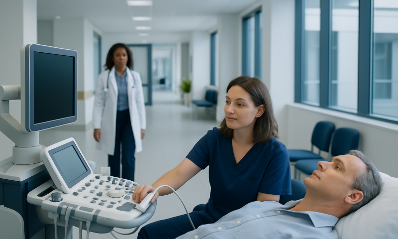
A tethered cord ultrasound is a painless scan of the lower spine that uses high-frequency sound waves to view the spinal cord and nearby structures. It is most often performed in newborns and young babies, when the bones of the spine have not yet fully hardened and still allow clear ultrasound images. The test does not use ionizing radiation and usually takes only a short time.
The purpose of a tethered cord spinal ultrasound is to look for signs that the spinal cord may be held lower than expected or attached in a way that limits its normal movement. This is called tethered cord syndrome when the tension contributes to neurological, bladder, bowel, orthopedic or pain-related symptoms. Ultrasound is a screening and assessment tool; it cannot always establish the diagnosis on its own.
A clinician may request the scan after noticing a skin marker over a baby’s lower back, such as a deep or unusual dimple, a fatty lump, a patch of hair, a birthmark or a split in the skin. It may also be used when there is concern about a spinal condition, including spina bifida, or when a child has symptoms that could involve the spinal cord.
How the scan works and who may need it
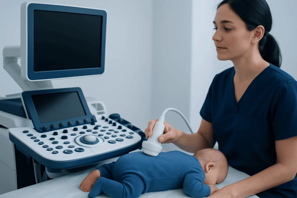
Ultrasound works by sending sound waves through a small handheld device called a transducer. The waves reflect differently from tissues and fluid, allowing a computer to create live images. In infant spinal ultrasound, the specialist places the transducer over the lower back and observes the spinal cord in different positions.
The test is generally most informative in infants younger than about 3 to 4 months, although the useful age range varies with the child’s anatomy and the imaging question. As the vertebral bones mature, they block sound waves more effectively. In older infants, children and adults, MRI is usually the preferred examination for suspected tethered cord syndrome.
Possible reasons for referral include a concerning lumbosacral skin finding, an atypical sacral dimple, known spinal dysraphism, leg weakness or asymmetry, abnormal foot shape, changes in bowel or bladder function, or a family and clinical history that raises concern. Not every lower-back dimple needs imaging; a clinician considers its position, size, appearance and any accompanying findings.
Tethered cord presentation can be subtle. Some children have no symptoms at the time of imaging, while others develop signs gradually as they grow. The scan is interpreted alongside the child’s examination, medical history and developmental progress.
Preparation and the step-by-step procedure
Little preparation is usually needed for tethered cord ultrasound. Parents or caregivers may find it helpful to feed and settle the baby shortly before the appointment, as a calm or sleeping infant can make imaging easier. Comfortable clothing that allows access to the lower back is practical. The imaging center can provide instructions if there are any local requirements.
During the examination, the baby is commonly placed on the stomach or side on a padded table. A sonographer applies a small amount of clear gel to the lower back, which helps the transducer maintain contact with the skin. The probe is moved gently over the area while images and measurements are recorded.
The scan typically does not hurt. The gel may feel cool, and the child may briefly object to positioning or being still, but sedation is not generally needed. A parent or caregiver can often stay nearby to provide comfort, depending on the imaging department’s policy.
After the examination, the gel is wiped away and normal feeding, bathing and activities can resume immediately. A radiologist reviews the images and sends a report to the requesting clinician, who explains what the findings mean in the context of the child’s individual situation.
What are the USG findings for tethered cord syndrome?
USG, another name for ultrasound, can identify anatomical findings that may raise concern for a tethered spinal cord. One important feature is a conus medullaris, the lower end of the spinal cord, that lies lower than expected for the child’s age. The report may also describe a thickened or fatty filum terminale, which is a thin structure that helps anchor the spinal cord.
Other potentially relevant findings include reduced pulsation or movement of the nerve roots, a spinal lipoma, a dermal sinus tract, a split cord malformation, a syrinx, or other forms of spinal dysraphism. The sonographer and radiologist assess the spinal canal, surrounding tissues and the relationship of the cord to nearby structures.
These ultrasound findings do not always mean that a child has symptomatic tethered cord syndrome. A low conus or another anatomical variation must be considered with symptoms, examination findings and often MRI. MRI provides more detailed views of the spinal cord, nerve roots and soft tissues and is especially useful when ultrasound is abnormal, limited or inconsistent with clinical concerns.
A normal scan can be reassuring, but it does not replace medical review if symptoms emerge later. The care team may recommend observation, neurological follow-up, urology assessment or MRI according to the child’s findings and development.
Results, benefits and limitations
A report may describe the ultrasound as normal, abnormal or limited. A normal result generally means the lower spinal cord appears to end at an expected level and no obvious tethering-related feature is seen. An abnormal result does not automatically mean surgery is necessary; it indicates that further clinical assessment, often including MRI, may be appropriate.
The main benefits of tethered cord ultrasound are that it is quick, radiation-free, widely available and comfortable for most infants. It can provide useful early information without exposing a baby to the risks associated with sedation or anesthesia that may sometimes be needed for MRI in young children.
Its main limitation is image detail. Ultrasound cannot see through mature spinal bone well, and it may not fully define complex spinal abnormalities. Body position, movement and the age of the infant can also affect image quality. For these reasons, MRI remains the standard detailed imaging test when tethered cord syndrome is strongly suspected.
There are no meaningful medical risks from the sound waves used in diagnostic ultrasound. The practical risk is mainly that an equivocal result may lead to more imaging or specialist review, which is intended to clarify rather than assume a diagnosis.
Treatment decisions and tethered cord release precautions
Management depends on the anatomy, symptoms and whether there is evidence of progressive nerve dysfunction. Some children with imaging findings but no symptoms are monitored by specialists, while others may benefit from surgery to release tension on the spinal cord. Decisions are individualized and commonly involve pediatric neurosurgery, neurology, urology, orthopedics and rehabilitation professionals.
Tethered cord release surgery aims to free the cord or divide the structure causing tension, when appropriate. A tethered cord release protocol varies between hospitals, but typically includes a preoperative neurological evaluation, review of MRI findings, anesthesia planning and discussion of bladder and bowel function when relevant.
How risky is tethered cord surgery? Like all spinal operations, it has risks, including infection, bleeding, leakage of cerebrospinal fluid, wound problems, nerve injury, changes in bladder or bowel function, and recurrence of tethering over time. However, the surgical team weighs these risks against the potential harm of ongoing or progressive spinal cord tension and explains the expected benefits and uncertainties for the individual child.
Tethered cord precautions before and after surgery are tailored to the patient. Families should follow guidance about medications, fasting before anesthesia, wound care, positioning, activity restrictions and follow-up appointments. New weakness, fever, wound drainage, severe headache, increasing pain, or changes in bladder or bowel function should be reported promptly to the treating team.
Is tethered cord surgery painful? Recovery and follow-up
Is tethered cord surgery painful? Pain is expected after an operation, particularly around the incision in the lower back, but it is managed with a planned approach that may include medications, positioning and age-appropriate comfort measures. During surgery, the patient is under general anesthesia and does not feel the procedure.
The immediate recovery timeline differs according to age, the type of tethering and the complexity of surgery. Patients are monitored in hospital after the procedure, and the team checks movement, sensation, pain control, wound healing and bladder or bowel function. Some may need to lie flat for a period, while others begin gentle movement as advised by the surgical team.
After discharge, recovery continues over days to weeks. Return to school, play, sports and other activities should follow the surgeon’s instructions, since restrictions are individualized. Follow-up visits help the team assess neurological function, review the incision and identify any signs of complications or retethering.
Acibadem International’s multidisciplinary specialists and JCI-accredited hospitals provide diagnostic assessment and treatment planning for international patients with suspected tethered cord and related spinal conditions.
Can spina bifida be seen on an ultrasound? When to seek medical care
Can spina bifida be seen on an ultrasound? Yes. Many forms of spina bifida, especially open spina bifida, can be identified during prenatal ultrasound through findings involving the spine and, in some cases, the developing brain. After birth, spinal ultrasound may help assess selected lower-spine abnormalities in young infants, although MRI may be needed to define the anatomy in detail.
Medical assessment should be arranged for a newborn or child with an unusual lower-back dimple, skin opening, lump, hair patch or vascular-looking birthmark, particularly when the finding is above the buttock crease, deep, large, draining or associated with other changes. A doctor should also assess leg weakness, altered walking, foot deformity, loss of sensation, persistent back or leg pain, recurrent urinary infections, new urinary accidents, constipation or loss of bowel control.
Urgent medical advice is important when new or rapidly worsening weakness, severe pain, fever with a possible spinal skin opening, or sudden changes in bladder or bowel control occur. These symptoms have several possible causes, but timely assessment helps clinicians determine whether the spinal cord or nerves may be involved.
Parents and caregivers do not need to apply special day-to-day tethered cord precautions unless a specialist has advised them. Keeping scheduled follow-up visits, noting changes in movement or toileting, and seeking medical advice for new concerns are practical steps while evaluation is underway.
Frequently asked questions
What age is best for a tethered cord ultrasound?
Spinal ultrasound is usually most useful in newborns and young infants, often before about 3 to 4 months of age. After the spinal bones mature, MRI generally provides clearer and more complete images.
Does a tethered cord ultrasound require sedation?
No, sedation is not usually required for a tethered cord ultrasound. The test is brief and painless, and feeding or comforting a baby before the scan may help them remain settled.
Can an ultrasound diagnose tethered cord syndrome with certainty?
Ultrasound can show features that suggest tethering, but it may not confirm symptomatic tethered cord syndrome on its own. MRI and specialist assessment are often needed, especially if the ultrasound is abnormal or the child has concerning symptoms.
What happens if the ultrasound result is abnormal?
The clinician may recommend MRI to obtain more detailed images of the spinal cord and surrounding tissues. Referral to pediatric neurosurgery or other specialists may also be appropriate, depending on the finding and the child’s symptoms.
What symptoms can occur with tethered cord syndrome?
Possible symptoms include leg weakness, changes in walking, foot deformities, back or leg pain, sensory changes and bladder or bowel difficulties. Symptoms vary widely, and some children with anatomical findings have no symptoms initially.
Can tethered cord syndrome return after surgery?
In some patients, scar tissue or ongoing growth-related tension can lead to retethering after surgery. Regular follow-up and prompt assessment of new neurological, pain or bladder and bowel symptoms are important.
References
- American Academy of Pediatrics
- Radiological Society of North America
- National Institute of Neurological Disorders and Stroke
- Spina Bifida Association
- International Society for Pediatric Neurosurgery
This article is for general information only and is not a substitute for professional medical advice. Please consult a qualified doctor about your individual situation.
Ultrasound in Turkey — costs, top hospitals & a free quote
JCI-accredited · board-certified surgeons · reply within 24h
Add us as a Preferred Source to see more of our trusted health content across Google Search, AI Overviews and Discover.

Check-up & Preventive Medicine
Comprehensive health screenings designed for early detection and prevention.
3 specialists in this unit