Ultrasound of Carotid Artery: Preparation, Procedure and Results
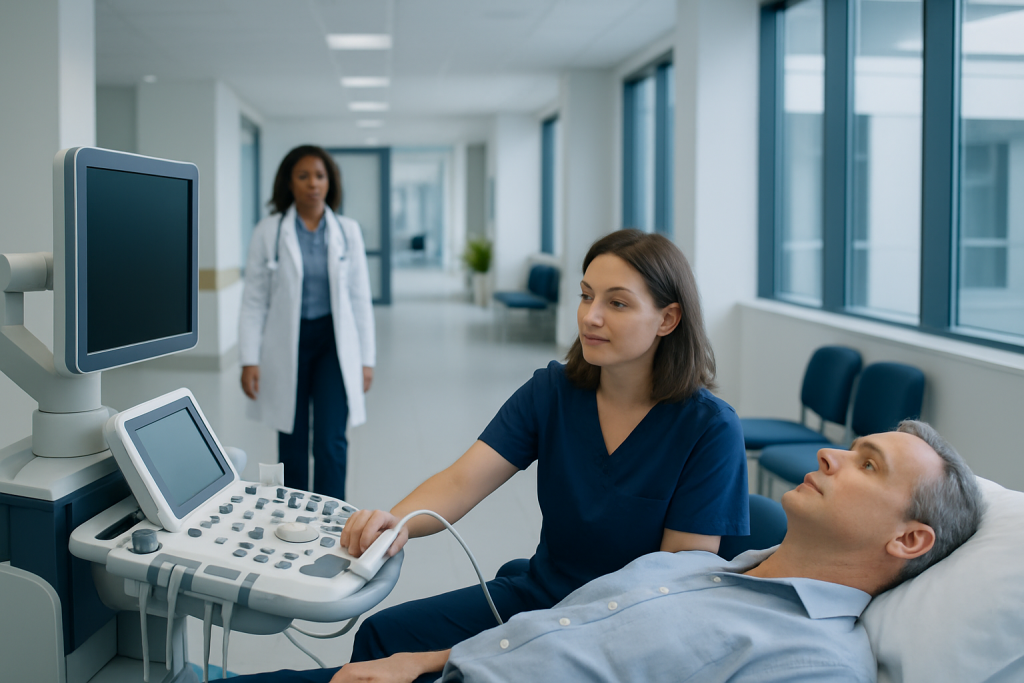
A carotid ultrasound is painless, uses no radiation and usually takes about 15 to 30 minutes. Most people do not need special preparation, although the imaging center’s instructions should always be followed.
Key Takeaways
- A carotid ultrasound is painless, uses no radiation and usually takes about 15 to 30 minutes.
- Most people do not need special preparation, although the imaging center’s instructions should always be followed.
- The test can show plaque, narrowing and changes in blood flow, but results must be interpreted alongside symptoms and overall cardiovascular risk.
- Carotid screening is most useful when a clinician recommends it because of symptoms, a bruit or specific risk factors; it is not routine screening for everyone.
- Sudden stroke-like symptoms require emergency medical attention, even if they disappear quickly.
An ultrasound of carotid artery is a non-invasive scan that uses sound waves to assess blood flow in the arteries on either side of the neck. It can help clinicians identify plaque buildup or narrowing that may increase stroke risk, while also guiding appropriate prevention and treatment decisions.
Overview: what is an ultrasound of carotid artery?
An ultrasound of carotid artery, also called a carotid Doppler ultrasound or carotid duplex scan, is an imaging examination of the two main arteries that run along the sides of the neck. These arteries deliver oxygen-rich blood to the brain. The scan uses high-frequency sound waves to create images of the artery walls and measure the speed and direction of blood flow.
The test is commonly used to look for fatty deposits, known as plaque, and for carotid artery narrowing, called stenosis. Plaque can reduce blood flow or contribute to a blood clot that travels to the brain. Carotid ultrasound does not use ionizing radiation, injections or surgical instruments, and it is generally comfortable for most people.
A clinician may order the examination after symptoms suggestive of a transient ischemic attack (TIA) or stroke, after hearing an unusual sound over the artery with a stethoscope, or when evaluating known vascular disease. It may also be part of follow-up for people with established carotid narrowing or after treatment.
How the scan works and who may need it
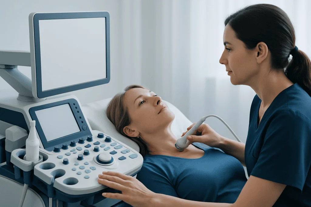
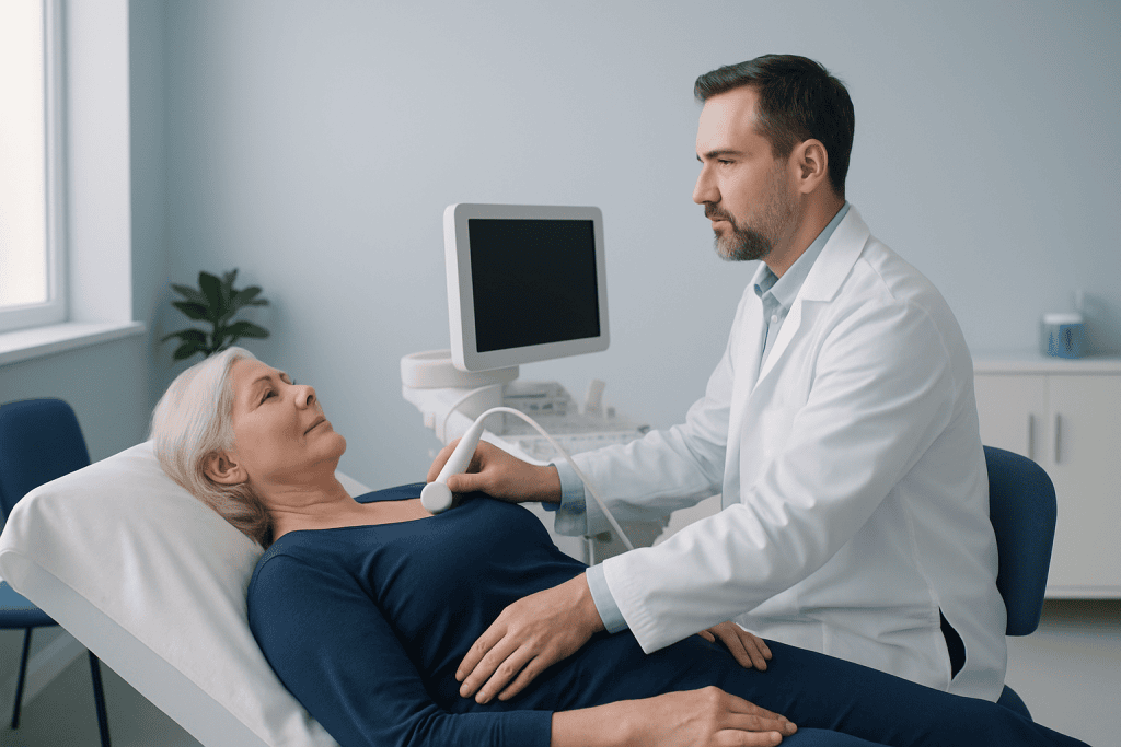
During a carotid duplex ultrasound, a handheld device called a transducer sends sound waves through the skin. Reflected waves are converted into pictures of the carotid arteries. Doppler technology adds information about blood flow, including whether it is moving unusually fast through a narrowed area.
The examination may be appropriate for people with a history of TIA or stroke, temporary loss of vision in one eye, unexplained weakness or speech difficulty, or known peripheral artery or coronary artery disease. It may also be considered for people with a carotid bruit, although a bruit alone does not confirm narrowing.
Risk factors that can contribute to atherosclerosis include smoking, high blood pressure, diabetes, elevated cholesterol, chronic kidney disease, increasing age and a family history of early cardiovascular disease. Having risk factors does not automatically mean a scan is needed; the decision should be individualized with a qualified clinician.
Carotid ultrasound is not usually a general screening test for people without symptoms or known vascular disease. In low-risk individuals, screening may identify minor findings that do not require treatment and can lead to additional testing. A clinician can explain whether the potential value of the scan outweighs these limitations in a particular situation.
Is there any prep for a carotid artery ultrasound?
In most cases, there is little or no preparation for a carotid artery ultrasound. People can usually eat, drink and take their regular medicines unless their healthcare team gives different instructions. The appointment letter or imaging center should be checked in advance, because local instructions may vary.
It is helpful to wear a loose, open-necked top or clothing that allows easy access to the neck. Necklaces, scarves and large earrings may need to be removed. Bringing a list of medicines, relevant medical records and previous imaging results can also help the clinical team compare findings over time.
People should tell the sonographer or doctor about recent symptoms, previous stroke or TIA, neck surgery, carotid procedures and allergies to gels or adhesives. Ultrasound gel rarely causes a reaction, but sharing this information helps the team provide comfortable care.
What not to do before a carotid ultrasound?
Unless specifically instructed otherwise, people should not stop prescribed medicines before a carotid ultrasound. This includes medicines used for blood pressure, diabetes, cholesterol or blood clot prevention. Stopping a medicine without medical advice may be harmful and does not improve the accuracy of the scan.
It is sensible to avoid wearing tight collars, heavy neck jewelry or products that could make access to the neck difficult. Smoking and nicotine use can temporarily affect blood vessels and are best avoided before the appointment when possible, particularly if the imaging team has asked for this.
People should also avoid assuming that a carotid ultrasound can replace emergency assessment. New facial drooping, arm weakness, speech difficulty, sudden vision loss, severe dizziness or loss of balance need urgent evaluation rather than waiting for a routine scan.
Can you drink coffee before a carotid artery ultrasound?
Most people can drink coffee before a carotid artery ultrasound because fasting is not routinely required. However, caffeine may temporarily affect heart rate and blood pressure, and some centers may ask patients to avoid caffeine, nicotine or other stimulants for a few hours before a Doppler examination.
The safest approach is to follow the instructions provided by the imaging center. If no instructions were given, a person may call ahead to ask whether coffee is permitted. Those with diabetes or other conditions that make fasting difficult should discuss preparation plans with their healthcare team rather than changing eating or medication routines on their own.
Water is generally suitable unless the center advises otherwise. Hydration can support comfort during the visit, but it does not change the need for careful medical interpretation of the results.
What happens during the procedure and after it?
At the appointment, the person usually lies on an examination table with the head slightly turned away from the side being scanned. A sonographer applies clear gel to the neck and moves the transducer gently over the skin. Mild pressure may be felt, but the test should not be painful.
The sonographer takes still images and records Doppler waveforms from several points along each carotid artery. The person may hear swishing sounds from the ultrasound machine; these represent blood flow measurements. Remaining still and following simple positioning instructions can improve image quality.
The scan commonly takes 15 to 30 minutes. Once it is complete, the gel is wiped away and normal activities can generally be resumed immediately. There is no recovery period, no sedation and no radiation exposure.
A radiologist, vascular specialist or another trained clinician reviews the images and sends a report to the referring doctor. Further tests, such as CT angiography or MR angiography, may occasionally be considered if the ultrasound findings are unclear or if treatment planning requires more detailed imaging.
Results, benefits and possible next steps
Results may describe normal arteries, plaque without important flow limitation, or a degree of stenosis estimated from the images and blood-flow velocities. The report may also note whether plaque appears calcified or irregular. Results are not interpreted in isolation: symptoms, examination findings and other cardiovascular risks all influence what they mean for the individual.
A normal scan is reassuring but does not eliminate every possible cause of neurological symptoms. Conversely, plaque or mild narrowing does not always require a procedure. Treatment often focuses on reducing stroke and cardiovascular risk through smoking cessation, blood pressure and cholesterol management, diabetes care, physical activity and appropriately prescribed medicines.
For more significant narrowing, especially in people who have had related neurological symptoms, a vascular or stroke specialist may discuss options such as intensive medical management and, in selected cases, a procedure to restore blood flow. Decisions about carotid endarterectomy or other interventions require individualized assessment of the anatomy, symptoms and expected benefits and risks.
The main benefits of ultrasound are that it is non-invasive, widely available and useful for monitoring blood flow over time. Risks are minimal and may include temporary discomfort from probe pressure or mild skin sensitivity to the gel. The more important limitation is that the test cannot by itself predict whether a person will have a stroke, so results should be discussed with the referring clinician.
Is carotid artery screening worth it? When to seek medical care
Carotid artery screening can be worthwhile when it is recommended for a specific medical reason, such as symptoms of TIA or stroke, a prior carotid procedure, known arterial disease or an abnormal clinical examination. For people without symptoms, routine screening is generally not recommended because the likelihood of finding clinically important disease is low and follow-up testing can sometimes cause unnecessary worry or treatment.
Emergency medical care is needed immediately for possible stroke symptoms: sudden weakness or numbness of the face, arm or leg, especially on one side; trouble speaking or understanding speech; sudden vision loss or double vision; severe unexplained imbalance; or a sudden, severe headache. Symptoms that resolve within minutes may be a TIA and still require urgent assessment.
A non-urgent medical appointment is appropriate for discussing cardiovascular risk factors, a family history of vascular disease, a newly detected neck bruit or questions about whether carotid imaging is indicated. Acibadem International’s multidisciplinary specialists and JCI-accredited hospitals can assess carotid artery disease and coordinate care for international patients when needed.
Long-term prevention is usually centered on managing modifiable risks rather than relying on imaging alone. A clinician can help develop a practical plan that addresses blood pressure, cholesterol, blood sugar, tobacco use, diet, exercise and prescribed medication follow-up.
Frequently asked questions
What does an ultrasound of carotid artery show?
The test shows the structure of the carotid arteries and how blood is flowing through them. It can identify plaque buildup, narrowing, blockage or altered flow patterns that may need further medical assessment.
How long does a carotid ultrasound take?
Most carotid ultrasound examinations take about 15 to 30 minutes. The exact time can vary depending on the images needed and whether both sides of the neck require detailed measurements.
Is a carotid ultrasound painful?
A carotid ultrasound is usually painless. Some people notice mild pressure from the transducer on the neck or a cool sensation from the ultrasound gel, but no needles or injections are normally used.
Do I need to fast before a carotid Doppler ultrasound?
Fasting is not usually necessary for a carotid Doppler ultrasound. Patients should follow the instructions from their imaging center, as preparation requirements can differ in certain situations or when other tests are scheduled at the same visit.
What happens if carotid ultrasound shows narrowing?
The clinician will consider the degree of narrowing, the appearance of plaque, symptoms and the person’s overall health. Management may include lifestyle measures and prescribed medication, follow-up imaging or referral to a vascular specialist to discuss whether an intervention is appropriate.
Can a carotid ultrasound detect a stroke?
A carotid ultrasound does not diagnose a stroke in the brain. It can identify carotid artery narrowing or plaque that may contribute to stroke risk, while brain imaging such as CT or MRI is typically used to assess a suspected stroke.
References
- American Heart Association
- United States Preventive Services Task Force
- Society for Vascular Surgery
- National Institute for Health and Care Excellence
- Radiological Society of North America
This article is for general information only and is not a substitute for professional medical advice. Please consult a qualified doctor about your individual situation.
Ultrasound in Turkey — costs, top hospitals & a free quote
JCI-accredited · board-certified surgeons · reply within 24h
Add us as a Preferred Source to see more of our trusted health content across Google Search, AI Overviews and Discover.
Check your numbers in seconds
BMI, calories, due date, blood pressure and 30+ more clinical calculators — free, instant, doctor-reviewed ranges.

Check-up & Preventive Medicine
Comprehensive health screenings designed for early detection and prevention.
3 specialists in this unit