Transcranial Ultrasound: Preparation, Procedure and Results

Transcranial Doppler ultrasound measures the speed and direction of blood flow in major brain arteries. The examination is painless, radiation-free, and commonly takes about 30 to 60 minutes.
Key Takeaways
- Transcranial Doppler ultrasound measures the speed and direction of blood flow in major brain arteries.
- The examination is painless, radiation-free, and commonly takes about 30 to 60 minutes.
- Preparation is usually minimal, but patients should follow their clinic’s instructions about medicines, caffeine, nicotine, and hair products.
- Results must be interpreted alongside symptoms, medical history, and sometimes other imaging tests.
- Urgent neurological symptoms such as sudden weakness, speech difficulty, or severe new headache require immediate medical assessment.
Transcranial ultrasound, often called transcranial Doppler (TCD) ultrasound, is a noninvasive test that uses sound waves to measure blood-flow speed in arteries inside the brain. It is usually quick, does not use radiation or injections, and can help a clinician assess certain stroke-related and blood-vessel conditions.
Transcranial Ultrasound Overview
Transcranial ultrasound is a noninvasive examination used to assess blood flow within arteries at the base of the brain. In clinical practice, it is most often performed as transcranial Doppler ultrasound (TCD). The test sends high-frequency sound waves through naturally thinner areas of the skull and analyzes the echoes reflected by moving red blood cells.
Unlike a standard brain scan, TCD does not create detailed pictures of brain tissue. Instead, it measures the speed and direction of blood flow. This information can help a neurology, stroke, or vascular team investigate narrowing, blockage, spasm, or unusual circulation patterns in selected brain arteries.
The test does not involve ionizing radiation, needles, contrast dye, or anesthesia. It is generally comfortable and can be repeated when ongoing monitoring is needed. A transcranial ultrasound result is one part of a wider clinical assessment and is interpreted together with symptoms, examination findings, and other tests when appropriate.
How Transcranial Doppler Works and Who May Need It
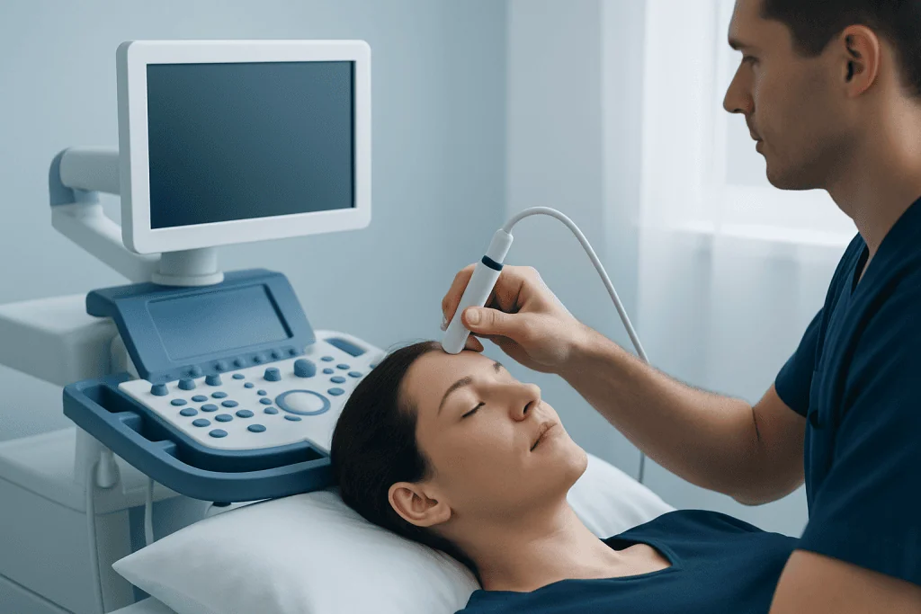
An ultrasound probe is placed against acoustic windows, which are areas where the skull bone is relatively thin. These commonly include the temple, the area behind the ear, the eye area with very low probe pressure, and the back of the head. Doppler technology detects changes in returning sound waves as blood cells move through an artery, allowing the system to calculate blood-flow velocity.
A doctor may order transcranial Doppler ultrasound when there is a need to evaluate cerebral circulation. It is used in the assessment and monitoring of some people with stroke or transient ischemic attack, known or suspected narrowing of intracranial arteries, sickle cell disease, subarachnoid hemorrhage, or suspected emboli. It may also be used during certain operations or procedures to monitor circulation.
Not every person with headache, dizziness, or memory symptoms needs this test. The choice depends on the clinical question and individual risk factors. In some people, especially older adults, the ultrasound signal may be difficult to obtain because the skull does not allow sound waves to pass through adequately. This is a technical limitation rather than a harmful finding.
When symptoms suggest a sudden interruption of blood supply to the brain, clinicians may also use CT, MRI, CT angiography, MR angiography, or other vascular tests. These investigations can provide anatomical information that complements blood-flow measurements from TCD.
How to Prepare for Transcranial Ultrasound

Preparation for transcranial ultrasound is usually simple. Patients can commonly eat, drink, and take their usual prescribed medicines unless their clinician gives different instructions. It is sensible to bring a current medication list and to tell the team about blood thinners, recent procedures, relevant medical conditions, and any new neurological symptoms.
Some laboratories ask patients to avoid smoking, nicotine products, caffeine, alcohol, or vigorous exercise for a period before the appointment because these can temporarily affect blood-vessel tone or heart rate. Requirements differ between hospitals and according to the reason for testing, so the appointment instructions should take priority.
Hair should be clean and free from heavy oils, sprays, gels, or other products that may prevent good contact between the probe and skin. Comfortable clothing is helpful because patients normally lie on an examination couch or sit with head support. No fasting is routinely required for an isolated transcranial Doppler study.
A transcranial Doppler is different from a transrectal ultrasound. For that reason, bowel emptying or an enema is not ordinarily needed before a transcranial test. Patients scheduled for a different type of ultrasound should follow the specific preparation guidance provided for that examination.
Step by Step: What Happens During the Procedure
At the start of the appointment, a trained sonographer, neurophysiology professional, radiologist, or clinician confirms the patient’s identity, reviews the reason for the test, and explains the procedure. The patient may be asked to lie comfortably on their back or side. The examination is generally performed in a quiet setting because attention to the Doppler sound signals can be useful.
A small amount of clear water-based gel is applied to the skin. The clinician gently places a handheld probe at the temple and may move it to other positions around the head and neck. The patient may hear rhythmic whooshing sounds that correspond to blood flow. The probe does not send radiation into the brain and should not be painful, although light pressure can be noticeable.
Measurements are taken from selected arteries, often on both sides of the head. Depending on the clinical purpose, the examiner may ask the patient to breathe normally, briefly hold their breath, or perform a simple task. In specialized studies, contrast-enhanced Doppler techniques or monitoring for microembolic signals may be used, but these are not part of every examination.
After the recordings are complete, the gel is wiped away and the patient can usually leave immediately. There is no recovery period, driving restriction, or routine activity limitation after a standard transcranial Doppler ultrasound.
How Long Does a Transcranial Doppler Ultrasound Take?
A standard transcranial Doppler ultrasound commonly takes about 30 to 60 minutes. The exact duration depends on the number of vessels being assessed, whether signals are easy to obtain, and whether specialized monitoring is required.
Some focused examinations may be shorter, while monitoring studies performed in hospital or during a procedure can last longer. Allowing extra time for check-in, explanation, and discussion is practical, particularly for a first appointment.
The clinician or diagnostic department can provide the most accurate estimate because the length of the examination is tailored to the reason it was requested. Patients should let staff know if they have neck discomfort, difficulty lying flat, or other needs that could affect positioning.
Results, Benefits, and Possible Limitations
A specialist reviews the Doppler waveforms and blood-flow velocities, usually comparing measurements from different arteries and both sides of the brain. A report may describe normal flow patterns, unusually high or low velocities, turbulence, poor acoustic windows, or signals that need correlation with another investigation. Results may be available the same day or sent to the referring clinician after formal review.
Higher-than-expected flow velocity can occur when an artery is narrowed or when blood flow is increased for another reason. Lower velocity may occur with reduced flow, technical factors, or normal variation. For this reason, a result does not confirm a diagnosis on its own. The treating clinician interprets it in the context of the person’s health history, symptoms, blood pressure, laboratory findings, and imaging where needed.
The main benefits are that the examination is painless, repeatable, portable, and free of radiation. It can provide real-time information about circulation and can be especially useful for follow-up in conditions where blood-flow changes over time. Its limitations include the inability to directly show all brain structures and the possibility that the skull may limit image quality.
If TCD suggests an important blood-flow abnormality, further assessment may be recommended. This can include vascular imaging or consultation with a stroke, neurology, neurosurgery, hematology, or cardiology specialist, depending on the suspected cause.
What Should You Not Do Before an Ultrasound?
Before a transcranial ultrasound, patients should not make assumptions based on preparation instructions for abdominal, pelvic, or transrectal ultrasound. Transcranial Doppler testing usually does not require fasting, a full bladder, bowel preparation, or stopping regular medicines without medical advice.
It may be helpful to avoid heavy hair products on the day of the test. If the clinic advises avoiding caffeine, nicotine, alcohol, or strenuous exercise beforehand, those instructions should be followed because they may influence circulation measurements. Patients should not stop prescribed medications, including blood-pressure medicines or blood thinners, unless the clinician specifically instructs them to do so.
For people having any type of ultrasound, the safest approach is to read the appointment letter and ask the diagnostic team if instructions are unclear. Preparation varies considerably according to the body area being examined and the medical reason for the scan.
Transrectal ultrasound has separate preparation requirements. Whether a person needs to empty their bowel before that test depends on the purpose of the examination and the facility’s protocol; the ordering team should provide clear directions. This does not apply to transcranial Doppler ultrasound.
Why Would a Doctor Order a Transcranial Doppler?
A doctor may order a transcranial Doppler to understand whether blood is moving through major brain arteries at an expected speed and pattern. The test can support evaluation of suspected narrowing in arteries inside the skull, assess circulation after a stroke or transient ischemic attack, and help guide follow-up when a vascular condition is already known.
In people with aneurysmal subarachnoid hemorrhage, repeated TCD studies may help monitor for vasospasm, a narrowing of brain arteries that can occur after bleeding around the brain. In children and adults with sickle cell disease, it may be used as part of stroke-risk screening or monitoring according to specialist guidance. It can also detect microembolic signals in selected patients when clinically indicated.
The test is not a general screening tool for everyone. A clinician selects it when its findings could help answer a focused clinical question or inform next steps. If symptoms raise concern for an acute stroke, urgent emergency evaluation is needed rather than waiting for an outpatient ultrasound appointment.
For patients whose evaluation identifies a stroke-related condition, coordinated care may involve neurological assessment and vascular imaging. Acibadem International’s multidisciplinary specialists and JCI-accredited hospitals provide diagnostic and treatment services for international patients, including care related to stroke.
Risks, Recovery, and When to Seek Medical Care
Standard transcranial Doppler ultrasound is considered very safe. The most common minor issue is temporary discomfort from probe pressure or cool gel. There is no radiation exposure, and there is generally no recovery time. Patients can return to their usual daily activities immediately unless they have been given separate instructions related to another test or treatment.
Patients should seek emergency medical care immediately for possible stroke symptoms, including sudden weakness or numbness of the face, arm, or leg, particularly on one side; sudden difficulty speaking or understanding speech; new loss of vision; sudden severe imbalance; or a sudden, severe headache unlike usual headaches. These symptoms can be time-sensitive and should not be assessed by waiting for a routine scan.
Non-urgent medical review is appropriate for recurring neurological symptoms, new headaches with concerning features, unexplained fainting, or questions about an abnormal test report. The referring clinician can explain whether further investigations or specialist review are needed and discuss the most suitable care plan.
Frequently asked questions
Is transcranial ultrasound painful?
No. Transcranial Doppler ultrasound is generally painless. A person may feel light pressure from the probe at the temple or other areas of the head, but the test does not involve needles, radiation, or surgery.
Do you need to poop before transrectal ultrasound?
Preparation for a transrectal ultrasound is different from preparation for transcranial ultrasound. Some transrectal examinations may involve instructions to empty the bowel or use an enema, while others may not. The patient should follow the instructions provided by the clinic performing that specific test.
Can transcranial Doppler diagnose a stroke?
Transcranial Doppler can identify blood-flow patterns that may be relevant to stroke evaluation, such as signs of narrowing or altered circulation in certain brain arteries. However, it does not replace emergency clinical assessment or brain imaging. CT or MRI and vascular imaging are often needed to diagnose and classify a stroke.
Do I need to fast before a transcranial Doppler test?
Fasting is not usually required for a standard transcranial Doppler ultrasound. However, the diagnostic team may give individualized instructions based on the reason for testing or other examinations scheduled on the same day. Patients should follow their appointment guidance.
What do abnormal transcranial Doppler results mean?
An abnormal result may show blood-flow velocity or waveform changes that suggest narrowing, spasm, reduced flow, or another circulation-related issue. Technical factors can also affect the study, including difficulty obtaining a signal through the skull. A clinician must interpret the result alongside symptoms and other tests.
Can I drive after transcranial ultrasound?
Most people can drive and return to normal activities immediately after a routine transcranial Doppler ultrasound. The test does not use sedatives or contrast dye. If the person has acute neurological symptoms or has undergone another procedure, separate advice may apply.
References
- American Heart Association
- American Stroke Association
- National Institute of Neurological Disorders and Stroke
- Radiological Society of North America
- World Health Organization
This article is for general information only and is not a substitute for professional medical advice. Please consult a qualified doctor about your individual situation.
Ultrasound in Turkey — costs, top hospitals & a free quote
JCI-accredited · board-certified surgeons · reply within 24h
Add us as a Preferred Source to see more of our trusted health content across Google Search, AI Overviews and Discover.
Check your numbers in seconds
BMI, calories, due date, blood pressure and 30+ more clinical calculators — free, instant, doctor-reviewed ranges.

Check-up & Preventive Medicine
Comprehensive health screenings designed for early detection and prevention.
1 specialists in this unit