Trephination — Explained by Medical Evidence, Not Myths

Trephination has legitimate modern medical uses, especially nail trephination and some neurosurgical procedures. Historical or self-help claims about trephination improving mental performance are not supported by medical evidence.
Key Takeaways
- Trephination has legitimate modern medical uses, especially nail trephination and some neurosurgical procedures.
- Historical or self-help claims about trephination improving mental performance are not supported by medical evidence.
- Nail trephination can quickly relieve pain from blood trapped under a nail after injury.
- Cranial trephination is a specialist procedure used only in selected emergency or surgical settings.
- Any suspected head injury, severe nail injury, or signs of infection should be assessed by a qualified clinician.
Trephination is a medical procedure that creates a small opening, most often in a fingernail or toenail to relieve trapped blood, or in the skull during carefully selected neurosurgical care. It is not a proven method to improve consciousness or wellbeing, and modern evidence supports its use only for specific medical reasons under proper clinical supervision.
Overview: what trephination really means
Trephination is the medical act of making a small opening in a hard structure to relieve pressure, drain trapped fluid or blood, or allow access for treatment. Today, the term is most commonly used in two very different situations: nail trephination, which creates a tiny hole in a nail to release blood trapped after an injury, and cranial trephination, which refers to making an opening in the skull as part of neurosurgical care.
The most important point is that trephination is a procedure with specific clinical purposes. It is not a general health practice, and it is not an evidence-based way to expand consciousness, improve mood, or enhance brain function. Historical accounts and myths have made the topic sound mysterious, but modern medicine treats trephination as a targeted intervention used only when there is a clear medical indication.
Because the word can refer to different procedures, context matters. A person with a painful black-blue discoloration under a fingernail may need a very simple office-based treatment. By contrast, an opening made in the skull is a serious procedure performed by trained specialists, often in emergencies or during carefully planned surgery.
Modern medical uses of trephination
Nail trephination is the most familiar and straightforward use. After a finger or toe is crushed, blood may collect under the nail, causing a dark patch called a subungual hematoma. If pain is significant and the nail is otherwise suitable, a clinician may make a tiny hole in the nail plate so the trapped blood can drain. This often brings prompt relief because it reduces pressure.
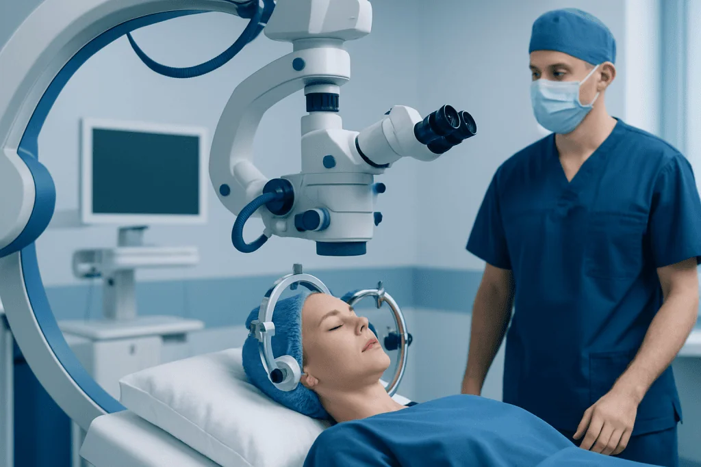
Cranial trephination is very different. In modern practice, opening the skull may be done as a burr hole or a larger neurosurgical opening to help diagnose or treat serious conditions. These can include some types of bleeding around the brain, raised pressure inside the skull, or the need to access brain tissue during surgery. This belongs to the field of neurosurgery and is performed under strict sterile conditions with imaging, anesthesia, and close monitoring.
In emergency care, a limited skull opening may sometimes be part of treatment for life-threatening brain compression. In elective care, it may be one step in a broader operation planned after scans and specialist review. People who are reading about trephination online often encounter dramatic claims, but in reality modern cranial procedures are carefully controlled medical interventions rather than lifestyle practices.
- Nail trephination: relieves pressure from blood under a nail
- Cranial trephination or burr hole: allows surgical access or pressure relief in selected brain conditions
- Dental or bone-related uses of similar drilling concepts exist, but they are usually described by other procedure names rather than simply “trephination”
Trephination myths versus medical evidence

Trephination has a long history, and archaeological findings show that openings in the skull were made in ancient societies. The reasons likely varied and may have included treatment of injuries, ritual practices, or attempts to address symptoms that were poorly understood at the time. Historical use, however, should not be confused with modern proof of safety or benefit.
Some contemporary myths claim that cranial trephination improves blood flow, raises consciousness, or offers mental or spiritual benefits. These claims are not supported by reliable medical evidence. Opening the skull without a valid medical indication carries serious risks, including bleeding, infection, brain injury, seizures, and potentially life-threatening complications.
Evidence-based medicine relies on anatomy, physiology, imaging, careful diagnosis, and outcomes research. Within that framework, there is no accepted role for self-performed or non-medical trephination. A person who has headaches, mood changes, concentration problems, or a desire for “brain optimization” needs proper medical assessment instead of an unsafe procedure based on internet myths.
It can also help to separate terms that sound similar. A clinically necessary opening in the skull for brain hemorrhage or pressure-related complications is not the same as the unproven idea of drilling the skull for wellness. The first belongs to emergency or specialist care; the second is not a recognized treatment.
Symptoms and situations that may lead to trephination
For nail trephination, the usual situation is a recent crush injury to a finger or toe. The person may notice throbbing pain, pressure, swelling, and a dark red, purple, or black area under the nail. The trapped blood itself can be very painful, especially when the collection is large.
For cranial trephination, the symptoms are related to the underlying condition, not to the procedure itself. These may include severe head injury, worsening headache, vomiting, confusion, weakness, changes in speech, drowsiness, seizures, or reduced consciousness. In such cases, the opening in the skull is not done because of the symptom alone but because brain imaging and specialist evaluation show a problem that may require urgent treatment.
One example is pressure caused by bleeding around the brain after trauma. Depending on the location and severity, care may range from close observation to emergency surgery. Patients with symptoms suggestive of a serious neurological condition may undergo CT or MRI scans and assessment by specialists in neurology or neurosurgery.
Not every painful bruised nail needs trephination, and not every brain bleed or head injury is treated with a burr hole. The decision depends on factors such as timing, severity, exam findings, imaging, associated injuries, and the person’s overall health.
How doctors evaluate whether trephination is needed
Assessment begins with the cause of the problem and a physical examination. In a nail injury, a clinician checks the amount of pain, the size of the blood collection, whether the nail is intact, and whether there may be a fracture or deep cut beneath the nail. Sometimes an X-ray is done, especially if the finger was crushed or the tip is very tender.
If nail trephination is appropriate, the procedure is usually quick. The clinician creates a tiny opening in the nail with a sterile heated device or needle designed for this purpose. Because the nail plate itself does not have sensation, many patients tolerate the procedure well, although the surrounding injured finger may remain tender. Once the blood drains, pressure often decreases almost immediately.
In suspected brain conditions, evaluation is more complex and urgent. Doctors perform a neurological examination and use imaging such as a CT scan to look for bleeding, swelling, a mass, or other structural problems. Blood tests and monitoring may also be needed, especially in people taking blood thinners or those with serious injury.
If surgery is considered, the risks and goals are reviewed as clearly as possible with the patient or family. In some cases, a burr hole is used to drain blood collections such as selected subdural hematomas; in others, a larger operation is necessary. Some patients may be managed without surgery if findings are stable and symptoms are mild. Conditions such as brain tumor or traumatic bleeding are assessed individually rather than with a one-size-fits-all approach.
Treatment, recovery, and possible risks
For nail trephination, aftercare usually includes keeping the area clean and dry for a short period, protecting the finger or toe, and watching for ongoing pain, drainage, or signs of infection. The underlying bruise may still take time to grow out with the nail. If the nail bed is badly torn or the nail is significantly disrupted, repair rather than simple trephination may be needed.
The risks of nail trephination are generally low when performed correctly, but they can include infection, persistent pain, incomplete drainage, or missing a deeper injury such as a fracture. Home attempts with heated paper clips, needles, or other non-sterile tools are not advised because they can cause burns, introduce infection, or worsen the injury.
Cranial trephination and burr hole procedures have more substantial risks because they involve the skull and structures around the brain. Potential complications include infection, bleeding, seizures, damage to nearby tissue, anesthetic complications, and recurrence of the underlying problem. Recovery depends largely on why the procedure was needed, how urgently it was done, and the patient’s neurological condition before treatment.
Follow-up may include hospital monitoring, repeat imaging, rehabilitation, and treatment of the underlying disease. For example, a patient treated for bleeding or pressure may also need ongoing care through brain surgery services, neurology, intensive care, or rehabilitation. Near the end of the care pathway, some international patients may seek evaluation at centers such as Acibadem International, where multidisciplinary specialists in JCI-accredited hospitals diagnose and treat complex neurological conditions.
Prevention and self-care
Most prevention advice relates either to avoiding nail trauma or reducing the chance of head injury. For hands and feet, practical steps include wearing proper footwear, using protective gloves when appropriate, taking care around heavy doors and tools, and supervising activities that commonly lead to crushed fingers in children.
Head injury prevention includes seat belt use, child car seats, helmets for cycling and contact sports, fall prevention measures at home, and attention to workplace safety. Older adults may benefit from medication reviews, vision checks, balance assessment, and home modifications to reduce tripping hazards.
Self-care has clear limits in this area. An ice pack, elevation, and rest may help a fresh finger injury, but severe throbbing pain, major nail discoloration, deformity, or suspicion of fracture should be assessed by a clinician. A person should never attempt cranial trephination outside a medical setting.
Anyone with persistent headaches, neurological symptoms, or concerns after a head injury should seek professional evaluation rather than relying on myths or social media advice. Safe care starts with identifying the real cause of symptoms and choosing evidence-based treatment.
When to seek medical care
Medical attention is advisable for a nail injury when pain is significant, the nail is largely discolored by blood, the finger or toe looks deformed, there is numbness, the skin is cut, or movement is difficult. Care is also important if the person has diabetes, poor circulation, immune suppression, or signs of infection such as spreading redness, warmth, pus, or fever.
Urgent or emergency care is needed after a head injury if there is loss of consciousness, worsening headache, repeated vomiting, confusion, unusual sleepiness, seizure, weakness, slurred speech, unequal pupils, or clear neurological decline. These can signal bleeding, swelling, or other problems inside the skull that require immediate assessment.
Even without a recent injury, new neurological symptoms such as persistent severe headache, sudden weakness, changes in vision, speech difficulty, or altered awareness should be evaluated promptly. Early diagnosis matters because some conditions become harder to treat if care is delayed.
Trephination is best understood not as a mythic practice but as a precise medical tool used only in selected situations. A qualified doctor can explain whether drainage, monitoring, imaging, or specialist treatment is the safest next step.
Frequently asked questions
Is trephination still done today?
Yes. Trephination is still used in modern medicine, but only for specific reasons. The most common example is nail trephination to drain blood trapped under a nail, while cranial trephination is a specialist neurosurgical procedure used in selected emergencies or operations.
Does trephination improve brain function or consciousness?
No reliable medical evidence shows that trephination improves intelligence, mood, awareness, or spiritual experience. Claims of this kind are not part of evidence-based medicine. Opening the skull without a medical reason is dangerous and can cause serious harm.
What is the difference between nail trephination and cranial trephination?
Nail trephination involves making a tiny hole in a fingernail or toenail to release painful trapped blood after an injury. Cranial trephination involves making an opening in the skull as part of neurosurgical care. They share a name because both create an opening, but they are very different procedures in purpose, setting, and risk.
Is nail trephination painful?
The injured finger or toe is often already painful because of pressure under the nail. The procedure itself is usually brief, and many patients feel relief once the blood drains. Tenderness from the original injury may continue for a while even after pressure is reduced.
Can trephination be done at home?
It should not be done at home. Trying to puncture a nail with non-sterile tools can cause burns, infection, and missed diagnosis of a deeper injury. Cranial trephination should never be attempted outside a hospital setting.
How do doctors decide if someone needs a burr hole in the skull?
Doctors base the decision on symptoms, neurological examination, and imaging such as a CT scan. A burr hole may be used when there is bleeding, pressure, or a need to access the brain safely for treatment. The exact approach depends on the cause, urgency, and overall condition of the patient.
This article is for general information only and is not a substitute for professional medical advice. Please consult a qualified doctor about your individual situation.
Explore treatments in Turkey — costs, top hospitals & a free quote
JCI-accredited · board-certified surgeons · reply within 24h
Add us as a Preferred Source to see more of our trusted health content across Google Search, AI Overviews and Discover.

More from the Health Library

Penile: A Complete Medical Overview

Green Tea — Explained by Medical Evidence, Not Myths
Klebsiella Pneumoniae: A Complete Medical Overview

Right Heart Catheterization: An Evidence-Based Guide for Patients
Bloody Discharge: What Patients Need to Know