Trimalleolar Fracture: An Evidence-Based Guide for Patients

A trimalleolar fracture involves the medial, lateral, and posterior malleoli of the ankle. It usually causes severe pain, swelling, bruising, and difficulty or inability to bear weight.
Key Takeaways
- A trimalleolar fracture involves the medial, lateral, and posterior malleoli of the ankle.
- It usually causes severe pain, swelling, bruising, and difficulty or inability to bear weight.
- Because the ankle joint may be unstable, prompt medical evaluation is important.
- Treatment may be non-surgical or surgical depending on fracture position, joint stability, and soft tissue injury.
- Recovery often includes immobilization, gradual weight-bearing, and physical therapy.
A trimalleolar fracture is a serious ankle injury that affects three bony parts of the ankle joint and can make the joint unstable. Patients often need urgent assessment, imaging, and a treatment plan that may include immobilization, surgery, and rehabilitation.
Overview: what a trimalleolar fracture means
A trimalleolar fracture is a type of broken ankle involving three parts of the joint: the inner ankle bone prominence (medial malleolus), the outer prominence (lateral malleolus), and the back part of the tibia at the ankle (posterior malleolus). Because these structures help keep the ankle aligned and stable, this injury is more complex than a simple ankle sprain or a minor fracture.
Many trimalleolar fractures happen after a fall, sports injury, twisting accident, or motor vehicle trauma. In some people, especially older adults or those with weaker bones, a lower-energy fall can still cause this injury. The exact pattern varies, but the main concern is whether the ankle joint remains stable and properly aligned.
Patients often hear the term “unstable ankle fracture.” That is important because a trimalleolar fracture frequently affects not only bone but also ligaments, cartilage, and nearby soft tissues. If the ankle is out of place or the bones are displaced, the joint can be damaged further without timely treatment.
Although this is a serious injury, outcomes are often good with appropriate care. Evaluation by an orthopedic specialist, proper imaging, and a structured recovery plan help support healing and reduce the risk of long-term problems such as stiffness, persistent pain, or post-traumatic arthritis.
Symptoms and how it differs from a sprain
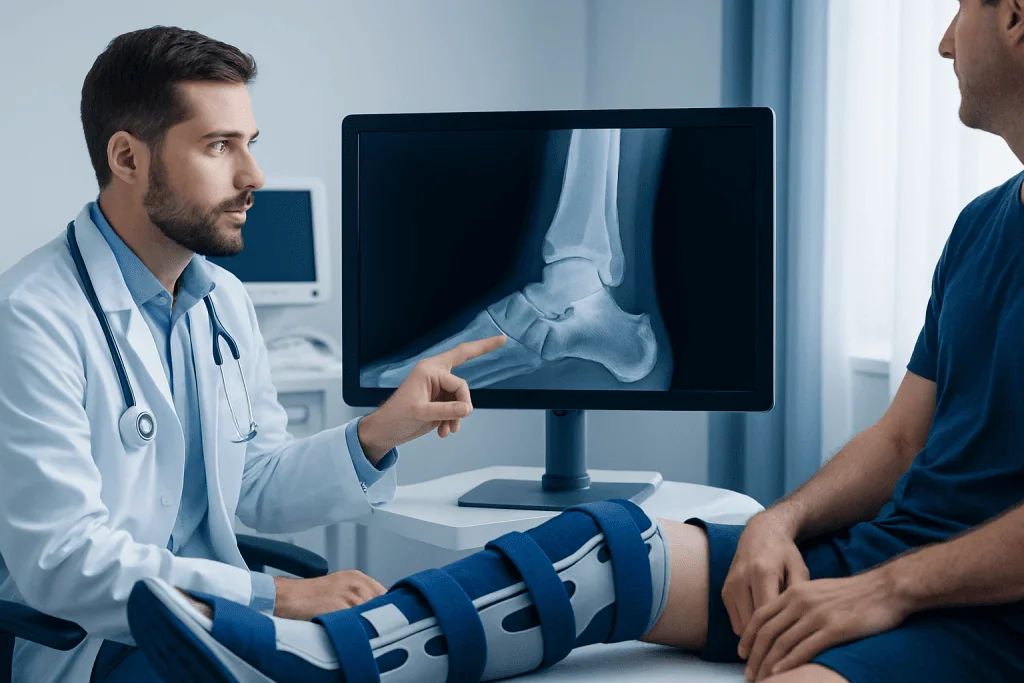
The most common trimalleolar fracture symptoms are sudden ankle pain, swelling, bruising, and difficulty standing or walking. Some people cannot bear weight at all. The ankle may look deformed, especially if the joint is displaced, and movement is usually very painful.
This injury can sometimes be mistaken for a severe sprain at first, particularly when there is no obvious deformity. However, a fracture is more likely when pain is intense, swelling develops quickly, weight-bearing is not possible, or tenderness is felt directly over the ankle bones. A clinician will look carefully for these signs and decide whether imaging is needed.
Additional symptoms can include a feeling that the ankle is unstable, numbness or tingling from swelling or pressure on nerves, and skin changes if the injury is severe. In an open fracture, the bone may communicate with the outside through a wound, which needs urgent treatment because of infection risk.
- Severe ankle pain after twisting or impact
- Rapid swelling and bruising
- Inability or major difficulty bearing weight
- Visible deformity or the ankle looking “out of place”
- Tenderness over the inner and outer ankle bones
Causes, risk factors, and associated injuries
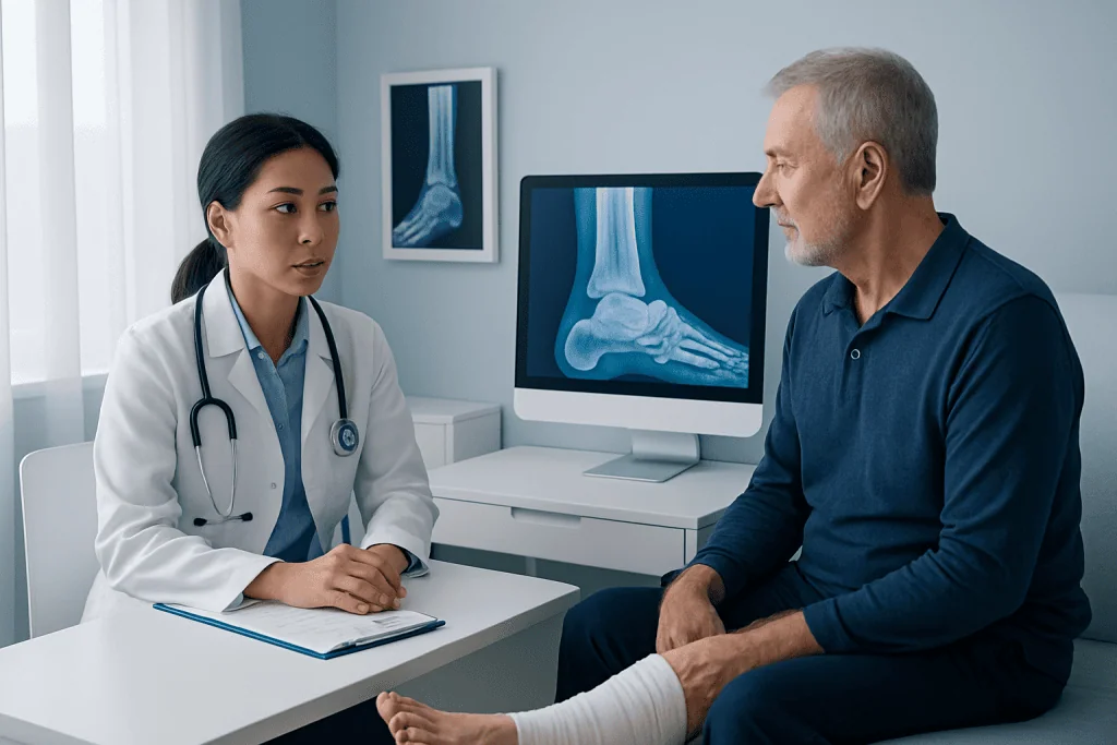
A trimalleolar fracture usually results from a force that twists, bends, or compresses the ankle joint. Common causes include slipping on stairs, missing a step, sports collisions, awkward landings, and road traffic injuries. The exact direction of force can affect which bones break and whether the ankle dislocates.
Risk factors include older age, reduced bone density, poor balance, previous ankle injury, and participation in activities with cutting, jumping, or rapid directional changes. Conditions that weaken bone, such as osteoporosis, can make fractures more likely after relatively minor trauma. In younger adults, high-energy injuries are a more common cause.
Doctors also consider associated damage beyond the fracture itself. Ligaments between the tibia and fibula can be injured, the ankle can become dislocated, and cartilage surfaces may be damaged. These related injuries matter because they influence treatment decisions and long-term joint function.
Some patients may also have other lower-leg or foot injuries at the same time. If pain extends beyond the ankle, clinicians may examine for related problems such as ankle fracture patterns that involve nearby structures or soft-tissue complications that need separate management.
How doctors diagnose a trimalleolar fracture
Diagnosis begins with a physical examination and a clear history of how the injury happened. The clinician will assess swelling, bruising, skin condition, circulation, sensation, and whether the ankle appears aligned. Checking the skin is especially important, because tense swelling or an open wound can affect the timing and type of treatment.
X-rays are the standard first imaging test and usually confirm the fracture pattern. They help show which malleoli are involved, whether the bones are displaced, and whether the ankle mortise, the space that reflects joint alignment, remains normal. Repeat X-rays may be needed after initial stabilization.
Computed tomography (CT) is often useful in trimalleolar fractures, especially when the posterior malleolus is involved. CT can show fragment size, joint surface injury, and subtle displacement more clearly than plain X-rays. This can be important for planning surgery and understanding the risk of later joint stiffness or arthritis.
If there is concern about blood vessel or nerve injury, the doctor may perform additional tests and urgent reassessment. The main goals are to define the fracture accurately, evaluate stability, and choose the safest route toward healing, whether that is casting, splinting, or orthopedic surgery.
Treatment options: immobilization, surgery, and rehabilitation
Initial treatment focuses on protecting the ankle, controlling pain, and preventing further displacement. In the emergency setting, the ankle may be placed in a splint rather than a full cast at first, because swelling can increase over the first few days. Elevation and ice may be advised, along with avoiding weight-bearing until a specialist review.
Some trimalleolar fractures can be managed without surgery, but this is usually considered only when the fracture is stable, well aligned, and unlikely to shift. In these cases, treatment may involve casting or a walking boot, close follow-up imaging, and a gradual return to movement and weight-bearing under medical supervision.
Many patients need surgery because the ankle joint is unstable or the bones are displaced. The goal is to restore alignment and joint congruity, often using plates, screws, or other fixation devices. In some injuries, operating is delayed until swelling improves and the skin is ready, which can reduce soft-tissue complications. Surgical decision-making is individualized and may involve trauma and orthopedic surgery expertise.
Rehabilitation is a key part of treatment whether surgery is needed or not. After a period of protection, patients usually begin range-of-motion exercises, then strengthening, balance work, and gait training. A structured plan through physical therapy and rehabilitation can help improve mobility, confidence, and function during recovery.
Recovery timeline and possible complications
Healing time varies depending on age, bone health, fracture complexity, and whether surgery was performed. Many patients need several weeks of protection before progressing to weight-bearing, and full functional recovery often takes months rather than days. Even after the bone has healed, swelling and stiffness may continue to improve gradually over time.
It is common for recovery to happen in stages. Early goals include pain control, swelling reduction, and protecting the repair. Later goals focus on regaining ankle motion, rebuilding strength in the calf and lower leg, and restoring balance for everyday activities such as walking on uneven ground or climbing stairs.
Possible complications include delayed union, nonunion, infection after surgery, wound healing problems, blood clots, nerve irritation, chronic instability, and post-traumatic arthritis. Not every patient develops these issues, but they are part of why follow-up care matters. Persistent pain or a sense that the ankle is not improving should be discussed with a doctor.
Long-term outlook is often better when the ankle joint is restored as accurately as possible and rehabilitation is followed consistently. Some patients may still notice weather-related aching, reduced flexibility, or limits in high-impact sports. These changes do not always prevent a return to daily life, but they may require patience and ongoing exercise.
Prevention and self-care after injury
Not all ankle fractures can be prevented, but some practical measures can lower risk. Supportive footwear, attention to slippery surfaces, strength and balance training, and sports techniques that reduce awkward landings may help. For older adults, fall prevention and bone health are especially important.
After the injury, self-care should follow the treating clinician’s advice. Patients are often told to keep the ankle elevated, avoid putting weight on it unless cleared, and watch for increasing swelling, numbness, severe pain, or skin problems around a cast or splint. Home safety measures, such as using mobility aids correctly and removing trip hazards, can also support recovery.
Bone health deserves attention, particularly for people who fracture after a low-energy fall. A clinician may review calcium and vitamin D intake, physical activity, medications, and whether testing for osteoporosis is appropriate. Addressing these factors may help reduce the chance of future fractures.
Near the end of treatment, some patients benefit from a broader orthopedic review if they have repeated injuries, persistent instability, or concerns about joint damage. In this context, related conditions such as ankle instability may be considered during follow-up planning.
When to seek medical care
Immediate medical assessment is important after a suspected trimalleolar fracture. A person should seek urgent care if there is severe pain, major swelling, inability to bear weight, visible deformity, numbness, cold toes, or a wound near the ankle. These signs can indicate a significant fracture, dislocation, or circulation problem.
Patients should also contact a doctor promptly if symptoms worsen after the initial visit. Warning signs include a cast or splint feeling too tight, increasing pain not improving with rest, fever, drainage from a wound, or new redness and swelling in the calf. These symptoms may suggest complications that need review.
For planned recovery, follow-up appointments are a routine and important part of care. Imaging may be repeated to check bone alignment and healing, and activity levels are adjusted over time. Individual advice matters, because returning to walking, driving, work, or sport too early can interfere with recovery.
For international patients who need specialist evaluation, Acibadem International’s multidisciplinary teams in JCI-accredited hospitals diagnose and treat complex ankle injuries and guide rehabilitation according to the individual fracture pattern and overall health needs.
Frequently asked questions
Is a trimalleolar fracture serious?
Yes, it is generally considered a serious ankle injury because it involves three parts of the joint and can make the ankle unstable. Many cases need urgent specialist assessment and some require surgery to restore alignment.
Can a trimalleolar fracture heal without surgery?
Some can heal without surgery if the bones are well aligned and the ankle remains stable. However, many trimalleolar fractures are displaced or unstable, so an orthopedic specialist usually evaluates whether surgery would offer a better outcome.
How long does trimalleolar fracture recovery take?
Recovery is different for each person, but it commonly takes several months. Bone healing may happen over weeks, while swelling, stiffness, strength, and confidence with walking can continue improving for a longer period.
When can someone walk again after a trimalleolar fracture?
Weight-bearing depends on the fracture pattern, the stability of the ankle, and whether surgery was performed. A doctor usually gives a step-by-step plan, and walking often resumes gradually rather than all at once.
What is the difference between a trimalleolar fracture and a bad ankle sprain?
A sprain affects ligaments, while a trimalleolar fracture involves broken bone in three parts of the ankle. Both can cause swelling and pain, but a fracture is more likely to cause severe tenderness over the bones, marked difficulty bearing weight, and changes in alignment on imaging.
Will the ankle be normal again after treatment?
Many patients recover well and return to daily activities, especially with proper treatment and rehabilitation. Still, some may have lingering stiffness, swelling, or discomfort, and a small number develop long-term joint changes such as arthritis.
This article is for general information only and is not a substitute for professional medical advice. Please consult a qualified doctor about your individual situation.
Joint and spine care in Turkey — expert assessment & treatment
JCI-accredited · board-certified surgeons · reply within 24h
Add us as a Preferred Source to see more of our trusted health content across Google Search, AI Overviews and Discover.

More from the Health Library

Below tongue bumps: Symptoms, Causes, and Treatment — Complete Patient Guide
Muscle of the Lower Arm: An Evidence-Based Guide for Patients

Chia Seeds and Digestive Health: A Complete Medical Overview

Large Breasts: A Complete Medical Overview

Pygeum — Explained by Medical Evidence, Not Myths

Shakeology: A Complete Medical Overview
Orthopedic Surgery & Traumatology Specialists at Acibadem

Dr. Bülent Özmen
Orthopedic Surgery & Traumatology
Dr. Bülent Yücel
Orthopedic Surgery & Traumatology
Prof. Dr. Burak Akan
Orthopedic Surgery & Traumatology
Dr. Burak Boynuk
Orthopedic Surgery & Traumatology