Twenty Week Scan: A Complete Medical Overview

The twenty week scan is a routine mid-pregnancy ultrasound, often called the anomaly scan. It looks closely at fetal anatomy, growth, the placenta, cervix, and amniotic fluid.
Key Takeaways
- The twenty week scan is a routine mid-pregnancy ultrasound, often called the anomaly scan.
- It looks closely at fetal anatomy, growth, the placenta, cervix, and amniotic fluid.
- Most scans are reassuring, but sometimes extra images, follow-up scans, or specialist review are needed.
- The scan can suggest possible concerns, but it does not diagnose every condition.
- A normal result is reassuring, while an unclear or abnormal finding should be discussed with a qualified obstetric specialist.
A twenty week scan is a detailed ultrasound usually performed between 18 and 22 weeks of pregnancy to assess the baby's development and the pregnancy environment. It helps check anatomy, growth, placenta position, and amniotic fluid, but it is a screening test rather than a guarantee that all conditions can be ruled out.
Overview: what a twenty week scan is
The twenty week scan is a detailed pregnancy ultrasound done most often between 18 and 22 weeks. It is designed to check how the baby is developing and to assess important features of the pregnancy, including the placenta, amniotic fluid, and the mother’s uterus and cervix. In many places, it is also called the anomaly scan or the mid-pregnancy anatomy scan.
This examination uses sound waves to create images of the baby in the womb. It is non-invasive and does not use radiation. A trained sonographer or fetal medicine specialist takes measurements and reviews visible structures such as the head, brain, face, spine, heart, stomach, kidneys, bladder, limbs, and abdominal wall.
One of the most helpful ways to understand this scan is to see it as a structured health check rather than a single yes-or-no test. It provides a broad picture of fetal anatomy and growth at a stage when many organs can be seen clearly. At the same time, it cannot detect every medical condition, and some findings may become clearer only later in pregnancy.
What the scan checks during the appointment
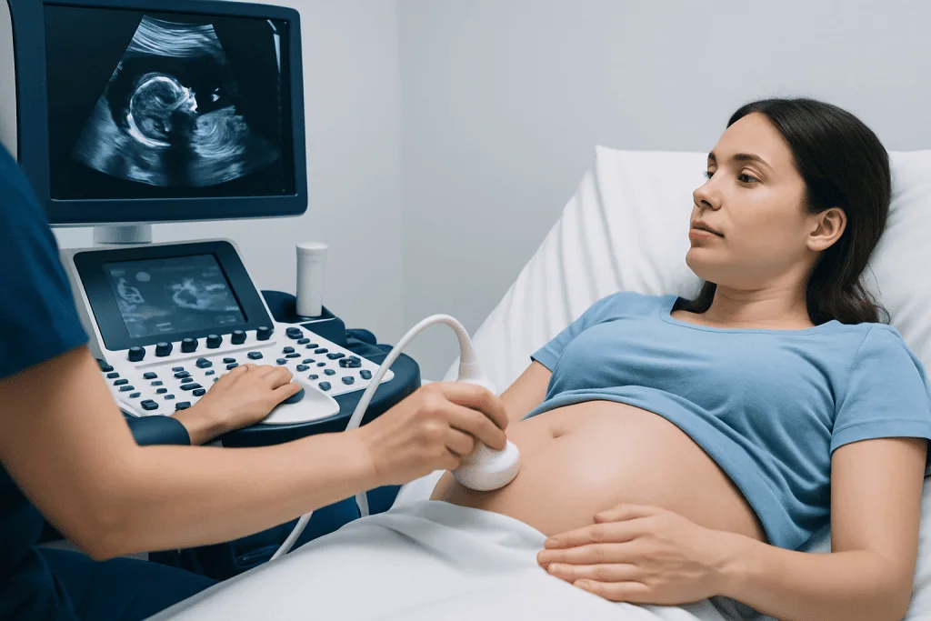
The main purpose of the twenty week scan is to look in detail at the baby’s anatomy. The healthcare professional usually checks the shape of the skull, parts of the brain, the face and lips, the spine, the chest, the four chambers and major outflow tracts of the heart as visible on ultrasound, the stomach, kidneys, bladder, abdominal wall, arms, legs, hands, and feet. Measurements are also taken to assess whether growth is in the expected range for the stage of pregnancy.
The scan also evaluates the pregnancy environment. This includes the position of the placenta, the amount of amniotic fluid, and often the length of the cervix when clinically appropriate. Placenta location can be especially important because a low-lying placenta may need follow-up later in pregnancy to see whether it moves upward as the uterus expands.
Depending on local practice and the baby’s position, the scan may also reveal the sex of the baby if the parents wish to know. However, the medical priority is the health assessment. If the baby is turned away, moving a lot, or lying in a position that blocks the view, some structures may be difficult to assess in one session, and a repeat scan may be recommended.
- Fetal anatomy and visible organ development
- Growth measurements such as head, abdomen, and femur length
- Placenta location and umbilical cord features that can be assessed
- Amniotic fluid volume
- Certain maternal structures relevant to pregnancy monitoring
What to expect before, during, and after the scan
For most people, the twenty week scan is performed through the abdomen. Gel is applied to the skin, and a handheld ultrasound probe is moved across the belly to capture images. The examination often takes longer than an early dating scan because it is more detailed, and the length can vary depending on the baby’s position, the mother’s body habitus, scar tissue, and how easily each structure can be seen.
Preparation instructions differ between hospitals and clinics. Some centers ask the patient to arrive with a moderately full bladder, while others do not. Comfortable clothing that allows easy access to the abdomen can make the appointment simpler. It is usually possible to bring a partner or support person, depending on local policy.
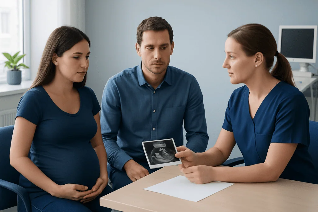
After the scan, the results may be discussed immediately or later by the obstetric team. In many cases, the findings are normal and no extra testing is needed. Sometimes the images are incomplete because the baby did not cooperate, and a return visit is arranged. In other cases, a finding may lead to more specialized imaging, fetal echocardiography, genetic counseling, or referral to maternal-fetal medicine if needed. If broader pregnancy concerns arise, further assessment may overlap with care for conditions such as high-risk pregnancy.
What the results can and cannot show
A normal twenty week scan is reassuring, but it does not guarantee that the baby has no health problems. Some conditions are too subtle to see on ultrasound, some develop later in pregnancy, and some can only be confirmed with other tests. Screening ultrasound works by identifying visible patterns that may suggest a concern, not by excluding every possible diagnosis.
When a possible abnormality is seen, it does not always mean there is a serious problem. Some findings are temporary, some turn out to be normal variations, and some need repeat imaging for clarification. The next step depends on what was seen and how certain the finding appears. Healthcare teams may recommend more detailed fetal imaging or discussion with specialists to explain what the result may mean for the pregnancy and birth plan.
Results can also raise questions about the placenta or cervix. A low-lying placenta or signs consistent with placenta previa may need follow-up later in pregnancy because these findings can affect delivery planning. If that issue is relevant, more information may be part of care related to placenta previa. In selected situations, doctors may recommend additional ultrasound-guided imaging assessment or other targeted evaluation, although invasive testing is only used when clinically justified.
Reasons a follow-up scan or extra tests may be recommended
Follow-up after a twenty week scan is common and does not automatically mean that something is wrong. The most frequent reason is simply that one or more parts of the baby’s anatomy could not be seen clearly enough in a single visit. The fetal heart, face, spine, or abdominal wall can be especially difficult to evaluate if the baby is facing the wrong way or if image quality is limited.
Extra tests may also be suggested if the scan shows a possible structural difference, unusual growth pattern, low or high amniotic fluid, or a placenta issue. Depending on the concern, options can include repeat ultrasound, fetal echocardiography, blood tests, genetic counseling, or specialist consultation. If symptoms suggest a change in the cervix or risk of early labor, assessment may be part of a wider review for preterm labor.
Parents often find this part stressful, but clear communication helps. It is reasonable to ask what was seen, how confident the team is, whether the finding is common, and what the follow-up aims to clarify. In specialized centers, multidisciplinary teams may include obstetricians, radiologists, neonatologists, genetic specialists, and pediatric surgeons when needed. If treatment planning becomes complex, advanced imaging and coordinated pregnancy follow-up and delivery care may be recommended.
Limits, accuracy, and factors that affect image quality
The twenty week scan is an important screening tool, but its accuracy depends on several factors. These include the baby’s position, gestational age, maternal body shape, the amount of amniotic fluid, the quality of the ultrasound equipment, and the experience of the professional performing and interpreting the scan. Some organs, especially the heart and brain, may require very careful views to assess as fully as possible.
Even in experienced hands, not every condition can be seen. Mild structural differences, some chromosome-related conditions, many genetic disorders, and functional problems that do not change visible anatomy may not appear on the scan. This is why ultrasound findings are interpreted alongside the pregnancy history, earlier screening results, blood tests, and any symptoms the mother may be experiencing.
It can also help to understand that ultrasound reports often use careful, technical wording. Terms such as “limited views,” “suboptimal visualization,” or “recommend repeat scan” are common and usually reflect the need for better images rather than an emergency. If a potentially significant finding is identified, the team explains what is known, what remains uncertain, and whether additional genetic testing or specialist review may be useful.
When to seek medical care
The twenty week scan is usually a planned routine appointment, but urgent medical advice should be sought if the pregnant person has symptoms such as vaginal bleeding, severe abdominal pain, fluid leaking from the vagina, fever, fainting, or a sudden severe headache. These symptoms are not diagnosed by the scan itself and should be assessed promptly by a qualified clinician or emergency service according to local guidance.
It is also important to contact the maternity team if there are concerns after the scan discussion, such as confusion about the results, advice to arrange follow-up that has not yet been scheduled, or emotional distress after an unexpected finding. Clear explanations and timely review can make a significant difference in understanding the next steps.
For ongoing pregnancy care, people should follow the schedule recommended by their obstetric team. Near the end of the care pathway, some international patients choose centers such as Acibadem International, where multidisciplinary specialists in JCI-accredited hospitals diagnose and manage complex pregnancy concerns with coordinated maternal and fetal care.
Frequently asked questions
Is the twenty week scan the same as the anomaly scan?
Yes. The twenty week scan is commonly called the anomaly scan or anatomy scan. It is a detailed mid-pregnancy ultrasound used to assess fetal development and the pregnancy environment.
How long does a twenty week scan usually take?
The appointment often takes longer than an early pregnancy scan because many structures must be checked carefully. The exact time varies depending on the baby's position and how easily clear images can be obtained.
Can the twenty week scan detect all birth defects?
No. The scan can identify many structural concerns that are visible on ultrasound, but it cannot detect every condition. Some problems are too subtle to see, develop later, or require other tests for diagnosis.
What happens if the sonographer cannot see everything clearly?
A repeat scan may be offered to obtain better views, especially if the baby is in an awkward position. This is common and does not necessarily mean there is a health problem.
Does a normal twenty week scan mean the baby is definitely healthy?
A normal result is reassuring, but it is not an absolute guarantee. Ultrasound is a screening tool, so some conditions may still not be visible at this stage.
Can parents find out the baby's sex at the twenty week scan?
Often yes, if local policy allows and the baby's position makes this possible. However, the main purpose of the scan is the medical assessment, so sex may not always be confirmed.
References
- World Health Organization
- American College of Obstetricians and Gynecologists
- International Society of Ultrasound in Obstetrics and Gynecology
- National Health Service
- Centers for Disease Control and Prevention
This article is for general information only and is not a substitute for professional medical advice. Please consult a qualified doctor about your individual situation.
Explore treatments in Turkey — costs, top hospitals & a free quote
JCI-accredited · board-certified surgeons · reply within 24h
Add us as a Preferred Source to see more of our trusted health content across Google Search, AI Overviews and Discover.