Ultrasonography vs Ultrasound: Differences Explained

Ultrasonography and ultrasound generally describe the same noninvasive imaging method. The test uses sound waves rather than ionizing radiation, so it is widely used during pregnancy and for many diagnostic purposes.
Key Takeaways
- Ultrasonography and ultrasound generally describe the same noninvasive imaging method.
- The test uses sound waves rather than ionizing radiation, so it is widely used during pregnancy and for many diagnostic purposes.
- Different ultrasound modes, including Doppler and internal ultrasound examinations, answer different clinical questions.
- Preparation depends on the body area being examined; many scans need little or no recovery time.
- Ultrasound has limitations and may need to be combined with blood tests, CT, MRI, or other investigations.
Ultrasonography and ultrasound are commonly used interchangeably. Ultrasound refers to high-frequency sound waves and the examination, while ultrasonography more specifically describes the medical technique of creating and interpreting images with those waves.
Overview: ultrasonography vs ultrasound
Ultrasonography vs ultrasound is usually a question of terminology rather than two separate tests. Ultrasound is the use of high-frequency sound waves to view structures inside the body. Ultrasonography is the medical imaging process that uses ultrasound waves to produce images and assess what they show.
In everyday healthcare conversations, clinicians may say “ultrasound,” “ultrasound scan,” “sonogram,” or “ultrasonography” when referring to the same examination. The images are often called sonograms. A specially trained sonographer commonly performs the scan, while a radiologist or another qualified clinician interprets the findings in the context of the person’s symptoms and medical history.
Ultrasound is used across many specialties. It can help assess pregnancy, abdominal organs, blood vessels, the heart, breasts, thyroid, muscles, joints, and pelvic organs. It may also guide procedures such as needle biopsies or drainage of fluid collections.
What is the difference between ultrasonography and ultrasound?
There is no meaningful difference in the underlying technology. “Ultrasound” describes sound waves with frequencies above the range of human hearing and is also the common name for the scan. “Ultrasonography” is the formal medical term for using those sound waves to create diagnostic images.
The wording can vary by country, hospital, specialty, and medical report. For example, an appointment may be called an abdominal ultrasound, while the report may describe an ultrasonographic examination of the liver, gallbladder, pancreas, kidneys, and spleen. Both phrases refer to the same imaging approach.
It is also helpful to distinguish ultrasound imaging from an ultrasound treatment device. Diagnostic ultrasound uses sound waves to make images. Some therapeutic technologies use sound energy for specific treatments, but these are different procedures with different aims and safety considerations.
How ultrasound works and what happens during the procedure

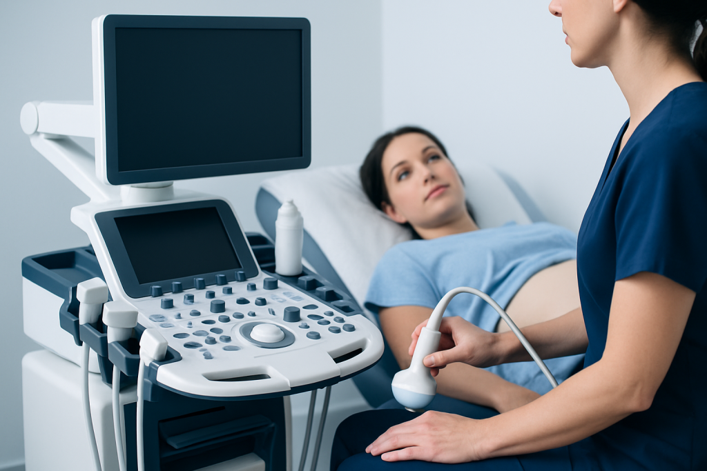
An ultrasound machine sends sound waves through a handheld device called a transducer. A clear gel is placed on the skin to remove air between the transducer and the body, allowing sound waves to travel effectively. As the waves encounter tissues and fluids, they reflect back in different ways. The machine converts these returning echoes into moving images.
For a standard external scan, the person usually lies on an examination couch while the sonographer moves the transducer over the area of interest. The examination is generally comfortable, although mild pressure may be needed to obtain clear images. Depending on the reason for the scan and the body area, it commonly takes around 15 to 45 minutes.
Some examinations use a slim, covered transducer inserted into the vagina or rectum to obtain closer images of pelvic or prostate structures. These are called transvaginal and transrectal ultrasounds. The clinician should explain why an internal examination is recommended, what it involves, and how privacy and comfort will be protected. A person can ask questions and discuss concerns before giving consent.
In selected settings, ultrasound can be combined with a small amount of contrast agent given through a vein. Contrast-enhanced ultrasound can help characterize blood flow or lesions in certain organs. It is not needed for most routine ultrasound examinations.
Are there two different types of ultrasounds?
There are more than two types of ultrasound. The most familiar is two-dimensional, or B-mode, ultrasound, which creates grayscale images of organs and tissues. This is often used for abdominal, pelvic, thyroid, breast, and pregnancy scans.
Doppler ultrasound is another important type. It displays and measures blood flow in blood vessels and the heart. Color Doppler adds color to show the direction and relative speed of blood flow, while spectral Doppler provides waveforms that help clinicians assess circulation. It may be used when evaluating suspected narrowing, blockage, abnormal blood flow, or certain pregnancy-related concerns.
Other types include three-dimensional and four-dimensional ultrasound, which can provide reconstructed or real-time moving 3D views in selected situations; echocardiography, which evaluates the heart; and musculoskeletal ultrasound, which examines tendons, muscles, joints, and superficial soft tissues. The best technique depends on the clinical question rather than personal preference.
- External ultrasound: the transducer moves over the skin.
- Internal ultrasound: the transducer is placed in a body opening for closer views.
- Doppler ultrasound: assesses blood flow as well as anatomy.
- Procedure-guiding ultrasound: helps clinicians position needles accurately during certain interventions.
Who may need an ultrasound and how to prepare
Ultrasound may be appropriate for people with symptoms, an abnormal physical examination, a follow-up need, or a screening indication recommended by their clinician. Common reasons include abdominal pain, pelvic symptoms, a lump or swelling, suspected gallstones, urinary concerns, thyroid changes, vascular symptoms, pregnancy monitoring, and evaluation of heart structure or function.
Preparation depends on the examination. An abdominal ultrasound may require avoiding food for several hours because a full gallbladder and reduced bowel gas can improve visibility. A pelvic ultrasound may require a comfortably full bladder. Other studies, including many thyroid, breast, soft-tissue, and vascular scans, usually need no special preparation.
People should follow the instructions supplied by the imaging department and let the care team know about relevant medications, recent procedures, allergies, pregnancy, or difficulties lying flat. They should not stop prescribed medication unless a clinician specifically advises it. Comfortable clothing and access to the area being scanned can make the visit easier.
Ultrasound can be especially useful when repeated imaging is needed because it does not use ionizing radiation. However, suitability still depends on the body part being assessed, the person’s symptoms, and whether another test could provide more complete information.
Why do doctors prefer ultrasound?
Doctors may prefer ultrasound when it can answer the clinical question safely and efficiently. It does not use X-rays or other ionizing radiation, can show real-time movement, and can assess flowing blood with Doppler methods. It is also portable, making it useful in outpatient clinics, emergency settings, operating rooms, and bedside care.
Real-time imaging is one of ultrasound’s particular strengths. It can show the movement of the heart, fetal activity, blood flow through vessels, and the way tendons or joints move. It can also help clinicians guide procedures, improving visualization of the intended target while avoiding nearby structures where possible.
Ultrasound is often a first imaging test rather than the final answer. If the images are unclear or an area requires more detailed assessment, the clinician may recommend CT, MRI, mammography, endoscopy, laboratory testing, or another investigation. This does not necessarily mean that anything serious has been found; it may simply reflect the strengths and limits of each test.
Results should be interpreted alongside symptoms, examination findings, and medical history. A normal or abnormal ultrasound result is only one piece of clinical information, and the requesting clinician can explain what it means for the individual.
Benefits, limitations, risks, and recovery
The main benefits of diagnostic ultrasound are that it is noninvasive, radiation-free, widely applicable, and able to provide immediate moving images. Most people can return to normal activities immediately after a routine external scan. There is usually no recovery period, no sedation, and no need for someone else to accompany the patient home.
Routine ultrasound is considered safe when performed by trained professionals for appropriate medical reasons. The gel may feel cool, and pressure from the transducer can be briefly uncomfortable over a tender area. Internal scans may cause temporary pressure or mild discomfort. Rare risks can be associated with a related invasive procedure, such as a biopsy, but these risks come from the procedure rather than the diagnostic ultrasound imaging itself.
Image quality can be affected by bowel gas, deep body structures, body habitus, wounds, dressings, or inability to hold a required position. Ultrasound also depends on the experience of the operator and the quality of the equipment. These practical limitations help explain why a clinician may recommend further testing even after an ultrasound has been performed.
At Acibadem International, multidisciplinary specialists in JCI-accredited hospitals can coordinate diagnostic imaging and follow-up care for international patients when ultrasound findings need further assessment.
What can ultrasound not detect?
Ultrasound cannot detect every condition, and its ability to identify a problem depends on the organ, location, size of a change, and image quality. Sound waves do not travel well through air or bone, so ultrasound has limited views of structures behind gas-filled bowel, inside the lungs, and within the adult brain through the skull. It may not be the best standalone test for deep structures in some parts of the body.
Small abnormalities, early disease, or subtle tissue changes may not always be visible. Ultrasound may identify a mass or an area of concern without being able to determine exactly what it is. In such cases, CT, MRI, mammography, blood tests, follow-up imaging, or a tissue sample may be needed for a more definitive diagnosis.
Ultrasound does not replace urgent assessment for severe symptoms. For example, sudden chest pain, signs of stroke, major injury, severe shortness of breath, or heavy bleeding need prompt medical evaluation using the tests most suitable for the situation. The right imaging choice is guided by clinical urgency and the suspected condition.
When to seek medical care
A person should contact a doctor if they have new, persistent, worsening, or unexplained symptoms that may need assessment, such as ongoing abdominal or pelvic pain, a new lump, swelling in one leg, urinary changes, unusual bleeding, or persistent jaundice. A clinician can determine whether ultrasound or another investigation is appropriate.
Urgent medical care is important for symptoms such as sudden severe pain, fainting, difficulty breathing, weakness or numbness on one side of the body, confusion, severe headache with neurological symptoms, heavy bleeding, or signs of a serious allergic reaction. Imaging should not delay emergency assessment.
After an ultrasound, patients should ask when and how results will be available, whether further tests are recommended, and what symptoms should prompt earlier review. If symptoms continue despite a normal scan, follow-up with a qualified clinician remains important because ultrasound may not show every possible cause.
Frequently asked questions
Is ultrasonography the same as ultrasound?
Yes. In most healthcare settings, ultrasonography and ultrasound refer to the same diagnostic imaging technique. Ultrasonography is the more formal term for producing images using ultrasound sound waves.
Does an ultrasound use radiation?
No. Diagnostic ultrasound uses high-frequency sound waves, not ionizing radiation such as X-rays. This is one reason it is commonly used for pregnancy and for repeated imaging when clinically appropriate.
Is an ultrasound painful?
Most external ultrasound examinations are painless. Pressure from the probe may be uncomfortable if the area is already sore, and internal ultrasound examinations can cause a temporary sensation of pressure.
How long does an ultrasound take?
Many ultrasound examinations take about 15 to 45 minutes, although timing varies by body area and the information needed. The imaging department can provide more specific guidance before the appointment.
Can ultrasound detect cancer?
Ultrasound can sometimes show a lump or other abnormal area that requires further assessment, but it cannot always confirm whether a change is cancer. Additional imaging, clinical evaluation, and sometimes a biopsy may be needed to establish a diagnosis.
Can I eat or drink before an ultrasound?
It depends on the type of scan. Some abdominal ultrasounds require fasting, while pelvic scans may require a full bladder; many other scans have no dietary restrictions. Patients should follow the preparation instructions from their imaging provider.
References
- World Health Organization
- Radiological Society of North America
- American College of Radiology
- National Institute for Health and Care Excellence
- U.S. Food and Drug Administration
This article is for general information only and is not a substitute for professional medical advice. Please consult a qualified doctor about your individual situation.
Ultrasound in Turkey — costs, top hospitals & a free quote
JCI-accredited · board-certified surgeons · reply within 24h
Add us as a Preferred Source to see more of our trusted health content across Google Search, AI Overviews and Discover.

Check-up & Preventive Medicine
Comprehensive health screenings designed for early detection and prevention.
2 specialists in this unit