Ultrasound of Trisomy 18: Preparation, Procedure and Results

Ultrasound can identify findings associated with trisomy 18 but cannot confirm or exclude it on its own. Some ultrasound findings are major structural differences, while soft markers are common and often nonspecific.
Key Takeaways
- Ultrasound can identify findings associated with trisomy 18 but cannot confirm or exclude it on its own.
- Some ultrasound findings are major structural differences, while soft markers are common and often nonspecific.
- A detailed anatomy scan, fetal echocardiography and genetic counseling may be recommended after concerning findings.
- Diagnostic testing such as chorionic villus sampling or amniocentesis can confirm whether the fetus has trisomy 18.
- Pregnancies affected by trisomy 18 usually benefit from coordinated care from maternal-fetal medicine, genetics, cardiology and neonatal specialists.
An ultrasound of trisomy 18 may show physical differences or growth concerns that raise suspicion for the condition, also called Edwards syndrome. Ultrasound findings are screening clues rather than a diagnosis, so results are usually considered alongside prenatal screening and, when appropriate, diagnostic genetic testing.
Overview: What an Ultrasound of Trisomy 18 Can Show
An ultrasound of trisomy 18 looks for fetal growth patterns and physical findings that can be associated with trisomy 18, a chromosome condition also known as Edwards syndrome. It may be performed during a first-trimester scan, a detailed mid-pregnancy anatomy scan, or later follow-up scans when there is a concern from screening or a prior ultrasound.
Ultrasound uses sound waves to create images of the pregnancy. It does not expose the pregnant person or fetus to ionizing radiation. While it can provide important information about fetal development, it cannot examine chromosomes directly. A normal scan does not fully rule out trisomy 18, and an abnormal scan does not establish a diagnosis.
When findings raise concern, clinicians usually review the pregnancy history, screening results and ultrasound images together. They may recommend genetic counseling, a targeted scan with a maternal-fetal medicine specialist, fetal heart assessment and an option for diagnostic genetic testing.
How the Scan Works, Who May Need It and How to Prepare
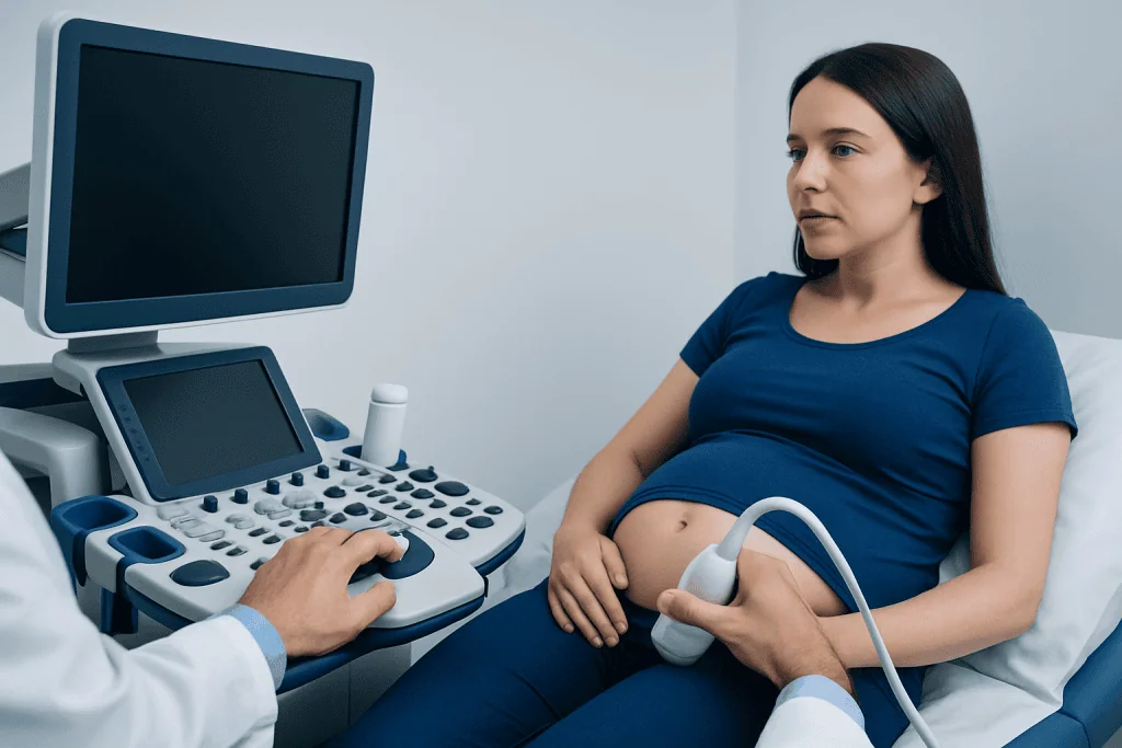
A standard prenatal ultrasound may be offered routinely during pregnancy. A more detailed examination may be advised if a screening test suggests an increased chance of trisomy 18, if an earlier scan shows a possible structural difference, if fetal growth is lower than expected, or if there is a relevant pregnancy or family history. The scan can also help clinicians plan appropriate follow-up care.
Most people do not need special preparation for a mid-pregnancy anatomy scan. The imaging center may give instructions about having a comfortably full bladder for an early pregnancy scan, particularly when the ultrasound is performed through the abdomen. Comfortable clothing that allows access to the abdomen is helpful.
Before the appointment, it can be useful to bring prior ultrasound reports and prenatal screening results. Patients may also wish to prepare questions, such as whether a finding is isolated, whether it changes the estimated chance of a chromosome condition, and which follow-up tests are available.
- First-trimester scans may assess fetal size, nuchal translucency and early anatomy.
- The detailed anatomy scan is commonly performed in the second trimester and evaluates multiple organ systems.
- Follow-up scans may monitor fetal growth, amniotic fluid and specific anatomical findings.
What Happens During the Procedure and Afterwards

For an abdominal ultrasound, the patient lies comfortably on an examination bed while a sonographer applies gel to the abdomen. A handheld device called a transducer is moved over the skin to obtain images of the fetus, placenta, umbilical cord and amniotic fluid. Pressure may be felt in some areas, but the examination is usually not painful.
Depending on fetal position and the amount of detail needed, a targeted examination may take longer than a routine scan. In early pregnancy, a transvaginal ultrasound may sometimes be used to obtain clearer images. This involves placing a slim covered probe into the vagina and is generally safe when clinically indicated.
There is no recovery period after a prenatal ultrasound. People can return to normal daily activities immediately. A clinician may discuss preliminary observations on the day, while a full report is reviewed by the responsible obstetric team. If images are limited by fetal position, a repeat scan may be arranged.
The main benefit is detailed, noninvasive information about fetal development. Ultrasound has no known harmful effects when used appropriately by trained professionals, but it has limitations: some findings may not be visible, and some findings can occur in babies without a chromosome condition.
What Are the Ultrasound Findings in a Baby With Trisomy 18?
There is no single ultrasound finding that proves trisomy 18. However, a pattern of growth concerns and structural differences can make the condition more likely. Findings vary widely, and some affected fetuses may have only subtle ultrasound changes, especially earlier in pregnancy.
Possible findings include fetal growth restriction; certain heart differences; abnormalities of the brain, kidneys or digestive tract; an omphalocele, where abdominal organs protrude into the base of the umbilical cord; and changes in the hands or feet. Clenched hands with overlapping fingers, a curved sole sometimes called a rocker-bottom foot, and a single umbilical artery may also be seen.
Other observations can include increased fluid behind the neck in the first trimester, choroid plexus cysts in the brain, changes in amniotic fluid volume, or differences in the placenta and umbilical cord. These findings do not have equal importance. The meaning depends on the specific finding, whether it is isolated or part of a pattern, gestational age and genetic screening results.
A detailed assessment may include fetal echocardiography because heart differences are common in trisomy 18. The purpose is to understand the fetal anatomy as clearly as possible and support informed discussions about testing, pregnancy monitoring and newborn care planning.
Should I Worry About Soft Markers on Ultrasound?
Soft markers are ultrasound features that are not usually harmful by themselves but can be linked with a somewhat higher chance of certain chromosome conditions. Examples may include a choroid plexus cyst, an echogenic intracardiac focus, mild enlargement of the kidney collecting system, or a short measurement of a long bone. Many are found in otherwise healthy pregnancies.
An isolated soft marker does not mean a baby has trisomy 18. Its significance depends on the exact marker, the quality and results of prior prenatal screening, and whether any structural differences are also present. In many cases, reassuring screening results and a normal detailed anatomy scan mean that no invasive testing is needed.
If more than one marker is present, or if a marker occurs with a structural difference or growth concern, a clinician may recommend consultation with maternal-fetal medicine and a genetic counselor. They can explain the possible meaning in the context of the individual pregnancy and discuss screening or diagnostic choices without pressure.
Diagnosis, Results and Pregnancy Care
Cell-free DNA screening, often called noninvasive prenatal testing (NIPT), can estimate the chance of trisomy 18 using a blood sample from the pregnant person. It is a screening test, not a diagnostic test. A higher-chance screening result should be confirmed with a diagnostic test before making major decisions about the pregnancy.
Chorionic villus sampling (CVS) and amniocentesis examine fetal genetic material and can diagnose trisomy 18. CVS is generally performed earlier in pregnancy, while amniocentesis is typically performed later. Both are invasive procedures with small procedure-related risks, which should be discussed carefully with the treating team.
If trisomy 18 is confirmed or strongly suspected, care is individualized. Maternal-fetal medicine, genetic counseling, fetal cardiology, neonatology, pediatric specialists and palliative care professionals may all contribute. Families can receive balanced information about expected medical needs, pregnancy monitoring, delivery planning and choices for comfort-focused or active newborn care.
Trisomy 18 is considered a high-risk pregnancy because it may involve fetal growth restriction, congenital differences, pregnancy complications and complex care needs around birth. High-risk does not mean that one outcome is certain; it means closer assessment and a tailored care plan are important.
Do Babies With Trisomy 18 Move a Lot in the Womb?
Fetal movement is not a reliable way to diagnose trisomy 18. Babies with trisomy 18 may move normally, less noticeably, or in a way that feels different to the pregnant person. Movement can also vary with gestational age, fetal sleep cycles, placental position and the individual pregnancy.
Some pregnancies affected by trisomy 18 have reduced fetal movement later in pregnancy, particularly if fetal growth restriction or other medical concerns are present. However, reduced movement has many possible causes and should never be interpreted as proof of a chromosome condition.
After the stage of pregnancy when regular movements are expected, a clear reduction or change in the baby’s usual movement pattern should be assessed promptly by the maternity team. Patients should follow the movement-monitoring advice provided by their own clinician rather than relying on comparisons with another pregnancy.
When to Seek Medical Care
Patients should contact their obstetrician, midwife or maternity unit promptly if they notice vaginal bleeding, leaking fluid, significant abdominal pain, fever, severe headache, vision changes, sudden swelling, or a reduction in the baby’s usual movements after movements have become established. These symptoms can occur for many reasons and deserve timely assessment.
It is also appropriate to request a discussion soon after an ultrasound report mentions soft markers, structural findings, low fetal growth or a possible chromosome concern. A clinician can explain the report in context and arrange referral to maternal-fetal medicine or genetic counseling when helpful.
Acibadem International’s multidisciplinary specialists and JCI-accredited hospitals support international patients requiring prenatal imaging, genetic evaluation and coordinated maternal-fetal care. A consultation can help families understand available testing and care pathways in a respectful, individualized way.
Frequently asked questions
Can ultrasound diagnose trisomy 18?
No. Ultrasound can identify findings that may be associated with trisomy 18, but it cannot confirm the chromosome condition. Confirmation requires diagnostic genetic testing, such as chorionic villus sampling or amniocentesis.
At what stage of pregnancy can trisomy 18 be seen on ultrasound?
Some possible signs can be seen in the first trimester, while many structural findings are assessed during the detailed second-trimester anatomy scan. The timing and visibility of findings vary between pregnancies. A later scan may be needed to clarify growth or anatomy.
Can a baby have trisomy 18 with a normal ultrasound?
Yes. A normal ultrasound reduces concern for major visible structural differences but does not rule out trisomy 18 completely. Some affected fetuses have subtle findings or findings that are not detectable on ultrasound.
What happens after an ultrasound suggests trisomy 18?
The care team usually reviews the images, pregnancy history and any prenatal screening results. They may recommend a targeted ultrasound, fetal echocardiography, genetic counseling and diagnostic testing. The patient can decide which testing and support options feel right for their situation.
Are soft markers always a sign of a genetic condition?
No. Soft markers are relatively common and often occur in babies without a chromosome condition. Their importance depends on whether they are isolated or occur with other findings, as well as prior screening results.
Is trisomy 18 inherited from a parent?
Most cases occur because of a random chromosome error during the formation of an egg or sperm and are not inherited from either parent. Less commonly, a chromosome rearrangement can be inherited. Genetic counseling can clarify recurrence considerations for an individual family.
References
- American College of Obstetricians and Gynecologists
- Society for Maternal-Fetal Medicine
- National Institutes of Health
- Centers for Disease Control and Prevention
- International Society of Ultrasound in Obstetrics and Gynecology
This article is for general information only and is not a substitute for professional medical advice. Please consult a qualified doctor about your individual situation.
Ultrasound in Turkey — costs, top hospitals & a free quote
JCI-accredited · board-certified surgeons · reply within 24h
Add us as a Preferred Source to see more of our trusted health content across Google Search, AI Overviews and Discover.

Check-up & Preventive Medicine
Comprehensive health screenings designed for early detection and prevention.
1 specialists in this unit