Upj Obstruction Surgery: Procedure, Recovery and Results

Pyeloplasty is the usual name for UPJ obstruction surgery and aims to preserve kidney drainage and function. Robotic, laparoscopic, and open approaches may be used, depending on anatomy, age, prior surgery, and surgical expertise.
Key Takeaways
- Pyeloplasty is the usual name for UPJ obstruction surgery and aims to preserve kidney drainage and function.
- Robotic, laparoscopic, and open approaches may be used, depending on anatomy, age, prior surgery, and surgical expertise.
- Most people have temporary discomfort, a urinary catheter or internal stent, and gradual recovery over several weeks.
- Follow-up imaging is important because symptom improvement alone does not fully confirm that drainage has improved.
- Urgent medical assessment is needed for fever with urinary symptoms, severe persistent flank pain, vomiting, or reduced urine output.
UPJ obstruction surgery is most often a pyeloplasty, a procedure that removes or reshapes a narrowed area where the kidney joins the ureter so urine can drain normally. It is used when obstruction causes symptoms, repeated infections, stones, or evidence that kidney function is at risk.
Overview: what UPJ obstruction surgery does
UPJ obstruction surgery treats narrowing or blockage at the ureteropelvic junction (UPJ), the point where urine leaves the kidney and enters the ureter. When this passage is too narrow, urine can collect in the kidney, causing swelling called hydronephrosis. The operation is designed to create a wider, dependable drainage pathway and reduce pressure on the kidney.
The operation is usually called pyeloplasty. During pyeloplasty, the surgeon removes the narrowed segment when appropriate and reconnects the healthy renal pelvis to the ureter. In some cases, a crossing blood vessel near the UPJ contributes to the blockage; the reconstruction can reposition the connection so urine can pass without compression.
UPJ obstruction can be present from birth, but it may be diagnosed later in childhood or adulthood. In adults, it is sometimes found during tests for flank pain, kidney stones, urinary infections, or unexpectedly enlarged kidneys on imaging. Surgery is not necessary for every person; decisions are based on symptoms, drainage studies, kidney function, and how these change over time.
Who may benefit from pyeloplasty
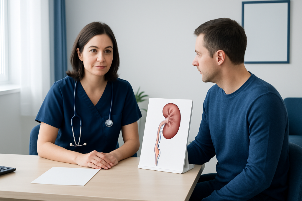
A urologist may discuss surgery when testing shows significant obstruction together with troublesome pain, recurrent urinary tract infections, stones, worsening hydronephrosis, or reduced function in the affected kidney. UPJ obstruction surgery in adults can also be considered when monitoring shows that kidney drainage or function is deteriorating, even if symptoms are limited.
Before recommending an operation, the care team considers the person’s overall health, previous abdominal or urinary surgery, kidney function, anatomy, and the likely cause of obstruction. Some mild, stable cases can be followed with repeat imaging and kidney function tests rather than treated immediately.
Assessment commonly includes ultrasound, CT or MRI urography in selected cases, urine testing, blood tests for kidney function, and a nuclear medicine renal scan. A renal scan can help show how each kidney functions and whether urine clears slowly after a tracer is given. These results help distinguish a true blockage from dilation that is not causing meaningful obstruction.
Children, adults, and people with a solitary functioning kidney require individualized planning. A urologist can explain whether observation, endoscopic treatment, or reconstructive surgery is most suitable for the person’s circumstances.
How pyeloplasty is performed, step by step
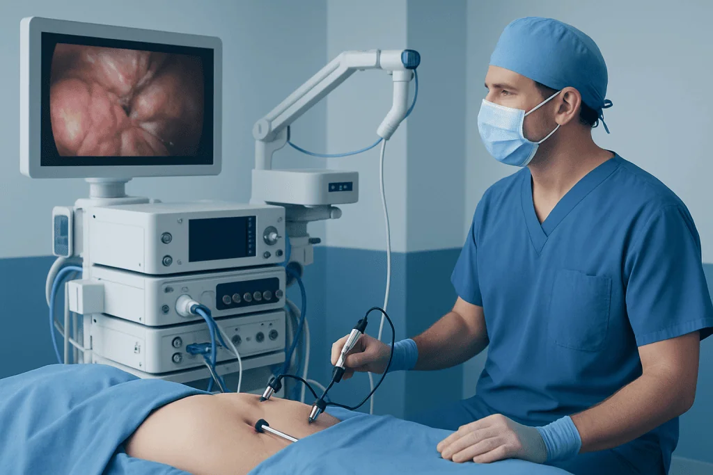
Pyeloplasty is performed under general anesthesia, so the patient is asleep and does not feel the procedure. The surgical approach may be open, laparoscopic, or robot-assisted laparoscopic. Minimally invasive approaches use several small incisions and specialized instruments, while an open operation uses one larger incision; both can provide effective reconstruction when appropriately selected.
After reaching the kidney and upper ureter, the surgeon identifies the narrowed area and any crossing vessel or scar tissue. The obstructed segment is commonly removed, the renal pelvis may be reshaped, and the healthy ureter is carefully reconnected to the kidney drainage system. This standard reconstruction is often called a dismembered pyeloplasty.
An internal ureteric stent is often placed across the repair to support healing and allow urine to drain. A bladder catheter may be used briefly after surgery, and some patients have a temporary drain near the incision. The exact plan varies with the approach and the findings during surgery.
Endoscopic procedures, such as an incision through the narrowed area, may be options in carefully selected situations but are not ideal for every anatomy or obstruction. A reconstructive urologist can discuss the expected advantages and limitations of each approach before treatment.
UPJ obstruction surgery recovery: timeline and aftercare
Recovery begins with monitoring in hospital for pain control, urine output, nausea, and early movement. Many people having laparoscopic or robotic surgery go home within a short hospital stay, while recovery after open surgery may require more time. The treating team gives instructions tailored to the operation, the patient’s health, and whether a drain or catheter remains in place.
At home, fatigue and incision tenderness are expected initially. Gentle walking, drinking fluids as advised, and avoiding constipation can support recovery. Heavy lifting, strenuous exercise, and driving while taking sedating pain medicine should be avoided until the surgeon confirms they are safe to resume.
An internal stent can cause temporary urinary frequency, urgency, mild burning, blood-tinged urine, or discomfort in the bladder or flank, especially with activity. These symptoms usually improve after the stent is removed, often several weeks after surgery. The team will arrange removal and explain whom to contact if symptoms become severe.
Follow-up visits and imaging are an essential part of UPJ obstruction recovery. Ultrasound and, when needed, a renal drainage scan help confirm that the kidney is draining well and that function is stable. The schedule differs between patients, so follow-up should continue for as long as the urologist recommends.
How painful is pyeloplasty recovery?
Pyeloplasty recovery involves some pain, but it is usually manageable with a planned combination of medicines and activity adjustments. Discomfort commonly comes from the incisions, deep tissue healing, gas-related shoulder discomfort after laparoscopic surgery, and sometimes the ureteric stent. Pain tends to be strongest in the first few days and should gradually improve rather than worsen.
Minimally invasive surgery often causes less incision-related discomfort than open surgery, although each person’s experience is different. The care team may use non-opioid pain relievers, local anesthetic techniques, and short-term stronger medicine when needed. Patients should use medicines only as directed and ask before adding over-the-counter products, particularly if they have kidney disease or take blood-thinning medication.
New severe pain, pain accompanied by fever, persistent vomiting, inability to pass urine, or rapidly increasing swelling around an incision needs prompt medical advice. These symptoms do not necessarily indicate a complication, but they should be assessed without delay.
Is pyeloplasty a major or minor surgery?
Pyeloplasty is a significant reconstructive operation because it involves repairing the connection between the kidney and ureter under general anesthesia. It is not considered a minor procedure, even when performed through small laparoscopic or robotic incisions. However, minimally invasive techniques may reduce incision size, hospital stay, and some aspects of recovery compared with an open approach.
The level of complexity depends on the anatomy and clinical context. Prior operations, scar tissue, a crossing blood vessel, stones, a very enlarged renal pelvis, or reduced kidney function can influence surgical planning. A surgeon should explain the planned approach, anesthesia considerations, expected recovery, and alternatives during informed consent.
Like all surgery, pyeloplasty carries risks. Possible complications include bleeding, infection, urine leakage from the repair, blood clots, injury to nearby structures, stent-related symptoms, recurrent narrowing, or need for further treatment. Serious complications are uncommon, but no procedure is risk-free.
How long does it take to recover from UPJ surgery?
UPJ obstruction surgery recovery time varies by surgical approach, age, baseline health, work demands, and whether complications occur. Many patients return to light daily activities within about one to two weeks after minimally invasive surgery, while more physically demanding activities and full energy levels may take several weeks. Open surgery generally involves a longer recovery period.
The internal stent, if used, may remain in place for a few weeks and can affect comfort during this period. A person may feel reasonably well before the stent is removed but still need to limit strenuous activity. Returning to work or school should be discussed with the surgeon, especially for jobs involving lifting, long travel, or physical exertion.
Internal healing and confirmation of improved drainage take longer than skin-incision healing. Follow-up tests may occur weeks to months after the operation. For this reason, recovery is best viewed as a process: early symptom improvement is encouraging, but scheduled imaging is needed to assess the repair properly.
What is the success rate of UPJ obstruction surgery?
Pyeloplasty has a high likelihood of successfully improving urine drainage and relieving obstruction, particularly when performed for a clearly documented UPJ blockage. Published outcomes often report durable success in more than 90% of appropriately selected patients, although results vary by surgical technique, definition of success, anatomy, previous treatment, and length of follow-up.
Success is measured by more than relief of pain. The urology team considers symptoms, ultrasound findings, renal scan drainage, kidney function, infections, and stone recurrence where relevant. Some people may have residual dilation of the kidney even after a successful repair, so imaging must be interpreted in context rather than treated as proof of ongoing obstruction by itself.
A small number of patients develop recurrent narrowing or persistent symptoms and may need further evaluation or treatment. Keeping follow-up appointments, reporting recurring flank pain or infections, and completing recommended imaging help identify concerns early.
Acibadem International’s multidisciplinary specialists and JCI-accredited hospitals diagnose and treat UPJ obstruction for international patients, with care plans guided by imaging, kidney function, and individual anatomy.
When to seek medical care
People with known or suspected UPJ obstruction should arrange a urology assessment if they have recurrent side or back pain, repeated urinary infections, nausea associated with pain episodes, blood in the urine, kidney stones, or imaging showing hydronephrosis. These symptoms can have several causes, so timely evaluation helps establish the correct diagnosis.
Urgent medical care is important for fever or chills with flank pain or urinary symptoms, severe or escalating pain, persistent vomiting, inability to keep fluids down, markedly reduced urine output, or feeling faint or very unwell. Infection behind an obstructed urinary system can require prompt treatment.
After surgery, patients should contact their surgical team for worsening pain, fever, heavy bleeding in the urine, pus or increasing redness at an incision, shortness of breath, leg swelling, or trouble urinating. Clear discharge instructions and access to the operating team are important parts of safe recovery.
Frequently asked questions
What is the name of UPJ obstruction surgery?
The most common UPJ obstruction surgery name is pyeloplasty. It reconstructs the narrowed connection between the kidney’s renal pelvis and the ureter to improve urine drainage. It may be performed using open, laparoscopic, or robot-assisted techniques.
Can UPJ obstruction surgery be done in adults?
Yes. UPJ obstruction may be diagnosed or become symptomatic in adulthood, and pyeloplasty is used in adults when obstruction is clinically significant. The choice of treatment depends on symptoms, kidney drainage, kidney function, anatomy, and overall health.
Will a stent be needed after pyeloplasty?
Many pyeloplasty procedures include a temporary internal ureteric stent to support the reconstructed area while it heals. It is commonly removed after several weeks, although the timing varies. Stents can cause temporary urinary urgency, frequency, or discomfort.
Can UPJ obstruction return after surgery?
Recurrent narrowing is possible but uncommon after a successful pyeloplasty. Follow-up imaging helps check drainage and kidney function, including when symptoms have improved. New flank pain, infections, or other urinary symptoms should be reported to a urologist.
How soon can someone exercise after pyeloplasty?
Gentle walking is usually encouraged soon after surgery, as advised by the care team. Strenuous exercise, heavy lifting, and contact sports are generally postponed until the surgeon confirms that healing is adequate. The timeline depends on the surgical approach and individual recovery.
Does pyeloplasty improve kidney function?
Pyeloplasty aims to protect kidney function by relieving pressure caused by impaired drainage. It may stabilize function and, in some cases, improve it, but the result depends on how much damage existed before treatment. Renal scans and blood tests are used to monitor the outcome over time.
References
- American Urological Association
- European Association of Urology
- National Institute of Diabetes and Digestive and Kidney Diseases
- Urology Care Foundation
- Mayo Clinic
This article is for general information only and is not a substitute for professional medical advice. Please consult a qualified doctor about your individual situation.
Explore treatments in Turkey — costs, top hospitals & a free quote
JCI-accredited · board-certified surgeons · reply within 24h
Add us as a Preferred Source to see more of our trusted health content across Google Search, AI Overviews and Discover.

More from the Health Library



Related Specialists

Asst. Prof. Dr. Ebru Kazancı
Neonatal Intensive Care Unit
Dyt. Simay Gülüş
Nutrition & Diet
Dr. Yalda Kargar
Inpatient Clinic Physicians
Prof. Dr. Gökhan Ergene
Thoracic Surgery