Valgus Stress Test: A Complete Medical Overview
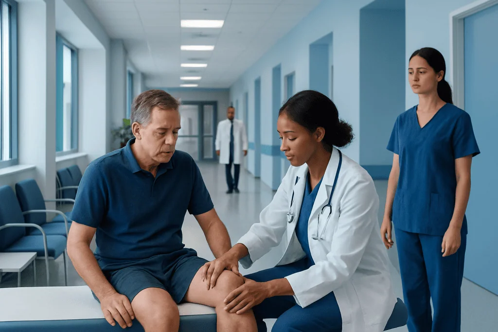
The valgus stress test is mainly used to evaluate the medial collateral ligament of the knee. A positive test may suggest an MCL sprain or tear, but it is not the only factor used to make a diagnosis.
Key Takeaways
- The valgus stress test is mainly used to evaluate the medial collateral ligament of the knee.
- A positive test may suggest an MCL sprain or tear, but it is not the only factor used to make a diagnosis.
- The test is usually performed with the knee both slightly bent and fully straight to assess different structures.
- Pain alone does not always mean a complete tear; abnormal looseness is also important.
- Further evaluation may include other knee exams, X-rays, ultrasound, or MRI depending on the injury.
The valgus stress test is a physical examination used to assess the inner side of the knee, especially the medial collateral ligament (MCL). It helps clinicians identify pain, looseness, or instability after a knee injury, but it is usually interpreted together with the person’s symptoms, history, and imaging when needed.
Overview: what the valgus stress test is
The valgus stress test is a hands-on knee examination that helps a clinician check the structures on the inner, or medial, side of the knee. In most cases, it is used to assess the medial collateral ligament, often called the MCL. This ligament helps keep the knee stable when force pushes the knee inward.
During the test, the examiner gently applies pressure to the outside of the knee while supporting the leg. This creates a controlled “valgus” force, meaning the knee is encouraged inward while the lower leg moves outward. The goal is to see whether this causes pain, a feeling of looseness, or more movement than expected compared with the other knee.
The test is commonly used after sports injuries, falls, twists, or direct blows to the outer side of the knee. It is not a treatment and it does not by itself confirm every detail of an injury. Instead, it is one part of a full clinical assessment that may also include medical history, swelling, range of motion, and additional ligament tests.
A clear understanding of the valgus stress test can help patients know what to expect during a knee examination. It can also make it easier to understand why a doctor may recommend rest, bracing, rehabilitation, or imaging if there is concern about an knee ligament injury.
How the test is performed
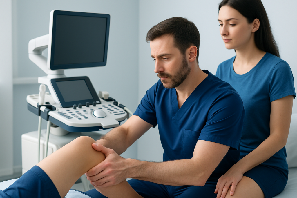
The valgus stress test is usually performed with the patient lying down and the leg relaxed. The examiner places one hand near the ankle and the other around the knee, then applies a gentle inward force at the knee while stabilizing the leg. The test is often done first at about 20 to 30 degrees of knee flexion and then again with the knee fully straight.
Testing the knee in slight flexion is especially useful because it reduces the contribution of some other stabilizing structures and focuses more on the MCL. If pain or excessive opening of the inner knee joint occurs in this position, it may suggest an MCL injury. Comparing the injured side with the uninjured knee can be very helpful, since normal joint looseness varies from person to person.
When the test is repeated with the knee fully extended, the findings may provide additional information. Abnormal laxity in full extension can suggest a more complex injury involving more than the MCL alone. For example, there may also be injury to deeper structures of the medial knee or to other major ligaments.
The exam is usually brief, but it may be uncomfortable if the knee is already painful or swollen. Clinicians generally perform it carefully and stop if discomfort is severe. In some cases, the initial exam is limited by pain, so reassessment later or imaging may be needed for a clearer picture.
What a positive test may mean
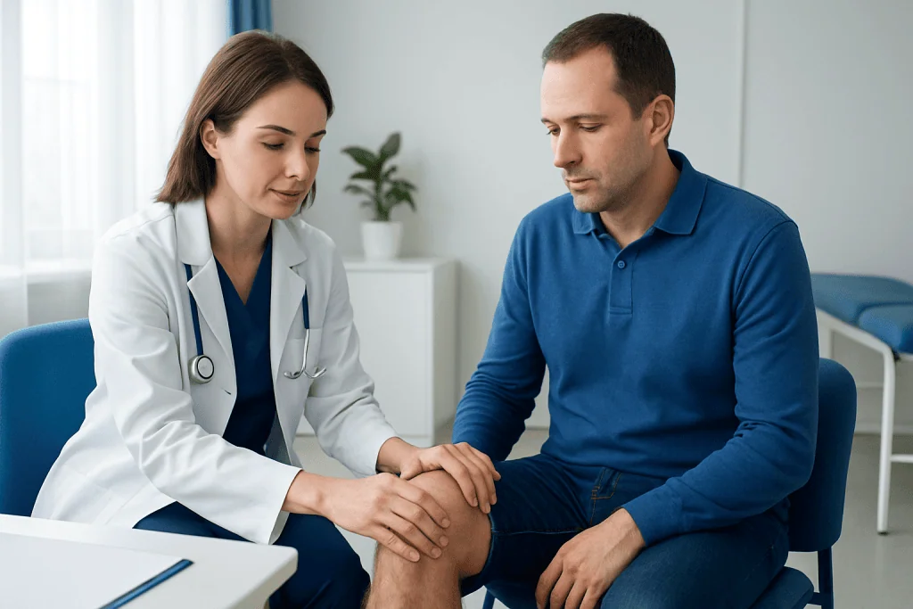
A positive valgus stress test usually means that the maneuver reproduces pain along the inner side of the knee, shows increased joint opening, or both. Pain can indicate a sprain or irritation of the MCL, while visible or palpable looseness may suggest a more significant partial or complete tear. The result is interpreted in the context of the person’s symptoms and injury mechanism.
Clinicians often think about MCL injuries in grades. A mild sprain may cause tenderness and pain with little or no instability. A more severe injury may allow noticeably greater movement compared with the opposite knee. However, grading can be difficult immediately after injury because swelling and muscle guarding may affect the exam.
It is also important to know what the test does not prove. A painful valgus stress test does not automatically mean surgery is needed, and a normal test does not always rule out every knee problem. Some injuries are subtle, and other sources of medial knee pain can mimic an MCL injury.
Because the knee functions as a unit, clinicians often combine this exam with other tests for the cruciate ligaments, menisci, and overall alignment. If there is concern for a more complex problem, the doctor may recommend imaging such as MRI to better define the injured tissues.
Symptoms and injuries linked to the test
The valgus stress test is most closely associated with suspected MCL injuries. These often happen when the outer side of the knee is hit during contact sports, or when the knee twists while the foot is planted. People may notice pain on the inner side of the knee, swelling, tenderness to touch, or a sense that the knee feels unstable.
Symptoms can range from mild soreness to significant difficulty walking. Some people can still bear weight, especially with lower-grade sprains, while others feel the knee may buckle during turning or side-to-side movement. Bruising may develop after a direct blow, but it is not always present.
Although the MCL is the main focus, other injuries may coexist. These can include damage to the anterior cruciate ligament, meniscus, or joint capsule. In those cases, symptoms may include popping at the time of injury, swelling inside the joint, catching, locking, or a stronger feeling of instability. Related problems may overlap with ACL tear patterns in some patients.
Doctors also consider other possible causes of medial knee pain, such as bone bruising, cartilage injury, tendon irritation, or less commonly a fracture. That is why the valgus stress test is most useful as part of a broader evaluation rather than as a stand-alone answer.
How doctors confirm the diagnosis
Diagnosis begins with the history. A clinician may ask how the injury happened, where the pain is located, whether swelling started quickly, and whether the knee gives way. The timing and mechanism of injury often provide important clues, especially in sports and trauma settings.
The physical examination usually includes inspection for swelling, bruising, and alignment; palpation of tender areas; and checks of motion, strength, and weight-bearing. The valgus stress test is then combined with other knee stability tests to build a more complete picture. Comparing both knees is a routine part of this process.
Imaging is not always needed for mild injuries, but it may be recommended if symptoms are severe, if another injury is suspected, or if recovery is not progressing as expected. Plain X-ray imaging may help rule out fracture or avulsion injury. MRI can show ligaments, cartilage, bone bruising, and meniscal damage in more detail.
In some settings, musculoskeletal ultrasound may also be used to evaluate soft tissues near the inner knee, particularly when performed by an experienced specialist. If a person has persistent instability, repeated swelling, or signs of multiple ligament injury, referral to an orthopedic or sports medicine specialist is often appropriate.
Treatment and recovery after an abnormal valgus stress test
Treatment depends on the underlying injury rather than the test result alone. Many MCL sprains improve with conservative care, including temporary activity modification, ice, compression, elevation, and a structured rehabilitation plan. A doctor may advise a brace for support, especially during walking or early return to sport.
Physical therapy is often central to recovery. Rehabilitation usually focuses on restoring range of motion, reducing swelling, improving quadriceps and hip strength, and retraining balance and movement patterns. A gradual return to activity helps lower the risk of repeat injury. Some people benefit from physical therapy and rehabilitation supervised by a specialist team.
Surgery is not required for most isolated MCL injuries, but it may be considered in select situations. These include severe instability, combined ligament injuries, certain avulsion patterns, or persistent symptoms despite appropriate non-surgical care. When a complex knee injury is present, treatment planning may involve sports medicine, orthopedics, imaging, and rehabilitation specialists.
Recovery time varies with injury severity, the presence of associated damage, and the person’s activity goals. A mild sprain may settle relatively quickly, while more significant injuries can take longer and may require a phased rehabilitation program. Following medical advice and not returning to high-risk activities too early are important for healing.
Self-care and when to seek medical care
After a possible medial knee injury, sensible early self-care can help. Rest from painful activities, use ice for short intervals, keep the leg elevated when possible, and consider a compression wrap if advised by a healthcare professional. Avoid movements that force the knee inward or cause it to give way. Supportive footwear and careful walking can also reduce strain.
It is reasonable to seek medical assessment if pain is moderate to severe, swelling is significant, or walking is difficult. Prompt care is especially important if the knee feels unstable, cannot fully straighten or bend, or if there was a clear twisting injury, direct impact, or audible pop. A clinician can determine whether the valgus stress test and further assessment are needed.
Urgent medical attention is appropriate if there is marked deformity, inability to bear weight, severe swelling right away, numbness, coldness of the foot, fever, or concern for fracture or dislocation. These features can point to injuries that should not be managed at home.
For people needing specialist evaluation, Acibadem International’s multidisciplinary teams in JCI-accredited hospitals diagnose and treat knee ligament conditions for international patients. Depending on the findings, care may include orthopedic assessment, advanced imaging, and rehabilitation, and in selected cases knee surgery.
Frequently asked questions
What does the valgus stress test check?
The valgus stress test mainly checks the medial collateral ligament, or MCL, on the inner side of the knee. It helps a clinician assess whether that part of the knee is painful, stretched, or unstable after injury.
Does a positive valgus stress test always mean an MCL tear?
No. A positive result can mean MCL irritation, a mild sprain, or a more significant tear, depending on the amount of pain and looseness found. Doctors usually combine the test with the injury history, other examination findings, and sometimes imaging.
Is the valgus stress test painful?
It can be uncomfortable if the inner side of the knee is already injured or inflamed. However, the test is usually brief and performed gently, and the examiner can stop if the pain becomes too strong.
Why is the test done with the knee bent and straight?
Testing at about 20 to 30 degrees of flexion helps focus more directly on the MCL. Repeating the test with the knee fully straight can show whether other stabilizing structures may also be involved.
Can imaging be needed after the valgus stress test?
Yes. X-rays may be used to check for fracture or bone injury, and MRI may be recommended if the injury seems more serious or if multiple structures may be affected. Imaging is especially useful when symptoms are severe or recovery is not progressing as expected.
How long does recovery take after an MCL injury?
Recovery depends on how severe the injury is and whether other knee structures are involved. Mild sprains may improve relatively quickly, while larger tears or combined injuries can take longer and may need bracing, rehabilitation, or specialist treatment.
References
- American Academy of Orthopaedic Surgeons
- American Orthopaedic Society for Sports Medicine
- National Institute of Arthritis and Musculoskeletal and Skin Diseases
- MedlinePlus
- BMJ Best Practice
This article is for general information only and is not a substitute for professional medical advice. Please consult a qualified doctor about your individual situation.
Joint and spine care in Turkey — expert assessment & treatment
JCI-accredited · board-certified surgeons · reply within 24h
Add us as a Preferred Source to see more of our trusted health content across Google Search, AI Overviews and Discover.

More from the Health Library
Sunken Eyes — Explained by Medical Evidence, Not Myths

Nad Injections: A Complete Medical Overview
Dream — Explained by Medical Evidence, Not Myths

Ejection Factor: A Complete Medical Overview

Heralopecia: What Patients Need to Know