Vascular Neurology Tests: MRI, CT, Angiography, and What Results Can Show
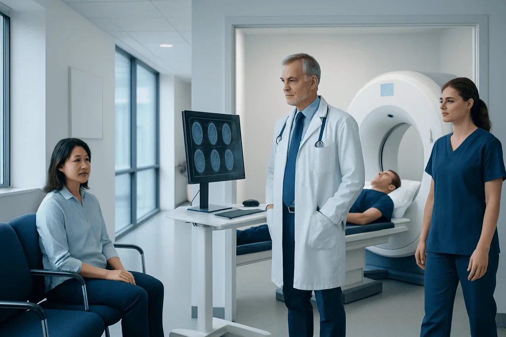
CT is often the first test in suspected stroke because it is fast and can quickly detect bleeding. MRI can show early brain injury and small areas of ischemia that may not be clear on CT.
Key Takeaways
- CT is often the first test in suspected stroke because it is fast and can quickly detect bleeding.
- MRI can show early brain injury and small areas of ischemia that may not be clear on CT.
- Angiography and vessel imaging help identify narrowed, blocked, or abnormal blood vessels in the brain and neck.
- Test results are interpreted together with symptoms, timing, medical history, and neurological examination.
- Early evaluation is important because some vascular brain conditions need urgent treatment.
Vascular neurology tests help doctors examine the brain and its blood vessels when stroke or other circulation problems are suspected. MRI, CT, and angiography can show bleeding, blocked arteries, narrowed vessels, tissue injury, and other findings that shape treatment decisions.
Overview of Vascular Neurology Tests
Vascular neurology focuses on conditions that affect blood flow to the brain and spinal cord. The most common reason for urgent testing is suspected stroke, but doctors also use these tests to investigate transient ischemic attack (TIA), brain hemorrhage, aneurysm, vascular malformations, artery narrowing, blood clots, and other circulation-related problems.
The main imaging tools include computed tomography (CT), magnetic resonance imaging (MRI), and studies that look directly at blood vessels, such as CT angiography, MR angiography, and catheter angiography. Each test provides different information. Some are best for speed, some for detail, and some for showing how blood moves through arteries and veins.
Doctors choose tests based on the person’s symptoms, how suddenly they began, whether bleeding or a clot is suspected, and whether treatment may be needed urgently. In many cases, imaging is done in stages. A person may first have a CT scan, followed by vessel imaging or MRI to clarify the cause and plan care.
When These Tests Are Used
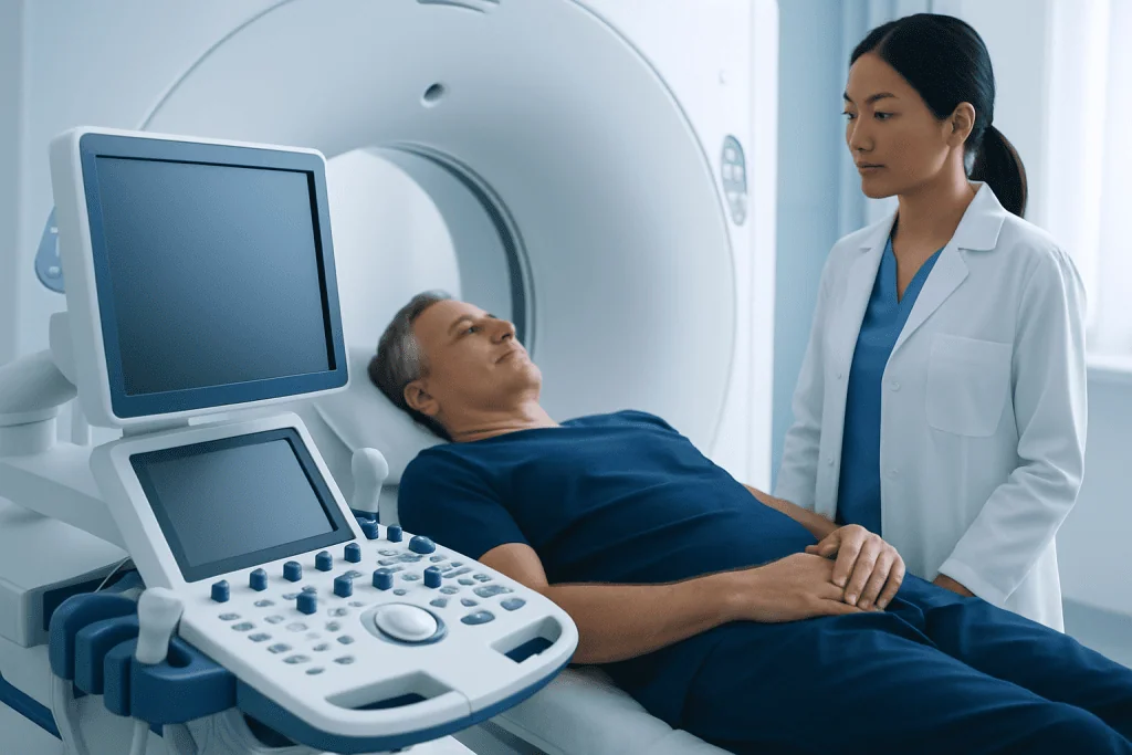
Vascular neurology tests are commonly ordered when someone has sudden neurological symptoms. These may include weakness on one side, numbness, trouble speaking, facial drooping, vision loss, severe dizziness, loss of balance, confusion, or a sudden severe headache. Because these symptoms can reflect reduced blood flow or bleeding in the brain, prompt testing is important.
Doctors may also use these tests after a brief episode of symptoms that has already improved. A TIA can be a warning sign of a future stroke, and imaging helps look for narrowed arteries, small areas of injury, or other causes. In addition, testing may be recommended when someone has risk factors such as high blood pressure, diabetes, smoking, high cholesterol, heart rhythm problems, or a history of vascular disease.
In some situations, testing is not done only in emergencies. It may be used to monitor a known aneurysm, follow up after stroke treatment, evaluate unusual headaches when a vascular cause is suspected, or investigate inherited or inflammatory conditions that affect blood vessels.
CT Scan: What It Shows
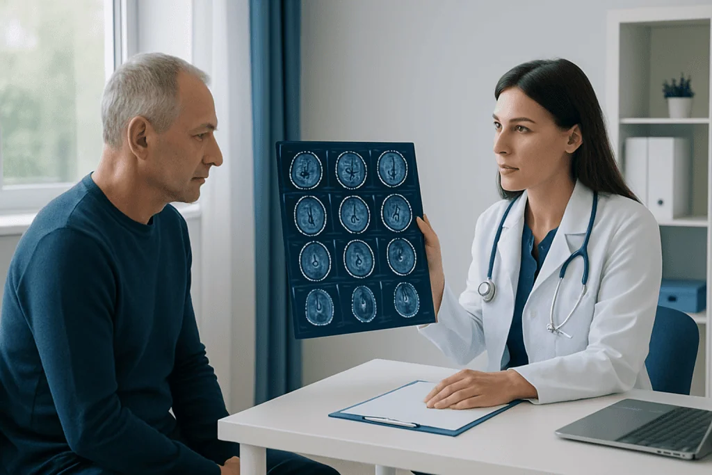
A non-contrast CT scan is often the first brain imaging test in suspected stroke because it is quick, widely available, and very useful for detecting bleeding. It can show whether symptoms are caused by a hemorrhagic stroke, bleeding around the brain, or another urgent problem such as swelling or a large mass effect. This is an important early step because treatment differs greatly between bleeding and clot-related stroke.
CT can also suggest signs of an ischemic stroke, which happens when blood flow is blocked. In the first hours, some changes may be subtle, but the scan can still help rule out bleeding and support rapid treatment decisions. Additional CT-based studies, such as CT angiography and CT perfusion, can provide more detail. CT angiography shows the arteries in the head and neck and may reveal a blockage, narrowing, dissection, or aneurysm. CT perfusion can help estimate which brain tissue is already injured and which tissue may still be at risk but potentially salvageable.
In practical terms, CT results may show:
- Bleeding inside or around the brain
- Signs of a large vessel blockage
- Narrowing of carotid or intracranial arteries
- Brain swelling or pressure effects
- Areas of reduced blood flow on perfusion imaging
Because of its speed, CT is a central part of emergency stroke evaluation and may be followed by treatments such as stroke treatment when appropriate.
MRI: What It Shows
MRI uses magnetic fields rather than radiation and provides highly detailed images of the brain. In vascular neurology, it is especially valuable for detecting ischemic stroke early. Diffusion-weighted MRI can show very small areas of recent brain injury that may not be visible on a standard CT scan, particularly in the early hours or in parts of the brain that are harder to assess with CT.
MRI can also help distinguish between old and new lesions, identify small strokes in the brainstem or cerebellum, and reveal patterns that suggest a specific cause, such as emboli from the heart, small vessel disease, or inflammation of blood vessels. Other MRI sequences can show bleeding products, vessel wall changes, swelling, and the effects of long-term vascular damage.
MR angiography, often performed during the same session, provides images of arteries without the need for a catheter in many cases. It can help detect stenosis, occlusion, aneurysm, or other abnormalities. MRI may be especially helpful when doctors are evaluating suspected stroke, a possible brain aneurysm, or symptoms that remain unexplained after initial CT imaging.
Although MRI offers excellent detail, it is not always the first test in emergencies because it may take longer and may not be suitable for everyone, such as some people with certain metal implants or severe claustrophobia. The care team weighs these factors when selecting the best study.
Angiography and Other Vessel Imaging
Angiography refers broadly to imaging that looks at blood vessels. In vascular neurology, this includes CT angiography, MR angiography, carotid ultrasound, and catheter cerebral angiography. These tests help doctors see whether blood vessels are narrowed, blocked, enlarged, twisted, inflamed, or leaking. They are often used when the question is not only whether a stroke occurred, but why it happened and where the vascular problem is located.
Catheter angiography is the most detailed vessel test. A specialist inserts a thin tube into an artery, usually through the wrist or groin, and guides it to the vessels supplying the brain. Contrast dye is then injected while X-ray images are taken. This test can show fine vessel anatomy and flow patterns very clearly. It is especially useful when evaluating aneurysms, arteriovenous malformations, vasculitis, dissections, or complex narrowing that may need treatment.
Because catheter angiography is more invasive than CT or MR angiography, it is generally reserved for situations where the extra detail matters or when a procedure may be performed at the same time. For example, the same approach may be used in interventional neuroradiology procedures to treat certain aneurysms, vessel malformations, or acute blockages.
Ultrasound of the carotid arteries may also be part of the workup. It is noninvasive and can show plaque buildup or narrowing in the neck arteries that supply the brain. This can be important in people with TIA or ischemic stroke, especially when doctors are looking for a preventable source of reduced blood flow.
How Doctors Interpret the Results
Imaging results are most useful when they are interpreted together with the person’s symptoms, physical examination, medical history, and the timing of symptom onset. For example, a small lesion on MRI may explain numbness or speech difficulty, while a large vessel blockage on CT angiography may indicate the need for urgent intervention. A normal scan does not always rule out a vascular event, especially very early in the course, so repeat or additional testing may sometimes be needed.
Doctors often look for several key questions in the results: Is there bleeding? Is there a blocked artery? How much brain tissue has been affected? Is there tissue at risk that might still recover? Is there an underlying cause, such as severe carotid narrowing, an aneurysm, an artery tear, or a heart-related embolic source? These answers help guide both emergency treatment and long-term prevention.
Results may also reveal chronic changes, such as white matter disease from long-standing high blood pressure, old silent strokes, or vessel calcification. These findings do not always explain current symptoms directly, but they can show an increased risk profile and support changes in treatment or lifestyle. In some cases, the interpretation leads to further evaluation with heart tests, blood work, or additional neurological assessment.
What to Expect During Testing
Most vascular neurology imaging tests are well tolerated. A CT scan is quick and painless, and the person lies still on a table that moves through the scanner. If contrast is used for CT angiography, there may be a warm sensation or a metallic taste for a brief time. MRI takes longer and can be noisy, but ear protection is provided, and staff give instructions throughout the exam.
Before contrast studies, the care team may ask about kidney function, allergies, pregnancy, and medications. People should mention any implanted medical devices before MRI. In emergencies, testing moves quickly, but the team still works to ensure safety and explain what is happening as clearly as possible.
Catheter angiography involves a more detailed preparation. Local anesthesia is usually used where the catheter is inserted, and some people receive medication to help them relax. After the procedure, the person is monitored for a period of time. The doctor will explain activity restrictions, what sensations are normal, and which symptoms should be reported promptly after going home.
After Diagnosis: Treatment, Prevention, and Follow-Up
Treatment depends on what the tests show. If imaging confirms an ischemic stroke, care may include medicines to restore or protect blood flow, or procedures to remove a clot in selected cases. If bleeding is found, treatment focuses on controlling the cause, reducing complications, and monitoring the brain closely. When tests identify carotid narrowing, aneurysm, or another vessel problem, doctors may recommend medication, surveillance imaging, or a targeted procedure.
Long-term care often aims to lower the risk of another event. This may include controlling blood pressure, cholesterol, and diabetes; stopping smoking; treating heart rhythm disorders; staying physically active; and following a balanced diet. Depending on the diagnosis, doctors may recommend antiplatelet or other blood-thinning medicines, but the right choice varies by person and should be discussed with a qualified clinician.
Follow-up imaging may be needed to confirm healing, monitor known vessel abnormalities, or assess recovery after treatment. People with a history of transient ischemic attack or stroke benefit from regular medical follow-up because risk reduction is an ongoing process. Near the end of the care pathway, some patients may also be referred for rehabilitation if they have speech, movement, or cognitive changes.
Anyone with sudden stroke-like symptoms should seek emergency care immediately rather than waiting for them to improve. For international patients who need evaluation or ongoing care, Acibadem International’s multidisciplinary specialists and JCI-accredited hospitals diagnose and treat vascular neurological conditions, including advanced imaging and procedures such as neurology care.
Frequently asked questions
Which test is usually done first in suspected stroke?
A non-contrast CT scan is often the first test because it is fast and can quickly show whether there is bleeding in the brain. This helps doctors decide on urgent treatment and whether further imaging is needed right away.
Is MRI better than CT for vascular neurology problems?
Neither test is always better in every situation. CT is often preferred first in emergencies because it is fast, while MRI can provide more detailed information about early ischemic injury and smaller strokes. Doctors choose based on the symptoms, timing, and clinical need.
What can angiography show that MRI or CT may not?
Angiography can provide a very detailed view of the brain’s blood vessels and blood flow patterns. It may show subtle narrowing, small aneurysms, vascular malformations, or vessel tears more clearly, especially when catheter angiography is used.
Do these tests always confirm the cause of symptoms?
Not always. Some scans can be normal early on, and some symptoms may have causes outside the blood vessels. Doctors often interpret imaging together with examination findings, blood tests, heart studies, and follow-up assessments.
Are contrast dyes safe?
Contrast agents are commonly used and are safe for many people, but they are not right for everyone. The medical team checks for kidney problems, prior reactions, pregnancy, and other factors before using contrast.
How long does it take to get results?
In emergencies such as suspected stroke, initial results are often reviewed very quickly so treatment decisions can be made without delay. A full formal report may follow, especially if several imaging studies are performed.
References
- World Stroke Organization
- American Heart Association
- National Institute of Neurological Disorders and Stroke
- Radiological Society of North America
- European Stroke Organisation
This article is for general information only and is not a substitute for professional medical advice. Please consult a qualified doctor about your individual situation.
Vascular Neurology in Turkey — costs, top hospitals & a free quote
JCI-accredited · board-certified surgeons · reply within 24h
Add us as a Preferred Source to see more of our trusted health content across Google Search, AI Overviews and Discover.
Check your numbers in seconds
BMI, calories, due date, blood pressure and 30+ more clinical calculators — free, instant, doctor-reviewed ranges.