Visual Field Defects: What Tunnel Vision or Missing Side Vision Can Mean
Visual field defects can appear as tunnel vision, blind spots, or missing areas of side or central vision. They may be caused by eye diseases, optic nerve problems, stroke, brain tumors, migraine, or other neurological conditions.
Key Takeaways
- Visual field defects can appear as tunnel vision, blind spots, or missing areas of side or central vision.
- They may be caused by eye diseases, optic nerve problems, stroke, brain tumors, migraine, or other neurological conditions.
- A visual field test helps map missing areas of vision and guides further evaluation.
- Sudden visual field loss needs urgent medical attention, especially if it occurs with weakness, headache, or speech changes.
- Treatment depends on the underlying cause and may involve eye care, neurological care, medication, or surgery.
Visual field defects are patterns of missing or reduced vision, often affecting side vision, central vision, or specific areas in one or both eyes. They are a symptom rather than a diagnosis and may be caused by conditions involving the eye, optic nerve, or brain.
Overview
Visual field defects are areas in which vision is reduced, blurred, or completely missing. A person may notice trouble seeing to the side, bumping into objects, difficulty reading parts of a page, or a dark or empty area in the visual scene. Some people describe this as tunnel vision, while others notice blind spots or a loss of vision in one half of what they see.
The visual field includes central vision, which is used for detailed tasks such as reading, and peripheral vision, which helps with orientation, walking, and detecting movement. A defect can affect one eye or both eyes, and the pattern often gives important clues about where the problem is located. It may arise in the retina, optic nerve, optic pathways, or visual areas of the brain.
Visual field defects are not a disease by themselves. Instead, they are a sign that something is affecting the visual system. Because causes range from common eye conditions to neurological disorders, a careful evaluation is important. In many cases, early diagnosis can help preserve vision and identify treatable medical conditions.
Symptoms and how visual field loss may feel
Symptoms vary depending on the location and size of the defect. Some people notice that side vision seems narrower than usual, making it harder to drive, walk in crowds, or detect objects approaching from the side. Others may miss words on a page, lose part of a face when looking at someone, or notice a patch of blurred or absent vision.
Visual field defects do not always cause obvious symptoms at first. The brain can sometimes compensate, especially if the change happens gradually. For this reason, disorders such as glaucoma may progress without early warning signs. A person may only become aware of the problem when the field loss becomes more advanced.
Common descriptions include:
- Tunnel vision, with reduced side vision
- Blind spots, also called scotomas
- Loss of vision on the right or left side
- Missing upper or lower areas of vision
- Difficulty reading or finding objects
- Bumping into door frames, steps, or people
Some people also have associated symptoms such as eye pain, headache, double vision, flashes of light, or sudden changes in vision. These accompanying features can help doctors identify whether the cause is mainly ocular, neurological, or both.
Causes and risk factors

Many different conditions can lead to visual field defects. In the eye itself, common causes include glaucoma, retinal disease, retinal detachment, and damage to the macula or other parts of the retina. The optic nerve can also be affected by inflammation, poor blood flow, compression, or injury. Problems farther back in the visual pathway, including the optic chiasm and occipital brain regions, can create characteristic patterns of field loss.
Neurological causes are especially important when the visual field defect affects both eyes in a similar pattern. Stroke can lead to loss of one half of the visual field, while pituitary tumors may affect side vision by pressing on the optic chiasm. Brain tumors, traumatic brain injury, multiple sclerosis, and infections or inflammation of the nervous system may also be involved. Migraine aura can sometimes cause temporary blind spots or shimmering areas, although this should be distinguished from more serious causes.
Risk factors depend on the underlying disorder but may include increasing age, diabetes, high blood pressure, smoking, family history of eye disease, vascular disease, autoimmune disorders, head injury, and certain medications. People with known retinal disease, optic nerve disease, or neurological conditions should report any new visual changes promptly.
Because the same symptom can have more than one explanation, self-diagnosis is not reliable. A specialist may need to evaluate whether the pattern suggests an eye condition such as retinal detachment or a neurological problem affecting the visual pathways.
How doctors diagnose visual field defects
Diagnosis begins with a detailed history. Doctors ask when the problem started, whether it came on suddenly or gradually, whether one or both eyes are affected, and whether there are related symptoms such as headache, weakness, pain, or flashing lights. They also review medical history, medications, and risk factors for eye and neurological disease.
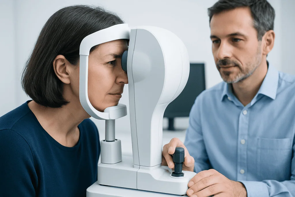
An eye and neuro-ophthalmic examination usually includes visual acuity, pupil assessment, eye movements, color vision, and examination of the optic nerve and retina. A visual field test is central to diagnosis because it maps the missing areas of vision. Automated perimetry is commonly used, but bedside confrontation testing may also provide useful early information.
Additional testing depends on the suspected cause. Imaging of the optic nerve or retina may help identify ocular disease, while brain and orbital imaging may be needed if a neurological lesion is suspected. Doctors may recommend MRI when the pattern of loss suggests a problem in the optic pathways or brain. In some cases, CT scan may be used, especially in urgent settings or when bone or acute bleeding must be assessed.
Blood tests or other neurological evaluations may also be appropriate in selected patients. The goal is not only to confirm that a visual field defect is present, but also to locate the source accurately so treatment can be directed to the underlying condition.
Treatment options
Treatment for visual field defects depends entirely on the cause. If glaucoma is responsible, care focuses on lowering eye pressure and protecting the optic nerve. If the defect is due to retinal disease, treatment may involve urgent retinal care. When a stroke, tumor, inflammation, or compressive lesion is involved, neurological or neurosurgical treatment may be needed.
Some conditions require prompt intervention to prevent further vision loss. For example, retinal detachment and certain optic nerve problems are time-sensitive. Compression of the visual pathways by a mass may need coordinated care with endocrinology, neurology, neurosurgery, and ophthalmology. Depending on the diagnosis, treatment may include medication, laser therapy, rehabilitation, or brain tumor surgery.
Even when the underlying cause has been treated, some visual field loss may persist. In these cases, vision rehabilitation can help people adapt more safely and confidently. Training strategies may improve scanning, reading, navigation, and home safety. Supportive care can be especially valuable for people with permanent field loss after stroke or optic nerve damage.
In complex cases, multidisciplinary assessment is important. Near the end of the diagnostic pathway or treatment planning, international patients may seek care at centers such as Acibadem International, where multidisciplinary specialists in JCI-accredited hospitals evaluate neuro-ophthalmic and neurological causes of visual field loss.
Prevention and self-care
Not all visual field defects can be prevented, but regular eye care and management of general health risks can reduce the chance of some causes progressing unnoticed. Routine eye examinations are especially important for people with diabetes, high blood pressure, a family history of glaucoma, or age-related eye disease. Early detection may identify changes before noticeable symptoms develop.
Healthy lifestyle measures also support eye and brain health. These include not smoking, controlling blood sugar and blood pressure, staying physically active, and following treatment plans for chronic conditions. Wearing protective eyewear during sports or hazardous work may help reduce trauma-related injuries.
People who already have a visual field defect can take practical steps at home and outdoors:
- Improve lighting in hallways, stairs, and reading areas
- Remove tripping hazards and keep walkways clear
- Turn the head more deliberately to scan the environment
- Use caution with driving until a doctor has assessed fitness to drive
- Attend follow-up appointments and repeat visual field testing as advised
Self-care should not replace medical evaluation. Any new, worsening, or unexplained loss of side vision or blind spots should be assessed by a qualified eye specialist or doctor.
When to see a doctor
A doctor should evaluate visual field defects whenever a person notices new tunnel vision, missing side vision, blind spots, or unexplained difficulty seeing in part of the visual scene. Gradual symptoms still deserve attention because they may reflect conditions that can worsen silently over time.
Urgent medical care is needed if vision loss starts suddenly, especially when it affects one side of vision in both eyes, or if it happens with severe headache, eye pain, weakness, numbness, confusion, trouble speaking, or imbalance. These symptoms may point to stroke, acute optic nerve disease, retinal detachment, or other emergencies.
Children, older adults, and people with known neurological or eye disease may not always describe symptoms clearly. Family members should pay attention to bumping into objects, reading difficulties, or unusual head turning. Timely assessment can help identify serious causes and support the best possible outcome.
Frequently asked questions
What are visual field defects?
Visual field defects are missing or reduced areas within the normal range of vision. They can affect central vision, side vision, or a specific section such as the upper or lower field. They are a symptom of an underlying eye, optic nerve, or brain problem rather than a diagnosis on their own.
Does tunnel vision always mean glaucoma?
No. Glaucoma is a well-known cause of peripheral vision loss, but tunnel vision can also occur with retinal disease, optic nerve disorders, neurological conditions, or rarely functional causes. A proper examination and visual field testing are needed to identify the reason.
Can visual field loss happen without affecting sharp central vision?
Yes. Some people still read an eye chart well but have significant side vision loss. This is one reason visual field testing is important, because standard vision tests alone may not detect the full extent of the problem.
Are visual field defects permanent?
Some are reversible or partly reversible if the cause is found and treated early, while others may be long-lasting. The outcome depends on what is causing the defect, how severe it is, and how quickly treatment begins. Follow-up care helps doctors monitor recovery or progression.
How is a visual field test performed?
A common visual field test asks the patient to look straight ahead and press a button when small lights appear in different areas. The test maps which parts of the visual field are seen clearly and which are missed. It is noninvasive and often repeated over time to monitor change.
When is side vision loss an emergency?
Sudden side vision loss should be treated as urgent, especially if it occurs with headache, weakness, trouble speaking, eye pain, or flashes and floaters. These symptoms can be linked to stroke, retinal detachment, or acute optic nerve problems. Prompt medical attention is important.
References
- American Academy of Ophthalmology
- National Eye Institute
- American Academy of Neurology
- National Institute of Neurological Disorders and Stroke
- Mayo Clinic
This article is for general information only and is not a substitute for professional medical advice. Please consult a qualified doctor about your individual situation.
Explore treatments in Turkey — costs, top hospitals & a free quote
JCI-accredited · board-certified surgeons · reply within 24h
Add us as a Preferred Source to see more of our trusted health content across Google Search, AI Overviews and Discover.
Check your numbers in seconds
BMI, calories, due date, blood pressure and 30+ more clinical calculators — free, instant, doctor-reviewed ranges.