What Bones Make Up the Knee Joint? Anatomy Explained

The knee joint is mainly formed by the femur, tibia, and patella. Cartilage, menisci, and ligaments help the bones move smoothly and stay stable.
Key Takeaways
- The knee joint is mainly formed by the femur, tibia, and patella.
- Cartilage, menisci, and ligaments help the bones move smoothly and stay stable.
- The knee is one of the body’s largest and most heavily used joints.
- Pain, swelling, locking, or instability can signal a knee problem that needs evaluation.
- Good muscle strength, healthy body weight, and safe movement habits help protect the knee.
The knee joint is built from three main bones: the femur, tibia, and patella. Together, they create a complex hinge-and-gliding joint that supports movement, balance, and body weight.
Overview
The knee is a large, complex joint that connects the thigh to the lower leg. When people ask what bones make up the knee joint, the short answer is that three bones work together there: the femur, tibia, and patella.
The femur is the thigh bone, the tibia is the larger shin bone, and the patella is the kneecap. These bones do not simply touch each other; they are shaped to fit together and are supported by cartilage, ligaments, tendons, and surrounding muscles to allow bending, straightening, and weight-bearing.
The knee must balance mobility with stability. It lets a person walk, climb stairs, squat, sit, and stand, while also absorbing force during daily activities and exercise. Because of this constant use, even small problems in the knee’s anatomy can affect comfort and function.
The Bones of the Knee
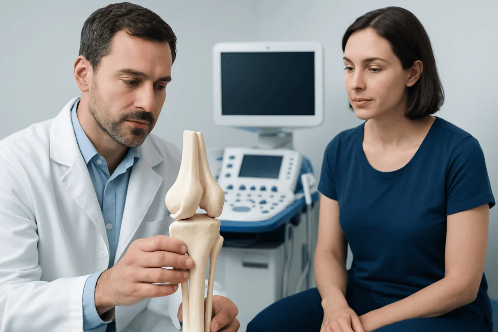
The knee joint includes three bones, each with a distinct role. The femur ends in rounded bony surfaces called the femoral condyles, which meet the top of the tibia. The tibia provides the main weight-bearing surface of the lower leg and forms the base of the joint.
The patella sits in front of the knee and moves within the groove of the femur as the knee bends and straightens. It improves the leverage of the thigh muscles, helping the leg extend more efficiently. Although the patella is often called a “floating” bone, it is firmly connected by tendons and plays an important mechanical role.
In addition to these bones, the knee also includes the fibula nearby. The fibula does not form the main knee joint surface, but it supports the lower leg and serves as an attachment point for some soft tissues. Understanding the relationship between these bones helps explain why the knee can be both strong and vulnerable.
How the Knee Works
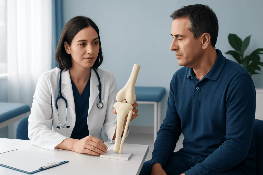
The knee is often described as a hinge joint, because it mainly bends and straightens. However, it also allows a small amount of rotation and gliding, especially when the knee is bent. This more complex movement is possible because the bone surfaces are shaped to fit together while soft tissues guide motion.
Articular cartilage covers the ends of the femur, tibia, and patella where they meet. This smooth tissue helps reduce friction and cushions the joint during movement. The menisci, which are two crescent-shaped pads of cartilage between the femur and tibia, further distribute weight and absorb shock.
Ligaments hold the bones in proper alignment, while muscles and tendons move and stabilize the knee. The quadriceps on the front of the thigh, for example, work with the patella to straighten the knee. When all of these structures function well together, the knee moves efficiently and safely.
Symptoms of Knee Problems
When the bones or surrounding structures of the knee are irritated or injured, symptoms may develop. Common signs include pain, swelling, stiffness, tenderness, or a feeling that the knee is giving way. Some people also notice clicking, catching, or reduced range of motion.
Symptoms can vary depending on the cause. A cartilage injury may cause locking or catching, while arthritis may lead to stiffness and aching that is worse after activity or after sitting for a long time. A kneecap problem may cause pain in the front of the knee, especially when going up or down stairs.
It is helpful to pay attention to whether symptoms started after an injury or developed gradually. That information can help a doctor determine whether the issue is related to bone, cartilage, ligament, tendon, or inflammation in the joint.
Causes & Risk Factors
Knee problems can come from many causes, and not all are related to the bones themselves. Injuries, overuse, arthritis, poor alignment, muscle weakness, and age-related wear can all affect how the knee bones and joint surfaces function.
Common risk factors include repetitive impact sports, sudden twisting movements, excess body weight, weak thigh or hip muscles, and previous knee injury. Certain occupations or activities that involve frequent kneeling, squatting, or climbing may also place extra stress on the knee.
Some conditions are more likely to affect one part of the knee than another. For example, osteoarthritis often affects the cartilage and bone surfaces in the joint, while patellofemoral pain is commonly related to how the kneecap tracks across the femur. A doctor may also consider inflammation, autoimmune conditions, or structural differences in the knee.
Diagnosis
Doctors usually begin with a medical history and a physical examination. They ask about symptoms, activity level, any injuries, and whether the knee feels unstable, swollen, or stiff. During the exam, they may check walking pattern, range of motion, tenderness, alignment, and ligament stability.
If needed, imaging tests can help show the bones and surrounding tissues. X-rays are commonly used to assess bone alignment, joint space, and signs of arthritis or fracture. In some cases, MRI may be recommended to evaluate cartilage, menisci, ligaments, or subtle bone injury.
Other tests are occasionally used depending on the suspected problem. The goal is not only to identify which bones are involved, but also to understand the soft tissues that help the knee function. This combined approach supports a more accurate diagnosis and treatment plan.
Treatment Options
Treatment depends on the cause of the knee problem, the structures involved, and how much the knee is limiting daily life. Many conditions start with conservative care such as rest from aggravating activity, ice, compression, elevation, and guided exercise or physical therapy. Pain relief medicines may be suggested by a doctor when appropriate.
Some knee issues benefit from supportive devices or targeted rehabilitation. Braces, taping, shoe changes, or strengthening exercises may improve alignment and reduce stress on the joint. For certain injuries or advanced joint conditions, a doctor may discuss injections or surgery. In some cases, knee arthroscopy is used to diagnose and treat internal joint problems through small incisions.
When arthritis or major joint damage is present, treatment may focus on reducing symptoms and preserving function for as long as possible. If structural damage is severe, joint replacement may be considered. A specialist can explain which option fits the person’s anatomy, symptoms, and activity needs.
Prevention & Self-care
While not every knee problem can be prevented, several habits may reduce stress on the joint. Regular strengthening of the quadriceps, hamstrings, glutes, and calf muscles helps support the bones and keep the knee aligned during movement. Gentle flexibility work can also improve comfort and mobility.
Maintaining a healthy body weight can reduce load on the knee, especially during walking, climbing stairs, and exercise. It also helps protect cartilage and other joint structures over time. Using proper technique in sports and gradually increasing activity levels may lower the risk of strain or overuse.
Practical self-care includes wearing supportive shoes, warming up before activity, avoiding sudden increases in training, and allowing time for recovery after heavy use. If a person has a history of knee pain, early attention to symptoms may prevent small problems from becoming long-lasting ones.
When to See a Doctor
A doctor should evaluate knee pain that lasts more than a few days, keeps returning, or interferes with walking, sleeping, or daily activities. Medical review is also important if the knee is swollen, unstable, difficult to bend or straighten, or painful after an injury.
People should seek prompt assessment if they cannot bear weight, the knee appears deformed, there is severe swelling, or the joint locks and will not move normally. These signs may suggest a fracture, significant soft tissue injury, or another condition that needs timely care.
For ongoing knee concerns, an orthopedic specialist can help identify which bones and supporting structures are involved and recommend the most appropriate treatment. Acibadem International’s multidisciplinary specialists and JCI-accredited hospitals diagnose and treat knee conditions for international patients in a coordinated, patient-centered way.
Frequently asked questions
What are the three bones that form the knee joint?
The knee joint is formed by the femur, tibia, and patella. The femur is the thigh bone, the tibia is the larger shin bone, and the patella is the kneecap. Together, they create the main structure of the knee.
Is the fibula part of the knee joint?
The fibula is close to the knee, but it is not one of the main bones that form the knee joint surface. It supports the lower leg and provides attachment points for muscles and ligaments. The primary knee bones are the femur, tibia, and patella.
Why is the kneecap important?
The kneecap helps the thigh muscles straighten the knee more efficiently. It also helps protect the front of the joint. Without the patella, knee movement would be less powerful and less stable.
What other structures support the knee besides bone?
The knee also depends on cartilage, menisci, ligaments, tendons, and muscles. These tissues cushion the joint, keep the bones aligned, and allow smooth movement. If one part is injured, the whole joint can be affected.
What does knee pain usually mean?
Knee pain can come from many causes, including overuse, injury, arthritis, or inflammation. The location and type of pain often give clues about which part of the knee is involved. A doctor can help determine the exact cause.
Can knee anatomy problems improve without surgery?
Many knee problems improve with conservative treatment such as rest, physical therapy, activity modification, and supportive care. Surgery is usually considered only when symptoms are severe, persistent, or caused by a problem that does not respond to non-surgical treatment.
References
- American Academy of Orthopaedic Surgeons
- National Institute of Arthritis and Musculoskeletal and Skin Diseases
- Mayo Clinic
- Cleveland Clinic
- Merck Manual Professional Edition
This article is for general information only and is not a substitute for professional medical advice. Please consult a qualified doctor about your individual situation.
Joint and spine care in Turkey — expert assessment & treatment
JCI-accredited · board-certified surgeons · reply within 24h
Add us as a Preferred Source to see more of our trusted health content across Google Search, AI Overviews and Discover.

More from the Health Library



Related Specialists

Prof. Dr. Mehmet Cihat Ünlü
Gynecology & Obstetrics
Assoc. Prof. Dr. Deniz Atasoy
General Surgery
Assoc. Prof. Dr. Cem Gün
Emergency Service
Dr. Sinem Altunyuva Usta
Pediatric Cardiology