What Does Breast Cancer Look Like on a Mammogram? Causes, Explanations, and Next Steps
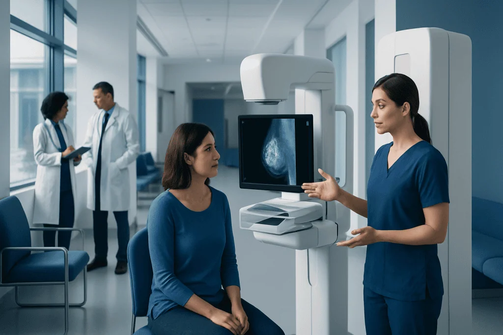
Many abnormal mammogram findings are benign and do not mean cancer is present. Breast cancer on a mammogram may appear as an irregular mass, suspicious calcifications, or architectural distortion.
Key Takeaways
- Many abnormal mammogram findings are benign and do not mean cancer is present.
- Breast cancer on a mammogram may appear as an irregular mass, suspicious calcifications, or architectural distortion.
- Radiologists use additional views, ultrasound, MRI, and sometimes biopsy to clarify uncertain findings.
- A mammogram can suggest cancer, but it cannot diagnose it with certainty on its own.
- Prompt follow-up after an abnormal mammogram helps rule out harmless causes or detect cancer early.
What does breast cancer look like on a mammogram? Often, suspicious areas appear as a dense irregular mass, clusters of tiny calcifications, or distortion of normal breast tissue—but many abnormal mammogram findings turn out to be noncancerous. The next step is usually more detailed imaging and, if needed, a biopsy to confirm the cause.
Overview: what a suspicious mammogram can show
When people ask what does breast cancer look like on a mammogram, the most helpful first answer is reassuring: many changes seen on screening mammograms are not cancer. Cysts, fibroadenomas, dense breast tissue, old injuries, benign calcifications, and normal overlapping tissue can all create findings that look unusual at first and need a closer look.
When breast cancer is visible on a mammogram, it often appears as one of a few patterns. These include a mass with irregular or spiculated edges, clusters of fine calcifications in a suspicious pattern, or architectural distortion, which means the normal tissue looks pulled or disrupted even if a clear lump is not obvious.
A mammogram is a very useful screening tool, but it is not the final diagnosis. Its role is to identify areas that deserve more attention. If something unusual is seen, the doctor usually recommends diagnostic mammography, ultrasound, and sometimes MRI or biopsy to find out exactly what it is.
How breast cancer may appear on a mammogram
Radiologists look for several visual clues when reviewing a mammogram. A suspicious breast mass may look denser than the surrounding tissue and may have uneven, irregular, or star-like borders. These spiculated margins can be more concerning than a round, smooth, well-defined lump, which is more often associated with a benign cause.
Another pattern involves calcifications, which are tiny white spots seen on the image. Many calcifications are harmless, especially when they are larger, scattered, or clearly related to aging or prior inflammation. However, tightly grouped, fine, irregular calcifications can sometimes be associated with early breast cancer, including ductal carcinoma in situ.
Architectural distortion is another red flag. In this pattern, the breast tissue looks distorted, as though it is being tugged inward, but there may not be a distinct mass. Mammograms can also show asymmetry, where one area looks denser than the matching area in the other breast. Some asymmetries are due to normal tissue overlap, while others need more evaluation.
Not all breast cancers are easy to see on a mammogram. In dense breasts, cancer can be harder to detect because both dense tissue and many cancers appear white on the image. That is one reason follow-up imaging is sometimes needed even when the original mammogram is only mildly abnormal.
Why abnormal findings are often not cancer
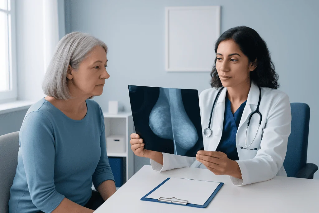
An abnormal mammogram result can be stressful, but it is important to remember that many findings are benign. Common noncancerous explanations include cysts, fibroadenomas, fat necrosis after trauma, benign calcifications, and overlapping tissue that creates the appearance of a shadow or asymmetry on a standard screening image.
Breast density is another important factor. Dense breasts are common and are not a disease, but dense tissue can make mammograms harder to interpret. A normal structure can sometimes look suspicious on one view and then appear clearly harmless on additional views. This is why call-backs after screening are relatively common.
Hormonal changes, prior breast surgery, breastfeeding history, infections, and aging can also affect mammogram appearance. Radiologists compare the current study with previous mammograms whenever possible because stability over time often suggests a benign process, while a new or changing finding may need further testing.
If a report mentions a lesion, asymmetry, calcifications, or distortion, that wording alone does not mean breast cancer has been found. It means the image needs interpretation in context. The next steps are designed to clarify the finding, not to assume the worst.
What doctors consider alongside the image
Doctors do not rely on the picture alone. They also consider age, breast density, personal history, family history, prior biopsies, hormonal factors, and whether there are symptoms such as a new lump, nipple discharge, skin dimpling, or focal breast pain. Together, these details help determine how likely a finding is to be benign or suspicious.
Radiologists classify mammogram findings using the BI-RADS system. This standardized system describes how concerning a finding appears and what should happen next. For example, some findings are clearly benign and only need routine screening, while others are probably benign and may be rechecked in a short interval. More suspicious findings usually lead to biopsy.
A patient with a normal mammogram but ongoing symptoms may still need additional evaluation. Mammography is excellent but not perfect, especially in dense breast tissue. If symptoms persist, a doctor may recommend ultrasound, MRI, or referral to a breast specialist to assess for conditions such as breast cancer or other breast disorders.
Next steps after an abnormal mammogram
The first follow-up step is often a diagnostic mammogram. Unlike a screening exam, this uses extra views, magnification, or spot compression to look more closely at the area in question. In many cases, these additional images show that the finding is due to normal tissue overlap or a benign pattern of calcification.
Breast ultrasound is commonly used when there is a lump or a localized abnormality on mammography. Ultrasound can help show whether an area is a fluid-filled cyst or a solid mass and can provide more information about margins and shape. In some situations, a breast MRI may be recommended, especially for high-risk patients or when mammogram and ultrasound results do not fully explain the finding.
If imaging remains suspicious, a biopsy is the only way to confirm whether cancer is present. This is often done with image guidance, using a needle to collect tissue from the area. Depending on the location and appearance, doctors may use breast biopsy, stereotactic guidance, or ultrasound-guided sampling. If cancer is diagnosed, treatment planning may include surgery and other therapies such as breast cancer surgery or medical oncology.
The timing of each step depends on the imaging findings and clinical picture. Most people are guided through this process in an organized, stepwise way, with the aim of avoiding unnecessary procedures while making sure significant abnormalities are not missed.
When to seek medical care
A person should contact a doctor promptly if they receive a call-back after a mammogram and are unsure what it means. Most call-backs are not emergencies, but they should not be ignored. Timely follow-up helps clarify whether the finding is harmless or needs further testing.
Medical review is also important if there is a new breast lump, nipple inversion that is new, bloody nipple discharge, persistent skin thickening, dimpling, redness that does not improve, or swelling in one breast or underarm. Even if a screening mammogram was recently normal, ongoing symptoms still deserve evaluation.
If a person has a strong family history of breast or ovarian cancer, known genetic risk, or previous breast cancer, they may need a personalized imaging plan. In these situations, doctors may recommend more frequent screening or additional imaging beyond standard mammography.
Near the end of the diagnostic pathway, some people may benefit from care in a specialized center. Acibadem International’s multidisciplinary specialists and JCI-accredited hospitals evaluate breast findings and provide coordinated care for international patients when more advanced testing or treatment is needed.
Prevention, screening, and self-care after results
While no test can prevent breast cancer, regular screening helps find changes earlier, when treatment is often more effective. The right screening schedule depends on age, risk factors, breast density, and local medical guidance. A doctor can help determine when to start mammograms and whether extra imaging is appropriate.
Keeping previous mammograms and bringing them to new appointments can be very helpful. Comparison with older images often allows radiologists to see whether a finding is new or unchanged. This may reduce unnecessary concern and avoid repeat testing.
General breast health habits support overall well-being, even though they cannot guarantee prevention. These include maintaining a healthy weight, limiting alcohol, staying physically active, and attending recommended checkups. Patients should also become familiar with their normal breast appearance and feel, so new changes are easier to notice.
After an abnormal result, self-care matters too. It can help to ask for a copy of the report, write down questions, and bring a trusted person to appointments. Clear information and prompt follow-up usually make the process more manageable and less overwhelming.
Frequently asked questions
Does an abnormal mammogram mean breast cancer?
No. Many abnormal mammogram findings are caused by benign conditions such as cysts, fibroadenomas, dense tissue, or overlapping breast structures. Additional imaging or biopsy may be needed to determine the exact cause.
What is the most concerning appearance of breast cancer on a mammogram?
Radiologists may be more concerned by an irregular dense mass with spiculated edges, suspicious clusters of fine calcifications, or architectural distortion. These patterns do not confirm cancer by themselves, but they often require further evaluation.
Can breast cancer be missed on a mammogram?
Yes, this can happen, especially in people with dense breast tissue. That is why ongoing symptoms such as a new lump or nipple changes should still be assessed even if a recent mammogram was normal.
What happens after a suspicious mammogram result?
The next step is usually a diagnostic mammogram with additional views and often a targeted ultrasound. If the finding remains unclear or suspicious, the doctor may recommend MRI or a biopsy to confirm the diagnosis.
Are calcifications on a mammogram always a sign of cancer?
No. Calcifications are common and are often benign. The radiologist looks at their size, shape, number, and pattern, because certain clustered or irregular calcifications are more suspicious than others.
Can benign lumps look like cancer on a mammogram?
Yes. Some benign breast changes can resemble cancer on initial imaging, which is why extra views and ultrasound are often needed. A biopsy may be recommended if imaging cannot confidently show the area is benign.
References
- American Cancer Society
- National Cancer Institute
- Centers for Disease Control and Prevention
- Radiological Society of North America
- American College of Radiology
This article is for general information only and is not a substitute for professional medical advice. Please consult a qualified doctor about your individual situation.
Breast Cancer in Turkey — costs, top hospitals & a free quote
JCI-accredited · board-certified surgeons · reply within 24h
Add us as a Preferred Source to see more of our trusted health content across Google Search, AI Overviews and Discover.

Medical Oncology Department
Medical treatment of cancer with chemotherapy, immunotherapy and targeted therapies under a multidisciplinary tumor board.
60 specialists in this unit