What Is a PET-MRI Scan and When Is It Used?
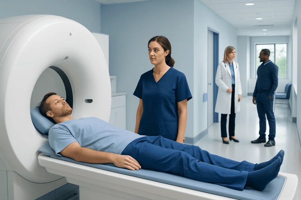
A PET-MRI scan combines PET and MRI images in one session. It shows both tissue structure and metabolic activity.
Key Takeaways
- A PET-MRI scan combines PET and MRI images in one session.
- It shows both tissue structure and metabolic activity.
- It is often used for selected cancer, brain, and heart evaluations.
- Preparation may include fasting and removing metal items before the MRI portion.
- The test is generally safe, but suitability depends on factors such as implants, kidney function, and pregnancy.
A PET-MRI scan is an advanced imaging test that combines positron emission tomography (PET) with magnetic resonance imaging (MRI) in a single exam. It helps doctors see both how tissues look and how they function, which can be useful in selected cancers, neurological disorders, and some heart conditions.
Overview: What Is a PET-MRI Scan?
A PET-MRI scan is a hybrid imaging test that combines two technologies in one examination. PET, or positron emission tomography, shows how tissues and organs are functioning by tracking a small amount of radioactive tracer in the body. MRI, or magnetic resonance imaging, uses magnetic fields and radio waves to create highly detailed pictures, especially of soft tissues such as the brain, liver, muscles, and pelvic organs.
By combining these methods, PET-MRI can provide both functional and anatomical information at the same time. This means doctors can see not only where an abnormality is located, but also how active it may be metabolically. In certain situations, this added detail can help with diagnosis, treatment planning, and follow-up care.
PET-MRI is not the right test for every patient or every condition. In many cases, other imaging methods such as standard MRI, CT, ultrasound, or PET-CT may be more practical or more widely available. A doctor chooses PET-MRI when its combined strengths are likely to answer an important clinical question more clearly.
How PET-MRI Works
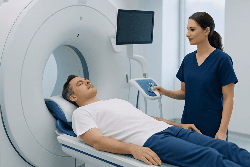
Before the scan, the patient usually receives a small amount of a radioactive tracer through a vein. One of the most commonly used tracers is a form of glucose, which tends to collect in tissues that are more metabolically active. After an uptake period, the scan begins. The PET component detects signals from the tracer, while the MRI component captures detailed images of the body’s structures.
The advantage of doing both studies together is that the images can be matched closely. This helps doctors identify whether an area that looks unusual on MRI is also metabolically active on PET. In cancer care, for example, this may help distinguish active disease from scar tissue in selected cases. In neurology, it can help compare brain structure and brain function in the same session.
Unlike CT, MRI does not use ionizing radiation to create anatomical images. The PET portion does involve radiation from the tracer, but the MRI portion does not. This can make PET-MRI an appealing option in some patients who may need repeated imaging, although the best test still depends on the medical reason for the scan.
When Is a PET-MRI Scan Used?
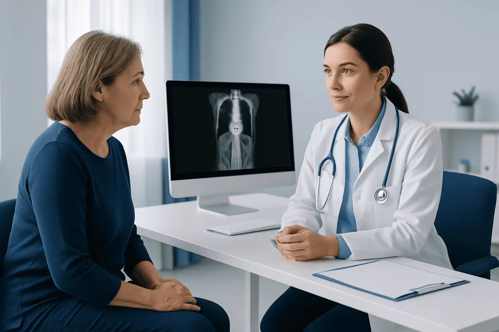
PET-MRI is used most often when doctors need especially detailed information about soft tissues and metabolism together. One common area is cancer care. For some patients, it may help with detecting tumors, assessing how far a disease has spread, planning treatment, or monitoring response after therapy. It can be particularly helpful in selected cancers involving the brain, head and neck, liver, pelvis, and certain children or younger adults when reducing radiation exposure matters.
The test also has important applications in brain and nervous system disorders. For example, doctors may use PET-MRI to evaluate certain types of epilepsy, dementia, brain tumors, or inflammatory conditions. In these settings, MRI can show anatomy in great detail, while PET can reveal changes in brain metabolism that may not be visible on structural imaging alone. Depending on the condition, other imaging pathways may also be used, including brain tumor assessment when a specialist needs more information.
In cardiology, PET-MRI may sometimes be used to assess heart muscle viability, inflammation, or more complex structural and functional questions. It can also support research and highly specialized care. However, PET-MRI is not a routine first-line test for most heart conditions, and its use is generally guided by the expertise of the treating team and the specific medical question.
- Selected cancer diagnosis, staging, and follow-up
- Brain tumors, epilepsy, and some neurodegenerative disorders
- Certain heart muscle and inflammatory heart evaluations
- Complex cases where both structure and function matter
How to Prepare and What Happens During the Scan
Preparation depends on the reason for the scan and the tracer being used. Many patients are asked not to eat for several hours before the PET portion, although drinking water may still be allowed. Strenuous exercise may also be discouraged beforehand because it can affect how the tracer is taken up by muscles. The imaging team will give specific instructions based on the condition being evaluated.
Because the test includes MRI, patients will be asked about metal implants, pacemakers, surgical clips, hearing devices, and other medical or metallic items. Some implants are MRI-compatible and some are not, so this safety review is very important. The patient should also remove jewelry, watches, and other metal objects before entering the scan room.
During the exam, the patient lies still on a scanning table. After the tracer is given and enough time has passed for it to circulate, the scan begins. PET-MRI studies can take longer than some other imaging tests, often from around 30 minutes to over an hour depending on the protocol. Remaining still is important because movement can reduce image quality. Some patients find the MRI space noisy or confined, so they should tell the team in advance if they are worried about claustrophobia.
In some cases, contrast material may be used for the MRI portion to improve detail. If so, the team may ask about kidney function, allergies, and previous reactions to contrast. After the scan, most patients can return to normal activities unless their doctor gives different instructions.
Benefits, Limitations, and Safety Considerations
The main benefit of PET-MRI is the ability to combine two types of information in one test. MRI offers excellent soft-tissue detail, while PET shows metabolic activity. Together, they may improve lesion characterization in selected cases and may help reduce the need for separate appointments. This can be especially useful in complex oncology and neurology assessments, including scenarios that may also involve broader oncology care planning.
PET-MRI also has limitations. It is a highly specialized technology, and it may not be available in all centers. The examination can be longer and more expensive than some other imaging studies. For some medical questions, PET-CT, standard MRI, or CT may provide the needed answer more efficiently. The choice depends on the organ being studied, the suspected condition, and what information the doctor needs.
From a safety perspective, the PET tracer exposes the patient to a limited amount of radiation, but MRI itself does not. MRI safety is especially important for people with certain implanted devices or metal fragments in the body. If MRI contrast is planned, kidney function and other risk factors may be reviewed. Patients who are pregnant, breastfeeding, or think they may be pregnant should tell their doctor and the imaging team so the safest approach can be chosen.
Children and people who have difficulty lying still may sometimes need additional support, and in some cases sedation may be considered. This decision is made carefully and depends on age, overall health, and the expected length of the scan. If the person has a known cancer diagnosis, PET-MRI may be one part of a broader diagnostic pathway that can also include MRI or PET-CT depending on the situation.
How Doctors Interpret Results
PET-MRI results are interpreted by radiologists and nuclear medicine specialists, often with input from the doctor who ordered the scan. They look at where the tracer accumulates and compare this pattern with the detailed MRI images. Some areas of high uptake may suggest active disease, but tracer uptake is not specific on its own. Inflammation, infection, healing tissue, and other non-cancerous processes can sometimes also appear active.
This is why imaging results are always considered alongside symptoms, examination findings, blood tests, and any previous scans or biopsy results. In cancer care, PET-MRI can help show whether a lesion is stable, responding to treatment, or suspicious for progression. In brain disorders, it may help identify areas of altered metabolism that support a diagnosis when combined with MRI findings and clinical evaluation.
Results are usually not explained fully at the moment of the scan. A formal report is prepared after the images are reviewed carefully. The treating doctor then discusses what the findings mean and whether any next steps are needed, such as more imaging, a biopsy, treatment, or simple follow-up.
When to See a Doctor and Questions to Ask
A PET-MRI scan is not usually something a person requests on their own without medical advice. It is ordered when a doctor believes it could answer a specific clinical question. A patient should speak with a doctor if they have ongoing symptoms that need investigation, if they are being monitored for a known condition, or if another imaging test has not provided enough information.
Before the scan, it is reasonable to ask why PET-MRI is being recommended instead of another test, whether fasting is required, how long the exam will take, and whether contrast will be used. Patients should also mention any implants, kidney problems, claustrophobia, pregnancy, or difficulty lying flat. These details help the team plan the safest and most useful examination.
After the scan, follow-up is important. The doctor can explain the results in context and discuss whether the images change the diagnosis or treatment plan. Near the end of the care pathway, some patients may seek evaluation at specialized centers; Acibadem International’s multidisciplinary specialists and JCI-accredited hospitals diagnose and treat complex conditions for international patients, including those who may need advanced imaging as part of their care.
Frequently asked questions
Is a PET-MRI scan the same as a PET-CT scan?
No. Both tests include PET imaging, which shows metabolic activity, but PET-MRI uses MRI for detailed anatomical images while PET-CT uses computed tomography. PET-MRI may be especially useful when soft-tissue detail is important, while PET-CT remains widely used in many clinical settings.
Why would a doctor choose a PET-MRI scan?
A doctor may choose PET-MRI when both structural detail and functional information are needed in the same exam. It can be helpful in selected cancers, brain disorders, and some heart conditions, especially when MRI offers advantages over CT.
How long does a PET-MRI scan take?
The total appointment may take more than an hour because it includes preparation, tracer uptake time, and the scan itself. The exact duration depends on the body area being studied and the imaging protocol.
Is a PET-MRI scan safe?
For most people, PET-MRI is generally safe when proper screening is done. The PET portion involves a small amount of radiation from the tracer, and the MRI portion requires careful review of implants, metal exposure, and other safety factors.
Do patients need to fast before a PET-MRI scan?
Often yes, but instructions depend on the tracer and the reason for the test. The imaging team will explain whether food, exercise, medications, or certain drinks should be avoided beforehand.
Can PET-MRI detect cancer?
PET-MRI can help detect, stage, or monitor certain cancers, but it is not a universal cancer screening test. Its value depends on the cancer type, body area, and the clinical question being asked.
References
- World Health Organization
- Radiological Society of North America
- American College of Radiology
- Society of Nuclear Medicine and Molecular Imaging
- National Cancer Institute
This article is for general information only and is not a substitute for professional medical advice. Please consult a qualified doctor about your individual situation.
Explore treatments in Turkey — costs, top hospitals & a free quote
JCI-accredited · board-certified surgeons · reply within 24h
Add us as a Preferred Source to see more of our trusted health content across Google Search, AI Overviews and Discover.

More from the Health Library



Related Specialists

Prof. Dr. Korhan Özkan
Orthopedic Surgery & Traumatology
Dr. Mustafa Göğebakan
Pediatrics
Gizem Gürzoğlu
Physical Medicine & Rehabilitation
Dr. İbrahim Keskin
Emergency Service