What Is an Arthrogram MRI: Preparation, Procedure and Results

An arthrogram MRI uses contrast placed directly into a joint before the MRI scan. It is commonly used for the shoulder, hip, wrist, elbow, knee and ankle.
Key Takeaways
- An arthrogram MRI uses contrast placed directly into a joint before the MRI scan.
- It is commonly used for the shoulder, hip, wrist, elbow, knee and ankle.
- Temporary pressure or soreness after the injection is common and usually improves within one to two days.
- Patients should tell the imaging team about pregnancy, allergies, kidney disease, implanted devices and possible infection.
- The scan helps guide diagnosis and treatment decisions but must be interpreted alongside symptoms and examination findings.
An arthrogram MRI, also called an MR arthrogram, combines a joint injection with magnetic resonance imaging to provide detailed pictures of structures inside a joint. It can help clinicians assess cartilage, ligaments, tendons and the joint labrum when standard imaging does not provide enough information.
Overview: What Is an Arthrogram MRI?
An arthrogram MRI, also known as magnetic resonance arthrography or an MR arthrogram, is a specialized imaging test in which contrast material is injected directly into a joint before an MRI scan. The contrast expands the joint space and outlines internal structures, helping radiologists see certain tears or abnormalities more clearly than on a standard MRI.
The test is often requested when a person has ongoing joint pain, clicking, instability, reduced movement or symptoms after an injury, but routine X-rays or a conventional MRI have not answered the clinical question. It is particularly helpful for assessing the shoulder labrum, hip labrum, cartilage surfaces, ligaments and some tendon attachments.
An MR arthrogram is not an operation. It is usually an outpatient procedure involving two parts: image-guided placement of contrast into the joint and an MRI examination. The healthcare team explains why it has been recommended and whether another test may be more appropriate for the individual.
How an MRI Arthrogram Works and Who May Need One
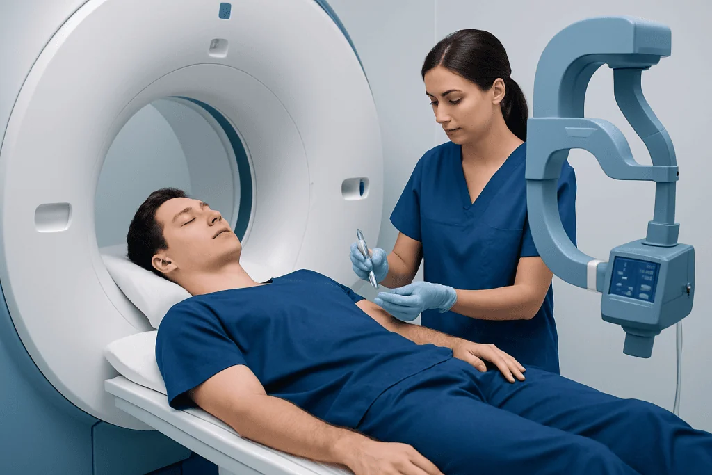
MRI uses a powerful magnet and radio waves to create detailed images without ionizing radiation. During an MR arthrogram, a small amount of contrast is placed inside the joint first. The fluid can enter small spaces created by a tear, making changes in the labrum, cartilage or joint capsule easier to identify.
Clinicians may consider this test for suspected shoulder instability or labral injury, a possible hip labral tear, recurrent joint symptoms after dislocation, unexplained pain in an active person, or evaluation after previous joint treatment. It may also help clarify whether a finding on a standard MRI is clinically important.
Not everyone with joint pain needs an arthrogram MRI. A clinician usually considers the medical history, physical examination, ordinary X-rays and previous scans first. For related assessment and care, patients may also benefit from information about shoulder pain and its possible causes.
People should notify the imaging provider in advance if they are pregnant or may be pregnant, have kidney disease, have had a prior contrast reaction, use blood-thinning medicines, have diabetes, or have metal or electronic implants. Many modern implants are MRI-compatible, but their details must be checked before the scan.
Preparation: What Not to Do Before an Arthrogram?

Preparation instructions can vary by joint, facility and medical history. In many cases, patients can eat and drink normally before an MRI arthrogram. However, they should follow the specific instructions provided by the imaging center, especially if sedation is planned or if they have a health condition that requires individualized preparation.
Before the appointment, a person should not ignore or withhold important health information. They should tell the team about a fever, skin infection, open wound or increasing redness near the joint, as an injection may need to be postponed. They should also report allergies, previous reactions to contrast, current medicines and any possibility of pregnancy.
Patients should avoid applying lotions, creams or metal-containing cosmetics near the area being scanned if instructed by the imaging team. Jewelry, watches, hearing aids, removable dental devices, hairpins and other metallic items generally need to be removed before entering the MRI room.
It is sensible to avoid scheduling strenuous sport, weight training or demanding manual work immediately after the examination when possible. Wearing comfortable clothing and arranging time to rest the joint afterward can make the experience easier.
Step by Step: What Happens During the Procedure?
First, the patient meets the imaging staff, reviews safety questions and gives consent. The skin over the joint is cleaned, and local anesthetic may be used to numb the area. A radiologist or another appropriately trained clinician uses imaging guidance, often fluoroscopy or ultrasound, to position a fine needle accurately in the joint.
Contrast material is then injected into the joint. Some people feel pressure, fullness or a brief stinging sensation. The needle is removed, a small dressing is applied and the patient is moved to the MRI scanner. Depending on the joint and imaging protocol, the complete visit commonly takes longer than the MRI scan alone.
During the MRI, the patient lies still on a padded table that moves into the scanner. The machine makes repeated loud tapping or knocking sounds, and ear protection is provided. The patient can communicate with the MRI staff throughout the scan, and the test is stopped if necessary.
The MRI images are reviewed by a radiologist, who sends a report to the clinician who requested the test. If a labral tear, cartilage injury or another joint problem is found, the next steps may include rehabilitation, medication, activity changes or referral for orthopedic evaluation and treatment.
How Painful Is an Arthrogram MRI in the Shoulder?
A shoulder MRI arthrogram is usually tolerable, but comfort levels differ from person to person. The local anesthetic can cause a brief pinch or burning feeling, followed by pressure or fullness while contrast is injected. Some people notice temporary ache or stiffness because the fluid distends the shoulder joint.
The MRI portion itself should not be painful, although remaining still with the arm positioned for imaging may be uncomfortable when the shoulder is already sore. Patients should tell the staff if they have significant pain, anxiety in enclosed spaces or difficulty lying flat, as adjustments or support may be possible.
Most discomfort settles with rest, ice wrapped in a cloth and simple pain relief if it is safe for the individual to use. Severe or worsening pain is not expected and should be discussed promptly with the imaging provider or a doctor.
What Is the Recovery Time After an MRI Arthrogram?
Most people return home shortly after an MRI arthrogram. A sense of fullness, mild swelling, stiffness or soreness in the injected joint can last for several hours and sometimes up to one or two days. The joint may feel temporarily less comfortable during movement because of the injected fluid.
For the first 24 hours, patients are commonly advised to rest the joint and avoid intense exercise, heavy lifting and contact sports. Light everyday activity may be possible depending on the joint involved and the person’s symptoms. The imaging center’s aftercare instructions should take priority.
A small adhesive dressing may be removed according to the instructions given. Keeping the injection site clean and observing it for increasing redness, warmth, drainage or swelling is appropriate. Results are not always available on the same day because the images need formal interpretation and clinical correlation.
When symptoms are related to a sports or overuse injury, a clinician may recommend a tailored rehabilitation plan after reviewing the scan. Physical therapy and rehabilitation may help restore movement, strength and confidence in joint use when it is clinically appropriate.
Can You Drive Home After an Arthrogram? Benefits and Risks
Many people can drive home after an arthrogram MRI if they have not received sedation, do not feel dizzy and can safely control the vehicle. However, the injected joint may be sore or stiff. Driving may be unsuitable after a shoulder, wrist, hip, knee or ankle injection if pain, limited movement or weakness affects safe vehicle control.
Anyone receiving sedating medicine should not drive, operate machinery or make important decisions for the time advised by their care team. In that situation, arranging a responsible adult to accompany them is important. When in doubt, patients should plan alternative transport before the appointment.
The main benefit of an MR arthrogram is improved visualization of selected internal joint structures, which may reduce uncertainty and support a more targeted care plan. Risks are uncommon but include bruising, bleeding, temporary pain, fainting, infection, allergic reaction and, very rarely, injury to nearby structures. MRI safety also requires careful screening for metal implants and devices.
For ongoing joint symptoms or imaging-confirmed injuries, multidisciplinary specialists at Acibadem International’s JCI-accredited hospitals can assess diagnosis and treatment options for international patients.
When to Seek Medical Care
Patients should contact the imaging provider or seek medical advice if they develop worsening pain rather than gradual improvement, marked swelling, redness, warmth, fever, chills, drainage from the injection site, numbness, weakness or a rash. These symptoms are uncommon but may need timely assessment.
Urgent medical care is appropriate for trouble breathing, swelling of the face or throat, chest pain, fainting that does not quickly resolve, or signs of a severe allergic reaction. People should also seek prompt care after a joint injury if the joint looks deformed, cannot bear weight, becomes cold or pale, or has new major weakness.
For less urgent but persistent joint pain, catching, repeated giving way, reduced range of motion or symptoms that interfere with sleep, work or sport, a clinician can decide whether an arthrogram MRI or another investigation is useful. Imaging results should always be discussed with the referring doctor in the context of the person’s symptoms and examination.
Frequently asked questions
Is an arthrogram MRI the same as a regular MRI?
No. A regular MRI creates detailed images without injecting contrast into the joint. An arthrogram MRI includes a joint injection before the scan, which can make small tears in structures such as the labrum or cartilage easier to see.
How long does an arthrogram MRI take?
The total appointment commonly includes time for safety screening, the contrast injection and the MRI scan. The exact duration varies by the joint being examined and the imaging protocol, so the imaging center can provide the most accurate estimate.
Do I need to fast before an MRI arthrogram?
Many patients do not need to fast, particularly when no sedation is used. Individual instructions vary, so patients should follow the directions from their imaging center and ask in advance if they are unsure.
Is the contrast used in an arthrogram MRI safe?
Contrast used for MR arthrography is generally used in a very small amount and is considered safe for most people. The imaging team should know about previous contrast reactions, kidney problems, pregnancy and other relevant health conditions before the procedure.
Can an MRI arthrogram show a rotator cuff tear?
It can show rotator cuff tendons and may help assess some tears, especially when other shoulder structures also need close evaluation. A standard MRI is often sufficient for many rotator cuff concerns, and the clinician selects the most suitable test based on the suspected problem.
When will MRI arthrogram results be available?
A radiologist reviews the images and prepares a report for the referring clinician. Timing differs between facilities, but the clinician will explain the findings and what they mean for treatment or follow-up.
References
- American College of Radiology
- Radiological Society of North America
- National Institute of Arthritis and Musculoskeletal and Skin Diseases
- U.S. Food and Drug Administration
This article is for general information only and is not a substitute for professional medical advice. Please consult a qualified doctor about your individual situation.
Joint and spine care in Turkey — expert assessment & treatment
JCI-accredited · board-certified surgeons · reply within 24h
Add us as a Preferred Source to see more of our trusted health content across Google Search, AI Overviews and Discover.