What Is an MRI Scan and How Should You Prepare?
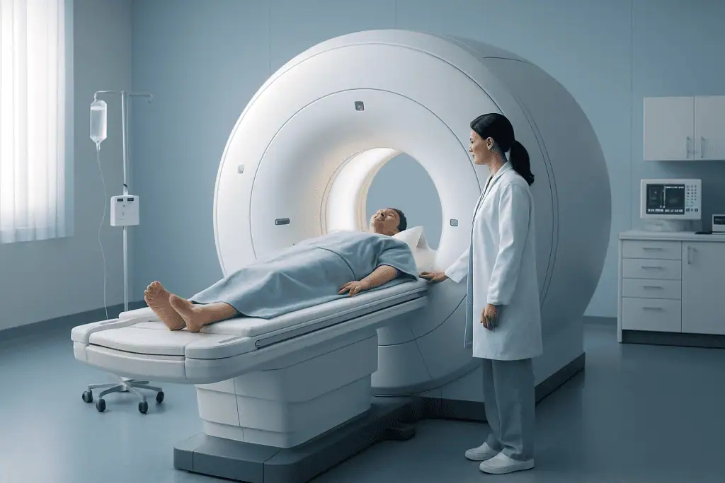
An MRI scan does not use ionizing radiation, but it does use a powerful magnetic field that requires careful safety screening. Patients should tell the healthcare team about any implants, devices, metal fragments, pregnancy, kidney disease, allergies, or claustrophobia before the appointment.
Key Takeaways
- An MRI scan does not use ionizing radiation, but it does use a powerful magnetic field that requires careful safety screening.
- Patients should tell the healthcare team about any implants, devices, metal fragments, pregnancy, kidney disease, allergies, or claustrophobia before the appointment.
- Preparation may include removing metal items, fasting for some types of scans, or receiving a contrast agent when clearer images are needed.
- The test is usually painless, although it can be noisy and requires the patient to lie still for the best image quality.
- MRI results are interpreted by a radiologist and shared with the referring doctor, who explains what the findings mean and discusses next steps.
An MRI scan is a non-invasive imaging test that uses a strong magnet and radio waves to create detailed pictures of organs, tissues, joints, and the nervous system. Knowing what to expect before, during, and after the scan can help patients feel more comfortable and prepared.
Overview: What Is an MRI Scan?
An MRI scan, or magnetic resonance imaging scan, is a diagnostic imaging test that creates detailed pictures of structures inside the body. It is commonly used to examine the brain, spine, joints, muscles, abdomen, pelvis, heart, blood vessels, and other organs. MRI can show soft tissues in great detail, which makes it especially helpful when doctors need information that may not be visible on a standard X-ray.
MRI does not use ionizing radiation. Instead, it uses a powerful magnetic field, radiofrequency pulses, and a computer to produce cross-sectional images. These images can be viewed in different planes and may help doctors diagnose injuries, inflammation, tumors, blood vessel problems, nerve conditions, or changes related to disease.
The scan is usually painless and non-invasive. Some patients find the machine noise or the enclosed space uncomfortable, but the imaging team can offer reassurance and practical support. For many people, understanding the process in advance makes the experience much easier.
Why Doctors Recommend MRI
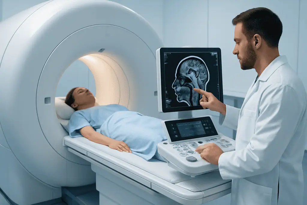
A doctor may request an MRI scan when detailed information is needed to confirm a diagnosis, plan treatment, or monitor a known condition. MRI is often chosen because it provides excellent contrast between different soft tissues. For example, it can help evaluate ligaments and cartilage in the knee, discs and nerves in the spine, or subtle changes in the brain.
Common reasons for MRI include persistent joint or back pain, suspected stroke-related changes, headaches with concerning features, multiple sclerosis evaluation, sports injuries, abdominal or pelvic conditions, heart or blood vessel assessment, and follow-up after treatment. In oncology, MRI may be used to help define the size and location of a tumor or to monitor response to therapy.
Sometimes MRI is used after another imaging test, such as ultrasound, X-ray, or CT, to provide additional detail. The choice of imaging depends on the patient’s symptoms, medical history, examination findings, and the clinical question the doctor wants to answer.
How MRI Works and What Types May Be Used
During an MRI scan, the patient lies on a table that moves into the scanner. The scanner’s magnetic field temporarily aligns hydrogen atoms in the body. Radio waves then disturb this alignment, and as the atoms return to their usual state, they release signals. A computer converts these signals into detailed images.
Different MRI techniques can highlight different tissues or processes. A musculoskeletal MRI may focus on bones, joints, tendons, ligaments, and muscles. A brain MRI may assess brain tissue, blood vessels, or nerves. An abdominal MRI may evaluate the liver, pancreas, kidneys, bile ducts, or other organs. In some cases, special sequences are used to study blood flow, inflammation, tissue composition, or organ function.
Some MRI scans are performed with contrast. Contrast agents, often gadolinium-based, are injected into a vein to help certain tissues, blood vessels, inflammation, or tumors appear more clearly. Not every MRI requires contrast, and the decision depends on the medical question, the body area being scanned, and the patient’s health status.
How to Prepare Before an MRI Scan
Preparation depends on the type of MRI and the area being examined. Many MRI scans require little special preparation, and patients may be able to eat, drink, and take usual medicines as normal. However, some abdominal, pelvic, or contrast-enhanced scans may require fasting for a few hours. Patients should follow the specific instructions given by the imaging center or doctor.
Because MRI uses a strong magnet, metal safety is essential. Before the scan, patients are usually asked to complete a screening form and remove jewelry, watches, hearing aids, removable dental work, hairpins, coins, keys, credit cards, and clothing with metal zippers or fasteners. The imaging team may provide a gown or metal-free clothing.
Patients should inform the team before the appointment if they have or may have any of the following:
- A pacemaker, defibrillator, neurostimulator, cochlear implant, infusion pump, or implanted medical device
- Aneurysm clips, metal fragments, shrapnel, or a history of metal work or eye injury
- Joint replacements, surgical plates, screws, stents, or heart valves
- Pregnancy or the possibility of pregnancy
- Kidney disease, prior reaction to MRI contrast, or severe allergies
- Claustrophobia, anxiety, or difficulty lying still
Many modern implants are MRI-conditional, meaning an MRI may be possible under specific conditions. The healthcare team may need device details, implant cards, or surgical records to confirm safety. Patients should never assume an implant is safe or unsafe without professional screening.
What Happens During the MRI
At the appointment, a radiographer or MRI technologist explains the procedure and confirms the safety checklist. The patient lies on the scanning table, and the team may place a special receiver coil around or near the area being examined. This coil helps collect the MRI signal and improve image quality. Cushions or straps may be used to help the patient stay comfortable and still.
The table then moves into the scanner. MRI machines make loud tapping, knocking, or humming sounds during image acquisition, so patients are usually given earplugs or headphones. The technologist can see and hear the patient from the control room, and most scanners have a call button so the patient can communicate if needed.
The scan may take from about 15 minutes to an hour or longer, depending on the body part and the number of image sequences needed. It is important to remain as still as possible because movement can blur the images. For some scans, the patient may be asked to hold their breath briefly, particularly during abdominal or chest imaging.
If contrast is needed, it is usually injected through a small intravenous line. Some patients notice a cool sensation in the arm or a brief metallic taste, but many feel nothing unusual. After contrast injection, additional images are taken to complete the examination.
Safety, Comfort, and MRI Contrast
MRI is considered a safe imaging method for many patients when proper screening is performed. The main safety concern is the strong magnetic field, which can attract certain metal objects or affect some implanted devices. This is why detailed screening before entering the MRI room is so important.
Patients who feel anxious in enclosed spaces should tell the medical team in advance. Options may include extra explanation, music through headphones, a support person in appropriate situations, scheduling extra time, or discussing mild sedation with the referring doctor when necessary. Some centers have wider-bore or open MRI systems, although image quality and suitability depend on the clinical need.
Gadolinium-based MRI contrast is widely used when clinically appropriate. Before contrast is given, the team may ask about kidney function, previous reactions, pregnancy, and breastfeeding. Serious reactions are uncommon, but any new symptoms during or after contrast should be reported to the healthcare team promptly.
Patients should also mention tattoos, permanent makeup, medication patches, glucose monitors, and body piercings. Most do not prevent scanning, but some may require special precautions or removal. The goal is to complete the MRI safely while obtaining images that are clear enough to answer the medical question.
After the Scan, Results, and When to See a Doctor
After most MRI scans, patients can return to normal activities immediately. If sedation was used, the patient will need monitoring until fully awake and should arrange for someone else to drive them home. If contrast was used, drinking fluids as normally allowed may help the body eliminate it through the kidneys.
The MRI images are reviewed by a radiologist, a doctor specially trained in medical imaging. The radiologist prepares a report for the referring doctor. The referring doctor then explains the results in the context of symptoms, examination findings, and other tests. Patients should ask when and how they will receive their results, because timing can vary.
Medical advice should be sought if symptoms worsen, if new symptoms develop, or if there are concerns after receiving contrast, such as rash, swelling, breathing difficulty, or feeling unwell. These reactions are not common, but prompt medical guidance is appropriate. Patients should also contact their doctor if they are unsure how to interpret the report or what the next step should be.
For international patients, Acibadem International’s multidisciplinary specialists and JCI-accredited hospitals provide diagnostic imaging services, including MRI, as part of individualized evaluation and treatment planning. Patients should bring previous imaging, reports, medication lists, and implant information to support a safe and efficient assessment.
Frequently asked questions
Is an MRI scan painful?
An MRI scan is usually painless. The main challenges are lying still, the loud scanner noises, and sometimes feeling confined inside the machine. The imaging team can provide ear protection and help patients stay as comfortable as possible.
Does MRI use radiation?
No. MRI uses a strong magnetic field and radio waves, not ionizing radiation. This is one reason MRI is often preferred for detailed soft tissue imaging when it is suitable for the patient and the medical question.
Can patients eat before an MRI scan?
Many MRI scans do not require fasting, and patients can eat and drink normally. However, some abdominal, pelvic, or contrast-enhanced MRI scans may require fasting for a few hours. Patients should follow the specific instructions given by their doctor or imaging center.
What should patients wear to an MRI appointment?
Patients should wear comfortable clothing without metal zippers, snaps, hooks, or decorative metal. Jewelry, watches, hairpins, piercings, and removable metal items usually need to be taken off. Many imaging centers provide a gown to reduce the risk of metal entering the MRI room.
Is MRI contrast safe?
MRI contrast is considered safe for many patients when used appropriately and after proper screening. The healthcare team will ask about kidney disease, previous contrast reactions, pregnancy, and other health factors before giving it. Patients should report any unusual symptoms during or after the scan.
Can someone with an implant have an MRI?
Some implants are safe for MRI, some are MRI-conditional, and some may not be compatible. Patients should tell the imaging team about all implanted devices, surgical clips, metal fragments, and previous operations. The team may need device information or medical records before deciding whether the scan can proceed.
How long does it take to get MRI results?
The timing varies by facility and clinical urgency. A radiologist reviews the images and sends a report to the referring doctor, who explains the findings and next steps. Patients should ask before leaving the imaging center when and how they can expect to receive their results.
This article is for general information only and is not a substitute for professional medical advice. Please consult a qualified doctor about your individual situation.
Joint and spine care in Turkey — expert assessment & treatment
JCI-accredited · board-certified surgeons · reply within 24h
Add us as a Preferred Source to see more of our trusted health content across Google Search, AI Overviews and Discover.

More from the Health Library

Diagnostic Tests for Breast Symptoms: Mammogram, Ultrasound, or MRI?

EMG and Nerve Conduction Studies: Diagnosing Neuromuscular Disease

Can You Drink Water Before a Blood Test or Scan?

Quest Diagnostics Hours: When Labs Open and How to Plan a Visit

Labcorp Appointment: How to Book, Prepare, and Avoid Delays