

What Is Fdg PET Scan: Preparation, Procedure and Results

FDG PET scanning maps how body tissues use a glucose-like tracer, rather than showing anatomy alone. The test is often performed as PET/CT, which combines metabolic and detailed anatomical images.
Key Takeaways
- FDG PET scanning maps how body tissues use a glucose-like tracer, rather than showing anatomy alone.
- The test is often performed as PET/CT, which combines metabolic and detailed anatomical images.
- Fasting and avoiding strenuous exercise before the appointment can improve image quality.
- Increased FDG uptake is not always cancer; infection, inflammation and normal tissues can also appear active.
- The injection is usually the only uncomfortable part, and most people return to normal activities shortly after the scan.
- A PET scan contributes to cancer staging but is interpreted alongside biopsy, CT or MRI, laboratory findings and clinical examination.
An FDG PET scan is a nuclear medicine imaging test that uses a small amount of radioactive glucose-like tracer to show areas of increased metabolic activity in the body. It is commonly combined with CT imaging and can help doctors diagnose, stage and monitor several conditions, especially cancer.
What is an FDG PET scan?
What is FDG PET scan? It is an imaging examination that shows how actively different tissues use sugar. A small amount of fluorodeoxyglucose (FDG), a glucose-like substance with a radioactive tracer, is injected into a vein before images are taken with a PET scanner, often combined with a CT scanner.
Cells use glucose for energy. Some cancers, infections and inflammatory conditions use more glucose than surrounding tissues and may take up more FDG. The scan creates a map of this uptake, helping the medical team identify areas that may need closer assessment. It does not diagnose a condition on its own; the findings are considered with symptoms, examination, other scans and, when needed, biopsy results.
FDG PET is widely used in oncology to assess known or suspected cancer, check whether treatment is working, look for recurrence and help plan further care. It may also be used in selected cases of infection, inflammation, some heart conditions and certain neurological disorders.
How FDG PET works and who may benefit
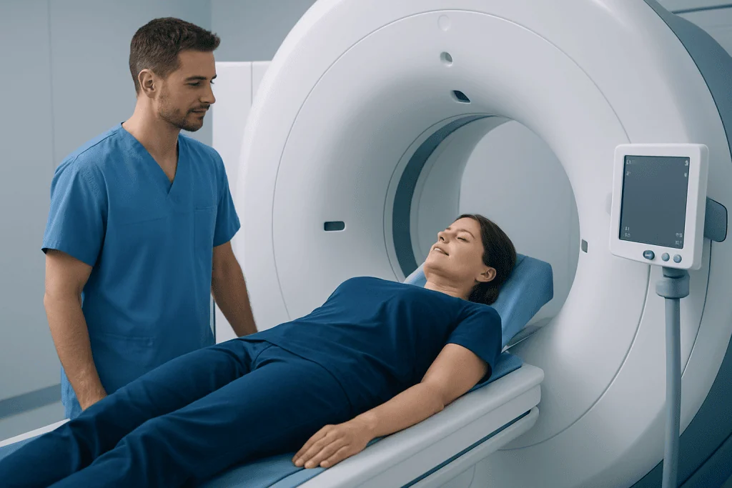
FDG is short for fluorodeoxyglucose. After injection, the tracer circulates in the bloodstream and is taken up by tissues. A PET scanner detects the small signals released by the tracer and converts them into images. When PET is paired with CT, the CT portion helps locate the area of uptake precisely within the body.
Doctors may recommend an FDG PET/CT when they need information beyond that available from structural imaging alone. For example, it can help clarify the extent of some cancers, assess whether a mass is metabolically active, distinguish possible scar tissue from active disease in some situations, or evaluate response after treatment. It may be part of the assessment of conditions such as lung cancer or lymphoma, depending on the person’s clinical situation.
Not every person or every type of cancer needs FDG PET. Some tumors do not take up FDG strongly, and scans may be less informative for them. The referring clinician and nuclear medicine specialist determine whether the test is appropriate and which scan protocol will best answer the clinical question.
What should I do to prepare for an FDG PET scan?

Preparation instructions can vary by hospital and by the reason for the scan, so patients should follow the directions provided by their imaging department. In many cases, people are asked to avoid eating for several hours before the appointment, while drinking plain water is usually encouraged. Fasting helps keep blood glucose and insulin levels more stable, which supports accurate FDG distribution.
Strenuous exercise is commonly avoided for at least a day before the scan because recently worked muscles can absorb more FDG and make images harder to interpret. Patients should also tell the team about diabetes, pregnancy, breastfeeding, allergies, current illnesses, recent surgery, all medicines and any difficulty lying still or being in enclosed spaces.
For people with diabetes, preparation requires individualized planning. Meals, insulin and other glucose-lowering medicines may need adjustment to achieve a safe blood sugar level and a useful scan. Patients should not change prescribed medication on their own; they should contact the imaging department or their treating clinician well before the appointment for specific instructions.
- Wear comfortable clothing without metal where possible, or be prepared to change into a gown.
- Bring relevant prior scan reports or images if requested.
- Ask about arrangements for young children, breastfeeding, sedation needs or language support in advance.
FDG PET scan procedure: step by step
At the appointment, staff confirm medical information and may check blood glucose. A technologist places a small intravenous line, usually in the arm or hand, and injects the FDG tracer. The injection feels similar to a routine blood test or intravenous medication injection.
After the injection, the person rests quietly, usually for about an hour, while the tracer distributes through the body. Keeping warm, relaxed and relatively still is important because muscle activity can affect tracer uptake. Depending on the scan type, patients may be asked to empty their bladder before imaging.
During scanning, the patient lies on a padded table that moves slowly through the PET/CT machine. It is important to remain still, but the test is not generally painful. Imaging time varies according to the body areas being examined and the protocol used. The team can communicate with the patient throughout the scan and monitor comfort.
The images are reviewed by a nuclear medicine physician or radiologist with relevant expertise. The report is sent to the referring doctor, who explains what the findings mean in the context of the person’s health and care plan.
What does FDG mean in a PET scan result?
In a PET scan result, FDG refers to the tracer fluorodeoxyglucose. Reports may describe “FDG uptake,” “avid” tissue or a “hypermetabolic” area. These terms mean that an area has taken up more tracer than expected or more than nearby tissue; they do not by themselves confirm cancer.
Normal organs can show FDG uptake. The brain, heart, kidneys and bladder often appear active because of normal glucose use or tracer excretion. Muscles can also show increased uptake after exercise, and inflammation from infection, healing or autoimmune disease may produce a similar pattern to some cancers.
Reports may include a semiquantitative measurement called SUV, or standardized uptake value. SUV can support interpretation and comparison over time, but it is influenced by technical and biological factors. A number alone cannot determine whether a finding is benign or malignant. The location, pattern, scan quality, previous images and clinical details all matter.
Can you tell what stage cancer is from a PET scan?
An FDG PET scan can provide important information for cancer staging, especially by showing whether there are metabolically active areas in lymph nodes or distant organs. This may help doctors determine the extent of disease and select the most appropriate next steps. However, a PET scan is only one part of staging.
Staging also depends on the cancer type, the primary tumor’s size and local extent, tissue diagnosis from biopsy, CT or MRI findings, laboratory tests and sometimes surgical assessment. A finding on PET may require additional imaging or biopsy before it changes a diagnosis or treatment plan. Conversely, a normal PET scan does not exclude every microscopic or low-FDG-uptake cancer focus.
For this reason, cancer results are commonly reviewed by a multidisciplinary team that may include oncologists, surgeons, radiologists, pathologists and radiation oncologists. When treatment is indicated, the diagnostic results can guide options such as cancer treatment tailored to the specific cancer and the person’s overall health.
Recovery, benefits and possible risks
Most people can leave soon after the scan and resume normal daily activities unless they received medicine for anxiety or sedation. Drinking extra water, if medically appropriate, and urinating regularly can help the body clear the tracer. The radioactive material used for PET decays quickly and is usually eliminated over a short period; the imaging team may provide practical instructions about close contact with infants or pregnant people for the rest of the day.
The main benefit of FDG PET is that it supplies functional information that can complement CT, MRI and other tests. This may help avoid uncertainty, define the extent of disease more accurately, monitor response or identify an appropriate site for biopsy. It can also help doctors plan radiation therapy or surgery in selected cases.
Risks are generally low. The test involves a small amount of radiation from the tracer and, if PET/CT is used, from the CT scan. Allergic reactions to FDG are very uncommon. Patients who are pregnant, could be pregnant or are breastfeeding should always inform the team so that the necessity and timing of the scan can be carefully considered.
Is a FDG PET scan painful? The scan itself is not painful. There may be brief discomfort when the intravenous line is placed, and some people find lying still or keeping their arms in position uncomfortable. Staff can help with positioning and can discuss concerns such as pain, claustrophobia or limited mobility before imaging begins.
When to seek medical care
Patients should contact their doctor promptly if they have new or worsening symptoms that led to the scan, such as an unexplained persistent lump, ongoing unexplained weight loss, prolonged fever, worsening shortness of breath, persistent pain or new neurological symptoms. These symptoms can have many possible causes, but timely assessment is important.
After an FDG PET scan, urgent medical care is appropriate for severe symptoms such as trouble breathing, swelling of the face or throat, fainting, chest pain or signs of a serious allergic reaction. Such reactions are rare, but any sudden or severe symptom should be assessed without delay.
It is also important to arrange follow-up with the referring clinician to discuss the report, even if the result appears reassuring. Acibadem International’s multidisciplinary specialists and JCI-accredited hospitals support international patients with diagnostic evaluation and treatment planning when needed.
Frequently asked questions
How long does an FDG PET scan take?
The whole visit often takes several hours because time is needed for preparation, tracer injection and a quiet uptake period before imaging. The actual scan commonly takes less time, but the duration depends on the protocol and body areas being examined. The imaging department can provide an estimated appointment length in advance.
Can I drink water before an FDG PET scan?
Plain water is often allowed and may be encouraged, but instructions vary by scan center and clinical reason for testing. Drinks containing sugar, calories or caffeine may not be permitted during the fasting period. Patients should follow the specific instructions given by their imaging department.
Why must exercise be avoided before a PET scan?
Exercise makes muscles use more glucose, so they may take up more FDG tracer. This can reduce image clarity or create uptake patterns that are more difficult to interpret. Avoiding strenuous activity before the scan helps improve accuracy.
Will I receive PET scan results on the same day?
Images usually require careful review by a nuclear medicine physician or radiologist before a final report is issued. The referring doctor then discusses the results in relation to the patient’s medical history and other tests. Timing varies by center, so patients can ask when and how results will be communicated.
Does high FDG uptake always mean cancer?
No. FDG uptake can increase in cancer, but also in infection, inflammation, healing tissue, active muscles and several normal organs. A specialist interprets the pattern of uptake together with CT findings, prior imaging and clinical information.
Is FDG PET safe after cancer treatment?
FDG PET can be useful after cancer treatment when the treating team needs to assess response or investigate possible recurrence. The appropriate timing depends on the cancer type and recent treatment, since surgery, radiation and inflammation can temporarily affect uptake. The oncology and imaging teams decide when scanning will provide the most meaningful information.
References
- National Cancer Institute
- Radiological Society of North America
- Society of Nuclear Medicine and Molecular Imaging
- American Cancer Society
- International Atomic Energy Agency
This article is for general information only and is not a substitute for professional medical advice. Please consult a qualified doctor about your individual situation.
Oncology care in Turkey — second opinion and treatment plan
JCI-accredited · board-certified surgeons · reply within 24h
Add us as a Preferred Source to see more of our trusted health content across Google Search, AI Overviews and Discover.

More from the Health Library


Related Specialists

Prof. Dr. Emre Toğrul
Orthopedic Surgery & Traumatology
Dr. Feride Pınar Doğru
Ophthalmology
Prof. Dr. Kazım Üzüm
Pediatric Cardiology
Assoc. Prof. Dr. Bahadır Topuz
Urology