Where Is the Heart Located? A Simple Guide to Heart Position and Anatomy

The heart sits in the middle of the chest in an area called the mediastinum. Most of the heart lies slightly left of center, but it is not entirely on the left side.
Key Takeaways
- The heart sits in the middle of the chest in an area called the mediastinum.
- Most of the heart lies slightly left of center, but it is not entirely on the left side.
- The heart is protected by the rib cage, breastbone, and a thin sac called the pericardium.
- Its four chambers and valves work together to pump blood to the lungs and the rest of the body.
- Chest symptoms do not always come from the heart, so persistent or severe symptoms should be assessed by a doctor.
The heart is located in the chest, between the lungs, behind the breastbone, and slightly to the left of the body's midline. Understanding its position and basic anatomy can help explain symptoms, medical tests, and how this vital organ works every day.
Overview: Where Is the Heart Located?
The heart is located in the chest, between the right and left lungs. More specifically, it sits in the central part of the chest called the mediastinum. It rests behind the breastbone, also called the sternum, and in front of the spine. Although many people think of the heart as being fully on the left side, its position is actually near the center of the chest, with a slight tilt to the left.
About two-thirds of the heart’s mass lies to the left of the midline and one-third lies to the right. The lower pointed part of the heart, called the apex, angles down and to the left. This is why people often place a hand on the left side of the chest when talking about the heart. The broader upper part, called the base, faces upward and slightly backward.
The heart is not fixed rigidly in one exact spot. Its position can look slightly different from person to person depending on body shape, age, lung size, and even breathing. Still, in most healthy adults, the heart remains centrally placed, slightly left of center, and well protected inside the chest.
What Surrounds and Protects the Heart?

The heart is protected by several important structures. The rib cage forms a strong outer shield, while the sternum protects the front of the chest. Behind the heart is the spine, and on both sides are the lungs. These surrounding structures help reduce injury and support the heart’s position during normal movement and breathing.
The heart itself is enclosed in a thin, double-layered sac called the pericardium. This sac helps hold the heart in place and reduces friction as it beats. A small amount of fluid between the pericardial layers allows the heart to move smoothly with each contraction.
The diaphragm, the main muscle used for breathing, sits just below the heart. The heart rests on this muscle, which helps explain why breathing and body position can sometimes affect how heartbeats or chest sensations are felt. In some people, lying down, taking a deep breath, or bending forward may make normal heart awareness more noticeable.
Basic Heart Anatomy: Chambers, Valves, and Major Blood Vessels
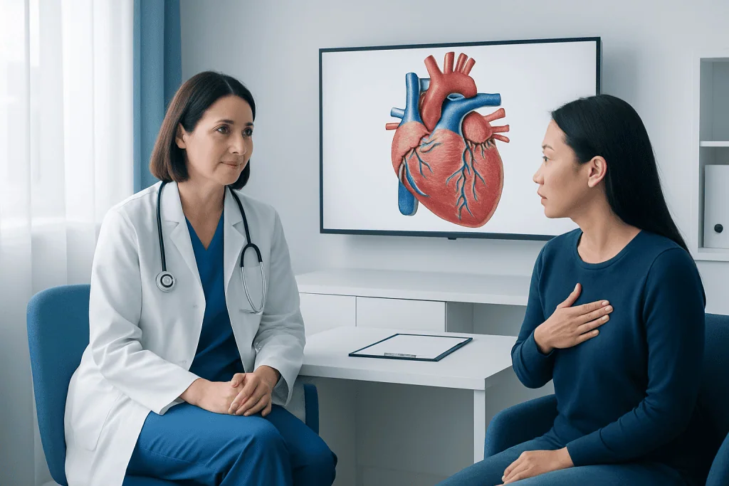
The heart is a muscular organ about the size of a clenched fist. It has four chambers: two upper chambers called the right atrium and left atrium, and two lower chambers called the right ventricle and left ventricle. The right side of the heart receives blood returning from the body and pumps it to the lungs. The left side receives oxygen-rich blood from the lungs and pumps it to the rest of the body.
Four valves keep blood moving in the correct direction. These are the tricuspid valve, pulmonary valve, mitral valve, and aortic valve. They open and close with each heartbeat to prevent backward flow. This coordinated movement is essential for efficient circulation and for supplying oxygen and nutrients to tissues throughout the body.
Several major blood vessels connect to the heart. These include the aorta, which carries oxygen-rich blood from the heart to the body; the pulmonary arteries, which carry blood from the heart to the lungs; the pulmonary veins, which return oxygen-rich blood from the lungs; and the vena cavae, which bring blood back from the body. Together, these structures make up the body’s central pumping system.
The heart also has its own blood supply through the coronary arteries. These arteries deliver oxygen to the heart muscle itself. When blood flow through these vessels becomes reduced or blocked, it can lead to problems such as coronary artery disease.
Why the Heart Feels Left-Sided
Many people ask, “Where is your heart exactly?” because heartbeats, chest sensations, or pain often seem to come from the left side. This is mainly due to the position of the heart’s apex, which points leftward. The left ventricle, the heart’s main pumping chamber, is also thick and strong, and its movement contributes to the sensation felt in the left chest.
In some people, a normal heartbeat can be felt by placing fingers lightly on the chest near the fifth intercostal space, just below the left nipple area in many adults. Doctors call this the point of maximal impulse. Its exact location can vary depending on anatomy, body size, pregnancy, lung conditions, or certain heart disorders.
It is also important to know that not all left-sided chest discomfort comes from the heart. Muscles, ribs, the esophagus, lungs, and even anxiety can cause symptoms in a similar area. On the other hand, heart-related discomfort may also be felt in the center of the chest, neck, jaw, shoulder, back, or arm. This is why symptoms should be interpreted carefully rather than by location alone.
Can the Heart Be in a Different Position?
In most people, the heart is slightly left of center. However, there are rare situations in which the heart may be positioned differently. One example is dextrocardia, a condition present from birth in which the heart points toward the right side of the chest. Some people with dextrocardia have no symptoms, while others may have associated structural heart differences that need medical follow-up.
The heart’s apparent position can also shift temporarily because of other conditions. For example, a very enlarged heart, severe lung overinflation, fluid around the lungs, pregnancy, or changes in the diaphragm can alter how the heart looks on an examination or imaging study. This does not always mean the heart itself has moved permanently, but it can affect how it appears and how nearby structures are displaced.
Doctors use physical examination and imaging tests to understand heart position accurately. If there is concern about the structure or function of the heart, an echocardiogram may be used to look more closely at the chambers, valves, and blood flow. In some cases, more detailed cardiac MRI or cardiology check-up evaluation may be recommended.
How Doctors Check Heart Location and Structure
Doctors learn about the heart’s location and condition in several ways. A physical examination includes listening to heart sounds with a stethoscope, checking the pulse, and sometimes identifying where the heartbeat is most noticeable on the chest wall. These simple steps can provide useful clues about heart rhythm, size, and function.
Imaging tests often give a clearer picture. A chest X-ray can show the general outline and position of the heart. An echocardiogram uses sound waves to show the heart beating in real time and can assess chamber size, valve movement, and pumping strength. An electrocardiogram records the heart’s electrical activity and can support the evaluation of rhythm problems or signs of strain.
If symptoms such as unexplained chest pain, shortness of breath, palpitations, fainting, or reduced exercise tolerance are present, a doctor may suggest more detailed testing. Depending on the situation, this may include stress testing, blood tests, echocardiography, or advanced scans. These tests help distinguish normal anatomy from conditions such as heart failure or other cardiovascular concerns.
Common Symptoms People Mistake for Heart Pain
Because the heart is located in the chest, many kinds of discomfort in that area may be mistaken for heart pain. Muscle strain, acid reflux, inflammation of the rib joints, lung irritation, and stress-related chest tightness can all cause symptoms near the heart. The location alone does not confirm the cause.
Heart-related symptoms are often described as pressure, heaviness, squeezing, or tightness in the center or left side of the chest. They may occur with shortness of breath, sweating, nausea, dizziness, unusual fatigue, or pain spreading to the arm, back, neck, or jaw. Some people, especially women, older adults, and people with diabetes, may have less typical symptoms.
Any new, severe, or persistent chest symptom deserves medical attention. It is especially important to seek urgent care if chest discomfort occurs with breathing difficulty, fainting, sudden weakness, or a feeling of pressure that does not go away. Early assessment can help identify whether symptoms are related to the heart, lungs, digestive system, or another cause.
When to See a Doctor and How to Support Heart Health
A doctor should be consulted if there are repeated episodes of chest pain, palpitations, breathlessness, swelling in the legs, unexplained fatigue, or reduced ability to exercise. These symptoms do not always mean there is a serious heart problem, but they should not be ignored. A proper evaluation can provide reassurance or allow timely treatment if needed.
Supporting heart health starts with everyday habits. Helpful steps include not smoking, staying physically active, eating a balanced diet, maintaining a healthy weight, managing blood pressure and cholesterol, sleeping well, and limiting excess alcohol. People with diabetes or a family history of heart disease may benefit from regular checkups and earlier screening.
For people who need specialized assessment, multidisciplinary heart teams can help diagnose both common and complex conditions. Acibadem International’s multidisciplinary specialists and JCI-accredited hospitals diagnose and treat heart conditions for international patients, including advanced evaluation and cardiovascular surgery when appropriate. The most suitable approach depends on the person’s symptoms, overall health, and test results.
Frequently asked questions
Is the heart on the left side of the body?
The heart is not completely on the left side. It sits mostly in the center of the chest, with a slight shift to the left. The lower tip of the heart points leftward, which is why it often feels left-sided.
Where is the heart located in relation to the lungs?
The heart lies between the two lungs in the middle part of the chest. It is surrounded by the lungs on both sides, with the left lung making room for part of the heart. This arrangement helps protect the heart while allowing both organs to work together during breathing and circulation.
Can a person feel their heart beating in the chest?
Yes, some people can occasionally feel a normal heartbeat, especially during exercise, stress, or when lying on the left side. This is not always a problem. However, frequent pounding, fluttering, skipped beats, or symptoms like dizziness should be checked by a doctor.
Can the heart be on the right side?
Yes, but this is uncommon. A congenital condition called dextrocardia causes the heart to point toward the right side of the chest. Some people have no major issues, while others may need further evaluation for associated structural differences.
What is directly under the heart?
The diaphragm is directly below the heart. This large breathing muscle separates the chest from the abdomen and supports the heart from underneath. Because of this close relationship, breathing and body position can sometimes change how chest sensations are perceived.
Does chest pain always mean a heart problem?
No, chest pain does not always come from the heart. It can also result from muscle strain, reflux, lung conditions, anxiety, or inflammation in the chest wall. Because some heart problems can feel similar, new or concerning chest pain should be assessed medically.
References
- American Heart Association
- National Heart, Lung, and Blood Institute
- MedlinePlus
- Mayo Clinic
- Cleveland Clinic
This article is for general information only and is not a substitute for professional medical advice. Please consult a qualified doctor about your individual situation.
Joint and spine care in Turkey — expert assessment & treatment
JCI-accredited · board-certified surgeons · reply within 24h
Add us as a Preferred Source to see more of our trusted health content across Google Search, AI Overviews and Discover.