X Ray: An Evidence-Based Guide for Patients

An x ray uses a small amount of ionizing radiation to create images of structures inside the body. It is commonly used to evaluate broken bones, chest symptoms, dental issues, joint problems, and some digestive concerns.
Key Takeaways
- An x ray uses a small amount of ionizing radiation to create images of structures inside the body.
- It is commonly used to evaluate broken bones, chest symptoms, dental issues, joint problems, and some digestive concerns.
- Most x ray exams are fast, painless, and require little or no preparation.
- Radiation exposure from a single standard x ray is generally low, but the test should still be used thoughtfully, especially during pregnancy.
- Results are interpreted together with symptoms, physical examination, and sometimes other imaging tests.
An x ray is a quick, widely used imaging test that helps doctors look inside the body without surgery. It is most often used to assess bones, the chest, teeth, and joints, and it usually involves a very small amount of radiation.
What an X Ray Is and Why It Is Used
An x ray is a medical imaging test that uses a small amount of radiation to create pictures of structures inside the body. It is one of the oldest and most commonly used diagnostic tools in medicine because it is fast, widely available, and helpful for many everyday health concerns. In many situations, an x ray gives doctors the information they need to confirm or rule out a problem quickly.
X rays are especially useful for showing bones, joints, and the chest. They can help identify fractures, joint changes, lung infections, fluid around the lungs, and some causes of abdominal symptoms. Dental x rays are also widely used to check teeth, roots, jawbone, and gum health. Because different body tissues absorb radiation differently, the resulting image can highlight certain structures clearly.
Although x rays are very useful, they do not answer every question. Soft tissues such as muscles, ligaments, nerves, and some organs may be seen less clearly on a standard x ray than on tests such as ultrasound, CT, or MRI. For that reason, doctors choose x ray imaging when it is the right first step and may recommend another test if more detail is needed.
How X Ray Imaging Works
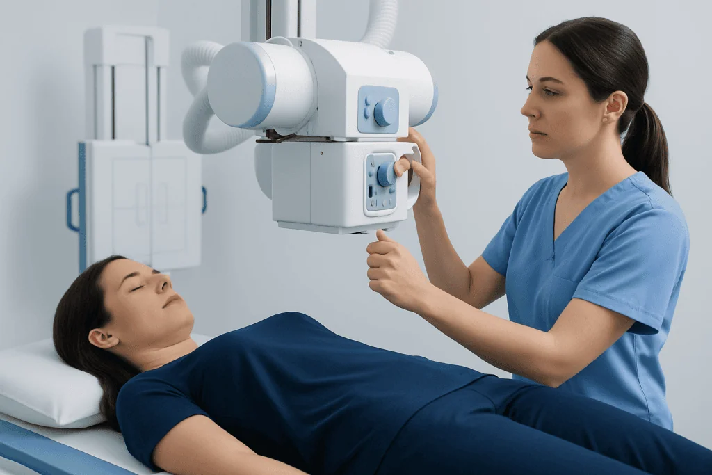
During an x ray, a machine sends a controlled beam of radiation through part of the body onto a detector. Dense materials such as bone block more of the beam and appear lighter on the image, while air-filled areas such as the lungs appear darker. This contrast helps radiologists and other doctors identify changes from normal anatomy.
There are several types of x ray-based exams. A plain x ray is the standard image many people know, such as a hand, chest, or knee x ray. Mammography is a specialized x ray for breast imaging. Fluoroscopy uses continuous or pulsed x rays to show movement in real time, such as swallowing or the passage of contrast through the digestive tract. CT scans also use x rays, but they combine many images to create cross-sectional views and more detailed pictures.
The exact position for the image depends on the body part being examined. A patient may be asked to stand, sit, or lie down and to hold very still for a few seconds. Sometimes images are taken from more than one angle because this provides a more complete view and can make abnormalities easier to detect.
Common Reasons Doctors Order an X Ray

Doctors often request an x ray when a symptom or injury suggests a structural problem. One of the most common reasons is suspected bone injury after a fall, sports injury, or accident. X rays can help confirm a fracture, assess bone alignment, and guide treatment planning. They are also frequently used to evaluate arthritis, joint wear, and some spinal changes.
Chest x rays are commonly used when a person has symptoms such as persistent cough, fever, shortness of breath, or chest pain. They may help identify pneumonia, an enlarged heart, fluid in or around the lungs, or other lung and chest conditions. If a doctor suspects a deeper or more complex problem, they may recommend further imaging such as a CT scan for additional detail.
Other reasons include checking dental problems, assessing some swallowed objects, and helping investigate certain abdominal symptoms such as obstruction or severe constipation. In orthopedics, x rays are often the first imaging step before advanced tests or procedures. If back or neck symptoms suggest a more complex issue, a doctor may also evaluate for related conditions such as herniated disc.
- Broken or dislocated bones
- Chest infections and some breathing problems
- Dental decay, jaw issues, and tooth development
- Joint pain, arthritis, and spine alignment
- Some abdominal complaints or foreign bodies
What to Expect Before, During, and After the Test
Most x ray exams need very little preparation. Patients are usually asked to remove jewelry, glasses, belts, or metal objects that could interfere with the image. Depending on the body area, they may change into a gown. For some specialized studies, such as contrast examinations of the digestive tract, fasting or other preparation may be required, and the imaging team explains this in advance.
The exam itself is usually quick and painless. A radiology technologist positions the body part carefully and may place shields when appropriate. The patient may be asked to hold their breath briefly or remain still to avoid a blurred image. Although the machine may make a small sound, most people do not feel anything during the exposure.
After a routine x ray, patients can typically return to normal activities right away. A radiologist reviews the images and sends a report to the referring doctor, who explains what the findings mean in the context of the patient’s symptoms and examination. If the images suggest a problem that needs more detail, the next step might include MRI imaging or another appropriate test.
Is an X Ray Safe? Understanding Radiation Exposure
For most people, a medically necessary x ray is considered safe. The radiation dose from a standard x ray is generally low, and imaging teams use the lowest amount needed to obtain a useful image. Modern equipment and well-established safety protocols are designed to keep exposure as limited as possible while still producing accurate results.
Even though the risk from a single x ray is small, radiation is not ignored. Doctors avoid unnecessary imaging and choose the most suitable test for each situation. This is especially important for children, who may be more sensitive to radiation over time, and for people who need repeated imaging. When appropriate, alternatives such as ultrasound may be considered because ultrasound does not use ionizing radiation.
Pregnancy is another important consideration. If a person is pregnant or may be pregnant, they should tell the imaging team before the test. In many cases, imaging can still be done safely if it is essential, but the team may modify the technique, shield the abdomen when useful, or choose another test. The goal is always to balance the medical benefit of the information against any potential risk.
Limits of X Rays and How Results Are Interpreted
An x ray can be very informative, but it has limits. Some injuries and illnesses may not appear clearly, especially in the very early stages. Small fractures, soft tissue injuries, certain infections, and subtle changes may require repeat imaging or a different test if symptoms continue. A normal x ray does not always mean there is no problem.
Radiologists interpret x rays by looking for patterns, alignment, density, and changes from what is expected in healthy tissue. Their report may describe findings such as a fracture, inflammation-related changes, fluid, or no acute abnormality. This report is then combined with the patient’s history, symptoms, physical examination, and sometimes blood tests or additional imaging.
Patients should feel comfortable asking what the result means in practical terms. Sometimes an x ray gives a clear answer right away. In other cases, it is only one piece of the puzzle. If symptoms involve long-term joint damage or other structural disease, the doctor may discuss related conditions such as osteoarthritis and whether follow-up care is needed.
When to Seek Medical Care
An x ray is not a treatment by itself, but it can be an important step in finding the cause of symptoms. Medical care should be sought promptly after a significant injury, especially if there is severe pain, visible deformity, swelling, inability to bear weight, or loss of movement. These symptoms can suggest a fracture or dislocation that needs urgent evaluation.
Medical assessment is also important for chest symptoms such as trouble breathing, persistent chest pain, high fever with cough, or coughing that does not improve. In children, older adults, and people with chronic illnesses, it is wise to seek care earlier because symptoms may worsen more quickly or be harder to interpret.
Routine, non-urgent medical review is appropriate for ongoing joint pain, repeated dental symptoms, chronic back pain, or a lingering cough that does not resolve. If an x ray is recommended, patients can ask why it is needed, what it may show, and whether another imaging test would be more suitable. Near the end of the care journey, multidisciplinary specialists at Acibadem International and its JCI-accredited hospitals diagnose and treat related conditions for international patients when imaging, specialist assessment, and follow-up are needed.
Frequently asked questions
What does an x ray show?
An x ray most clearly shows bones, joints, teeth, and the chest. It can also help detect some lung conditions, certain abdominal problems, and swallowed foreign objects. Soft tissues are usually less detailed than they are on MRI, ultrasound, or CT.
How long does an x ray take?
Most routine x ray exams take only a few minutes, although the full visit may be a little longer because of check-in and positioning. More specialized studies can take longer, especially if contrast or multiple images are needed.
Is an x ray painful?
The x ray itself is painless because the patient does not feel the radiation. Some discomfort can happen if the body part is injured and needs to be positioned for the image, but the technologist tries to make this as gentle as possible.
Can an x ray detect cancer?
An x ray can sometimes show a mass, bone change, or chest abnormality that raises concern, but it usually cannot confirm cancer on its own. If something unusual is seen, doctors often recommend further imaging or biopsy to make a clear diagnosis.
Should a pregnant person avoid x rays?
Pregnancy should always be mentioned before any x ray so the medical team can consider the safest approach. Some x rays may still be appropriate if the medical benefit is important, but the team may adjust the technique or choose a different test when possible.
What is the difference between an x ray and a CT scan?
A standard x ray creates a simple image of one area and is often the first test used for bones and chest problems. A CT scan also uses x rays, but it takes many images and combines them into more detailed cross-sectional views. CT usually provides more information but involves more radiation than a plain x ray.
References
- World Health Organization
- Radiological Society of North America
- American College of Radiology
- U.S. Food and Drug Administration
- National Institute of Biomedical Imaging and Bioengineering
This article is for general information only and is not a substitute for professional medical advice. Please consult a qualified doctor about your individual situation.
Joint and spine care in Turkey — expert assessment & treatment
JCI-accredited · board-certified surgeons · reply within 24h
Add us as a Preferred Source to see more of our trusted health content across Google Search, AI Overviews and Discover.