Xanthelasma: A Complete Medical Overview

Xanthelasma appears as yellow, flat or slightly raised plaques near the eyelids. It is usually painless and not dangerous on its own, but it can be associated with abnormal blood lipids.
Key Takeaways
- Xanthelasma appears as yellow, flat or slightly raised plaques near the eyelids.
- It is usually painless and not dangerous on its own, but it can be associated with abnormal blood lipids.
- Diagnosis is often clinical, with blood tests used to assess cholesterol, triglycerides, glucose, and related risk factors.
- Treatment may include lifestyle changes, management of underlying lipid disorders, and removal procedures for cosmetic or functional concerns.
- Even after treatment, xanthelasma can recur, especially if underlying metabolic risks are not addressed.
Xanthelasma is a soft, yellowish plaque that forms most often on or around the eyelids. It is usually benign, but because it can be linked with cholesterol disorders and cardiovascular risk factors, a medical evaluation may be recommended.
Overview
Xanthelasma is a yellowish, cholesterol-rich deposit that develops in the skin around the eyelids, most commonly near the inner corners of the upper or lower lids. It often looks like a flat or slightly raised soft plaque. In many people, it causes no pain, itching, or change in vision, and the main concern is cosmetic appearance.
Although xanthelasma itself is usually harmless, it can sometimes be a visible clue to an underlying lipid disorder, diabetes, thyroid disease, liver conditions, or a broader pattern of cardiovascular risk. For that reason, the condition is best viewed not only as a skin finding but also as an opportunity to check overall metabolic health.
Xanthelasma can occur in people with normal cholesterol levels as well, so its presence does not automatically mean a serious disease is present. Still, a clinician may recommend blood tests and a general health assessment, especially if the plaques appear at a younger age, grow over time, or are accompanied by a family history of early heart disease or abnormal cholesterol.
What xanthelasma looks and feels like
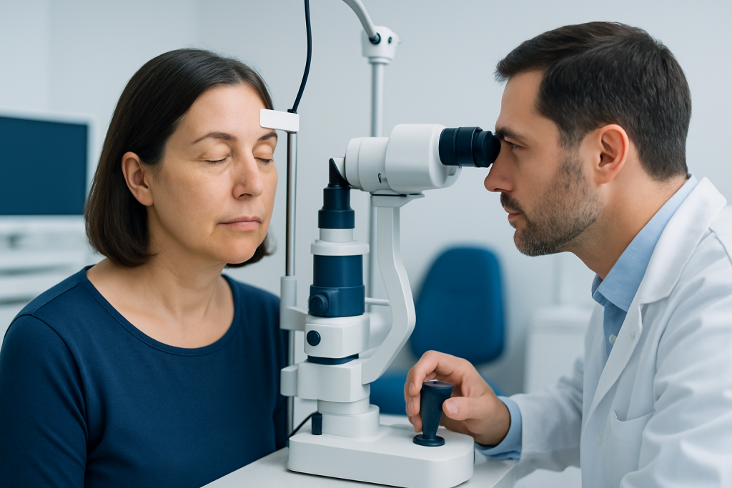
The typical lesion is a yellow, cream-colored, or slightly orange patch on the eyelids. It may begin as a small spot and slowly enlarge over months or years. Some people have one plaque, while others develop several on both sides of the face in a fairly symmetrical pattern.
Xanthelasma is usually soft, smooth, and painless. The skin over it is not inflamed, and the area generally does not bleed or crust. Vision is usually unaffected unless the plaques become unusually large and interfere mechanically with eyelid movement.
People may confuse xanthelasma with other eyelid or skin changes. A doctor may consider conditions such as milia, syringomas, sebaceous hyperplasia, or other cholesterol-related lesions such as high cholesterol-associated skin findings. Any new eyelid growth that changes quickly, ulcerates, or has an irregular appearance should be assessed to rule out other causes.
- Common location: upper and lower eyelids, especially near the inner corners
- Common color: yellow to pale orange
- Common symptoms: usually none apart from visible plaques
- Common concern: cosmetic appearance or worry about cholesterol levels
Causes and risk factors
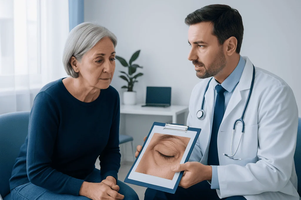
Xanthelasma forms when lipid-laden cells collect within the superficial layers of the skin. In simple terms, fats such as cholesterol accumulate in certain skin cells, creating the characteristic yellow plaque. The exact reason this happens in one person and not another is not always clear, but lipid metabolism plays an important role.
Many people with xanthelasma have abnormal blood lipids, including elevated total cholesterol, LDL cholesterol, or triglycerides. It may also be seen in inherited lipid disorders, obesity, insulin resistance, diabetes, hypothyroidism, and some liver diseases. In other cases, blood lipid levels are normal, suggesting that local skin factors and genetics may also contribute.
Age is a factor, as xanthelasma is more common in middle age and older adulthood, though it can appear earlier. Family history, smoking, high blood pressure, and other cardiovascular risk factors may increase the importance of a broader evaluation. Because xanthelasma may be part of a larger metabolic picture, doctors sometimes use its appearance as a prompt to review heart health, weight, diet, activity level, and screening for cardiovascular diseases.
How doctors diagnose it
Diagnosis is often made during a physical examination. The appearance and location of the plaques are usually characteristic enough for an experienced clinician to identify xanthelasma without a biopsy. The doctor will ask when the lesions appeared, whether they are changing, and whether there is a personal or family history of cholesterol disorders, diabetes, thyroid disease, or early cardiovascular disease.
Because the condition can be associated with metabolic abnormalities, evaluation often includes blood tests. These may include a lipid profile, fasting glucose or HbA1c, and sometimes thyroid or liver function tests. The goal is not only to confirm the skin diagnosis but also to look for risk factors that may benefit from treatment or closer follow-up.
If the diagnosis is uncertain, or if a lesion looks atypical, further assessment may be needed. That can include referral to dermatology, ophthalmology, or another specialist. Rarely, a small tissue sample may be examined if there is concern that the lesion is not xanthelasma. In some cases, an overall health workup may include assessment for diabetes or other conditions that can influence lipid metabolism.
Treatment options
Treatment depends on two separate questions: whether there is an underlying medical issue to address and whether the plaques themselves should be removed. Managing abnormal cholesterol, diabetes, or other metabolic risk factors is important for general health, but it does not always make existing xanthelasma disappear. Still, treating those conditions may help reduce future risk and may lower the chance of recurrence after removal.
If the main concern is appearance, several removal methods may be considered. Options can include careful surgical excision, chemical cauterization with selected agents, laser therapy, or other energy-based treatments. The best choice depends on the size, depth, location, skin type, and whether the plaque is close to important eyelid structures. For selected patients, a specialist may discuss approaches used in eyelid surgery planning when lesions are extensive or when excess eyelid skin is also present.
Each treatment has potential benefits and limitations. Possible issues include recurrence, pigment changes, scarring, or contour changes of the delicate eyelid skin. That is why treatment around the eyes should be performed by clinicians experienced with this area. In some situations, removal may overlap with techniques used in dermatology care or plastic and reconstructive surgery to preserve both safety and appearance.
- Observation may be reasonable if the plaques are small and not bothersome.
- Medical treatment targets underlying lipid or metabolic conditions, not the plaque itself.
- Procedural removal is mainly for cosmetic or functional reasons.
- Recurrence can happen even after successful treatment.
Prevention and self-care
There is no guaranteed way to prevent xanthelasma, especially when genetics play a role. However, improving overall metabolic health is a sensible strategy. A clinician may recommend regular cholesterol screening, blood sugar monitoring when appropriate, and attention to blood pressure, weight, and smoking status.
Heart-healthy lifestyle measures can be helpful whether or not cholesterol is elevated. These include a balanced diet rich in vegetables, fruits, whole grains, legumes, and healthy fats; limiting excess saturated and trans fats; staying physically active; and avoiding tobacco. People already diagnosed with lipid disorders should follow their care plan and take prescribed medicines as directed.
It is also important not to squeeze, scrub, or try to remove xanthelasma at home. The eyelid skin is thin and sensitive, and self-treatment can cause irritation, burns, infection, or scarring. If the appearance is distressing or if the lesions are changing, professional assessment is the safest next step.
When to seek medical care
Medical care is appropriate when a new yellow eyelid lesion appears, especially if the diagnosis is uncertain or if there is a personal or family history of high cholesterol or early heart disease. A doctor can confirm whether the lesion is xanthelasma and decide whether blood tests or further evaluation are needed.
Prompt assessment is also advisable if the lesion grows rapidly, changes color, bleeds, becomes painful, affects eyelid movement, or is associated with visual symptoms. These features are not typical of xanthelasma and may point to another condition that deserves attention.
For people considering removal, consultation with an experienced specialist helps clarify realistic expectations, the chance of recurrence, and the risks of scarring or pigment change. Near the end of the care pathway, some patients may choose treatment at centers such as Acibadem International, where multidisciplinary specialists in JCI-accredited hospitals diagnose and treat eyelid and skin conditions for international patients.
Frequently asked questions
Is xanthelasma dangerous?
Xanthelasma itself is usually not dangerous and is generally a benign skin finding. However, it can sometimes be associated with high cholesterol, diabetes, or other cardiovascular risk factors, so a medical review may be useful.
Can xanthelasma go away on its own?
Xanthelasma usually does not disappear on its own. Some people find that the plaques remain stable for a long time, while others notice gradual enlargement over time.
Does xanthelasma mean a person has high cholesterol?
Not always. Many people with xanthelasma do have abnormal lipid levels, but some have normal cholesterol and triglyceride results, which is why testing is recommended rather than assuming the cause.
What is the best treatment for xanthelasma?
There is no single best treatment for everyone. The most suitable option depends on the lesion's size, depth, location, skin type, cosmetic goals, and whether there are underlying metabolic conditions that also need attention.
Can xanthelasma come back after removal?
Yes, recurrence is possible even after successful treatment. The risk may be higher when lipid abnormalities or other metabolic factors remain present.
Should xanthelasma always be removed?
No. Removal is not always medically necessary if the lesions are clearly diagnosed, painless, and not affecting eyelid function. Many people choose treatment for cosmetic reasons, while others prefer monitoring.
This article is for general information only and is not a substitute for professional medical advice. Please consult a qualified doctor about your individual situation.
Heart care in Turkey — expert evaluation and treatment
JCI-accredited · board-certified surgeons · reply within 24h
Add us as a Preferred Source to see more of our trusted health content across Google Search, AI Overviews and Discover.
Check your numbers in seconds
BMI, calories, due date, blood pressure and 30+ more clinical calculators — free, instant, doctor-reviewed ranges.

More from the Health Library

Jojoba Oil Benefits — Explained by Medical Evidence, Not Myths

Frequency Specific Microcurrent: An Evidence-Based Guide for Patients

Rutabaga — Explained by Medical Evidence, Not Myths

Lime Powder — Explained by Medical Evidence, Not Myths

Balance Diet for Hair Growth: Procedure, Recovery and Results