Xray Fractured Ankle: Preparation, Procedure and Results

An ankle X-ray is quick, painless and uses low-dose radiation to assess suspected fractures. Most examinations include several views of the ankle to assess the bones and alignment of the joint.
Key Takeaways
- An ankle X-ray is quick, painless and uses low-dose radiation to assess suspected fractures.
- Most examinations include several views of the ankle to assess the bones and alignment of the joint.
- A normal X-ray does not always rule out a small, stress-related or very early fracture; repeat imaging or MRI may sometimes be needed.
- Recovery depends on the fracture pattern, stability, treatment and rehabilitation, and often takes several weeks to months.
- Urgent assessment is important when the ankle looks deformed, pain is severe, or the foot is numb, cold or pale.
An xray fractured ankle examination is the standard first imaging test after an ankle injury when a fracture is suspected. It can identify broken bones, their position and joint alignment, helping clinicians decide whether protection in a boot or cast, reduction, surgery or further imaging is needed.
Overview: what does an X-ray for a fractured ankle show?
An xray fractured ankle assessment is usually the first test used when a clinician suspects that one or more ankle bones may be broken. It produces images of the tibia and fibula at the lower leg and the talus at the top of the foot. These images help show whether there is a fracture, where it is located, whether bone pieces have moved out of position and whether the ankle joint remains stable.
Ankle fractures can happen after a twist, fall, sports injury, road traffic injury or direct blow. They range from small, stable breaks that can heal with protection to unstable fractures involving more than one bone or the joint surface. An X-ray is an important starting point, but it is interpreted alongside symptoms, physical examination and the mechanism of injury.
Receiving an X-ray does not automatically mean surgery is needed. Many fractures are managed without an operation. The key question is whether the ankle joint is aligned and likely to remain stable while the bone heals.
Who may need an ankle X-ray?
A clinician may recommend an ankle X-ray when pain, swelling or tenderness follows an injury, particularly when pain is focused over the ankle bones or the person cannot take four steps comfortably. Decision tools such as the Ottawa ankle rules may help healthcare professionals determine when X-ray imaging is appropriate, although clinical judgement remains essential.
Features that can raise concern for a fracture include rapid swelling, bruising, difficulty bearing weight, pain that worsens with movement, tenderness directly over bone, or a visible change in ankle shape. Children, older adults and people with osteoporosis may need particularly careful assessment because their fracture patterns and risks can differ.
Soft-tissue injuries such as sprains can cause similar symptoms. An X-ray does not show ligaments as clearly as it shows bone, but it can reveal signs of joint widening or a small bone fragment that may suggest a significant ligament injury.
Preparation and how the ankle X-ray works
There is usually very little preparation for an ankle X-ray. The person may be asked to remove shoes, socks, jewellery or other objects around the ankle that could interfere with the image. Comfortable clothing that can be moved above the ankle is helpful. It is important to tell the radiographer about possible pregnancy, as imaging staff can use the appropriate precautions and decide whether the examination should be adapted.
X-rays use a small amount of ionising radiation to create an image. Bone absorbs more X-rays than soft tissue, so it appears lighter on the resulting image. The radiation exposure from a standard ankle examination is low, and the test is generally considered safe when clinically indicated.
The examination itself is not painful, although positioning the injured ankle can be uncomfortable. The radiographer will support the leg and aim to avoid unnecessary movement. Pain control and gentle positioning can make the process more manageable.
Step by step: what happens during the procedure?
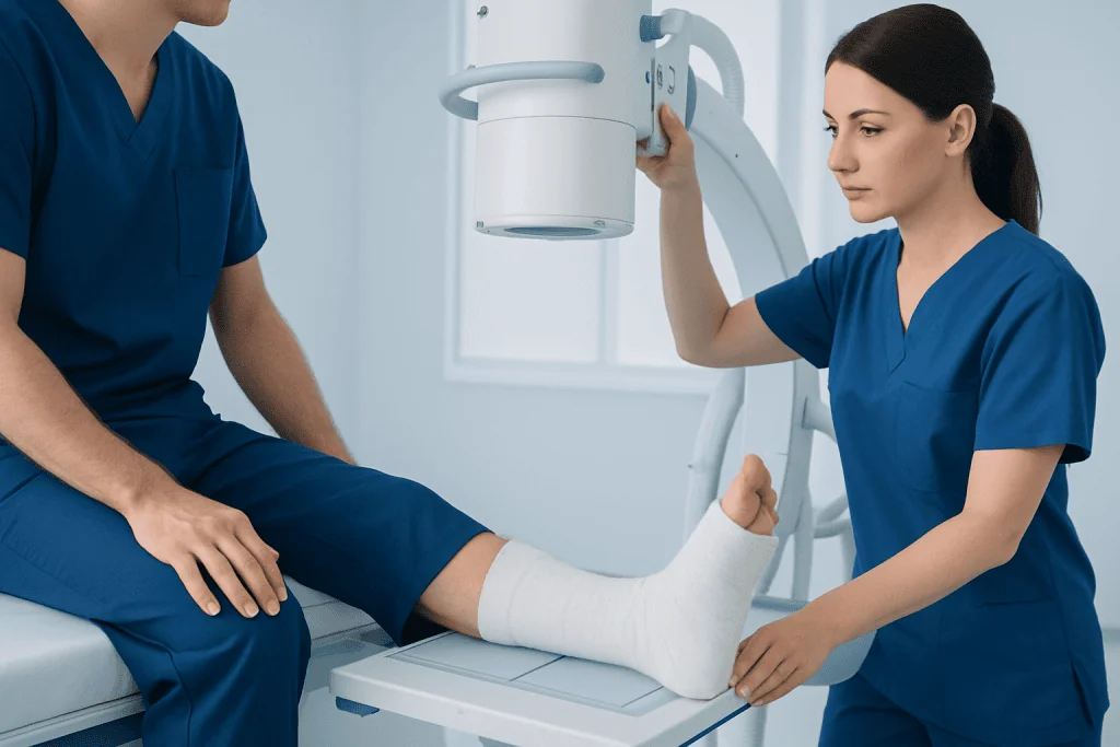
On arrival, a radiographer confirms the person’s identity, checks the area to be examined and asks relevant safety questions. The injured leg is positioned on an X-ray table or chair, depending on comfort and the severity of the injury. In many cases, images are taken without removing a temporary splint if this is necessary to protect the ankle.
Standard imaging generally includes views from the front, side and an angled position. The person needs to keep still for a few seconds while each image is taken. The radiographer may move the X-ray machine around the ankle, but does not enter the radiation beam during exposure.
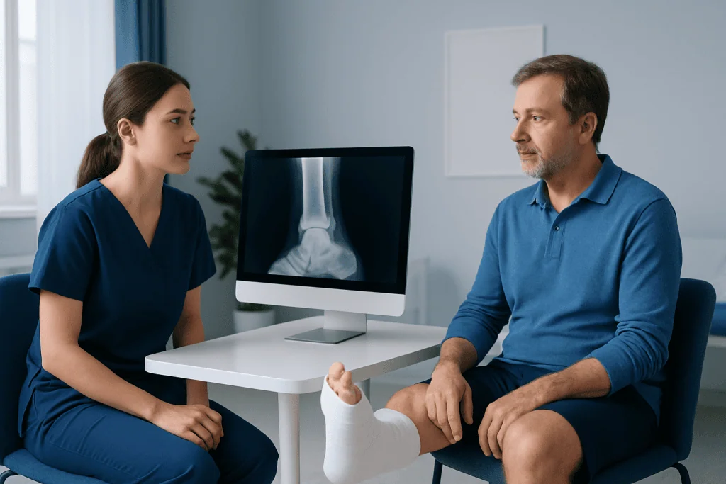
The image-taking portion commonly takes only a few minutes. Allowing time for safe positioning, discussion and review, the overall visit may take longer. A radiologist or treating clinician reviews the images, and the findings are combined with the examination before a treatment plan is discussed.
- Before: remove items around the ankle and report pregnancy or mobility concerns.
- During: remain as still as possible while several images are taken.
- After: normal activities can usually resume unless the clinician advises immobilisation, crutches or urgent treatment.
Understanding the results, benefits and limits
An ankle X-ray may show a fracture of the outer ankle bone (lateral malleolus), inner ankle bone (medial malleolus), back of the tibia (posterior malleolus), or more than one area. The report may describe whether a fracture is displaced, meaning the bone fragments are no longer in their usual alignment. It may also comment on joint space, swelling and other visible changes.
The main benefit is that X-ray imaging is fast, widely available and highly useful for identifying many acute bone fractures and checking alignment. It helps clinicians plan appropriate immobilisation, determine whether an urgent reduction is needed, and decide if orthopaedic review is required.
Not every fracture is visible on the first X-ray. Small fractures, stress fractures and injuries obscured by swelling or overlapping structures can occasionally be missed. If symptoms and examination remain strongly suggestive of a fracture despite normal images, a clinician may arrange repeat X-rays, CT or MRI. CT is particularly useful for defining complex fracture patterns and planning surgery, while MRI can assess hidden fractures and soft-tissue injury.
Treatment and recovery after a confirmed ankle fracture
Treatment depends on the fracture’s location, displacement, stability, skin condition and the person’s overall health and activity needs. Stable fractures may be treated with a boot, cast or splint, along with guidance on weight-bearing and follow-up imaging. Rest, elevation and clinician-approved pain relief can help early symptoms, while keeping pressure off the ankle may be important initially.
If the joint is unstable or bones are displaced, a clinician may need to realign the ankle promptly. Some fractures require an operation to restore joint alignment using plates, screws or other fixation devices. This is usually followed by a period of protection and a structured rehabilitation plan to restore movement, strength, balance and confidence with walking.
Recovery is individual. Bone healing often takes around six weeks or longer, but swelling, stiffness and reduced endurance can continue for several months. Follow-up appointments are important because the clinician may change weight-bearing instructions as healing progresses. Acibadem International’s multidisciplinary specialists and JCI-accredited hospitals diagnose and treat ankle injuries for international patients.
When to seek medical care
Medical assessment should be sought promptly after an ankle injury if there is significant pain, inability to bear weight, marked swelling or bruising, or tenderness over the ankle bones. An X-ray may be needed to distinguish a fracture from a sprain and to identify injuries that need urgent stabilisation.
Emergency care is appropriate if the ankle is visibly deformed, bone is visible, there is an open wound near the injury, or the foot becomes numb, pale, blue or cold. These signs can indicate a serious fracture, circulation problem or nerve involvement and should not be managed at home.
Even after a diagnosis, medical advice is needed if pain or swelling suddenly worsens, a cast or boot feels excessively tight, toes change colour, fever develops, or new numbness occurs. Follow-up should not be delayed when advised by the treating team.
How long does it take to recover from a broken ankle bone?
Many broken ankle bones take at least six weeks to heal, although the full recovery period often extends beyond this. A simple, stable fracture may allow a gradual return to everyday activities sooner than a complex fracture, but swelling and stiffness can remain noticeable for several months.
Recovery is affected by the number of bones involved, whether the joint was displaced, whether surgery was needed, smoking status, diabetes, circulation, nutrition and adherence to weight-bearing instructions. Rehabilitation exercises are often introduced gradually after the clinician confirms that movement and loading are safe.
Returning to running, jumping, demanding work or sports should be guided by the treating clinician or physiotherapist. It is usually based on healing, strength, range of motion, balance and ability to perform activity without significant pain or swelling, rather than a fixed date alone.
How painful is an ankle fracture?
An ankle fracture can be quite painful, especially immediately after injury. People often describe sharp pain, throbbing, swelling and difficulty putting weight through the foot. However, pain levels vary widely, and some fractures can feel similar to a severe sprain.
Pain alone cannot confirm how serious an injury is. Some people can still walk with a fracture, while others cannot bear weight with a sprain. An examination and, when indicated, an X-ray provide a more reliable assessment.
Until medical advice is obtained, it is sensible to avoid forcing the ankle to move or bear weight. Protecting the area, elevating it if comfortable and using wrapped cold packs for short periods may reduce swelling. Pain medicines should be chosen with advice from a pharmacist or clinician, particularly for people with kidney disease, stomach ulcers, blood-thinning medication or other health conditions.
How long does an ankle fracture operation take?
The length of ankle fracture surgery varies according to the fracture pattern, swelling, number of bones involved and whether the joint needs complex reconstruction. A straightforward fixation procedure may take around one to two hours, while more complicated injuries can take longer. Preparation before anaesthesia and recovery after the operation add additional time to the hospital visit.
In some injuries, surgery is performed once swelling has reduced enough for the skin to be safer for an incision. In others, urgent realignment or temporary stabilisation may be needed sooner. The orthopaedic surgeon explains the recommended timing, the type of anaesthesia and the expected hospital plan for each individual.
After surgery, patients commonly need a splint, cast or boot and specific instructions about wound care, weight-bearing and follow-up. The aim is to restore stable joint alignment so that the ankle has the best opportunity to heal and function well.
What not to do after an ankle fracture?
After an ankle fracture, a person should not walk on the injured leg unless their clinician has clearly said weight-bearing is safe. Putting weight through an unstable fracture can move bone fragments, worsen pain or affect healing. Crutches, a walker or other mobility aids may be advised.
It is also important not to remove or alter a splint, cast or boot without clinical guidance, and not to insert objects inside a cast to scratch the skin. Avoid smoking and nicotine products, as they can impair bone healing. Alcohol should also be limited, especially when taking pain medicines or recovering from anaesthesia.
People should not return to sport, driving or physically demanding work before they can do so safely and have been cleared by their healthcare professional. Skipping follow-up appointments or rehabilitation can delay recognition of healing problems and may prolong stiffness or weakness.
Frequently asked questions
Is an X-ray enough to diagnose a fractured ankle?
An X-ray is usually the first and most useful test for suspected ankle fractures. It can show many breaks and assess joint alignment, but some small or hidden fractures may not appear immediately. If symptoms remain concerning, a clinician may recommend repeat X-rays, CT or MRI.
Can a fractured ankle be mistaken for a sprain?
Yes. Both injuries may cause pain, swelling, bruising and difficulty walking. Because symptoms overlap, an examination and, when appropriate, an X-ray are important after a significant ankle injury.
Do I need to fast before an ankle X-ray?
No fasting is required for a routine ankle X-ray. A person can usually eat, drink and take regular medicines as usual unless another test or procedure is planned.
Can I walk on an ankle fracture?
Some people can take steps with certain stable fractures, but this does not mean walking is safe. Weight-bearing advice depends on the exact fracture and treatment plan. Until assessed, it is safer to avoid putting weight on an ankle that may be broken.
When will I get my ankle X-ray results?
In emergency or urgent care settings, a treating clinician may review the images the same day. A formal radiology report may also be issued, depending on local practice. The clinician should explain what the findings mean and what follow-up is needed.
Will an ankle fracture always need a cast?
No. Some fractures are treated in a removable walking boot or splint rather than a traditional cast. The choice depends on fracture stability, swelling, healing progress and the clinician’s assessment.
References
- American Academy of Orthopaedic Surgeons
- American College of Radiology
- National Institute for Health and Care Excellence
- Radiological Society of North America
- OrthoInfo
This article is for general information only and is not a substitute for professional medical advice. Please consult a qualified doctor about your individual situation.
Joint and spine care in Turkey — expert assessment & treatment
JCI-accredited · board-certified surgeons · reply within 24h
Add us as a Preferred Source to see more of our trusted health content across Google Search, AI Overviews and Discover.